Demyelinating diseases of the CNS are acquired conditions characterized by preferential damage to myelin, with relative preservation of axons. The clinical deficits are due to the effect of myelin loss on the transmission of electrical impulses along axons. The natural history of demyelinating diseases is determined, in part, by the limited capacity of the CNS to regenerate normal myelin and by the degree of secondary damage to axons that occurs as the disease runs its course.
Several disease processes can cause loss of myelin. These include destruction of myelin by immunological reactions, as in multiple sclerosis, and by infections. In progressive multifocal leukoencephalopathy, JC virus infection of oligodendrocytes results in loss of myelin (described above). In addition, inherited disorders may affect synthesis or turnover of myelin components; these are termed leukodystrophies and are discussed with metabolic disorders.
Multiple sclerosis (MS) is an autoimmune demyelinating disorder characterized by distinct episodes of neurologic deficits, separated in time, attributable to white matter lesions that are separated in space. It is the most common of the demyelinating disorders, having a prevalence of approximately 1 per 1000 persons in most of the United States and Europe. The disease may become clinically apparent at any age, although onset in childhood or after age 50 years is relatively rare. Women are affected twice as often as are men. In most individuals with MS, the clinical course takes the form of relapsing and remitting episodes of variable duration (weeks to months to years) marked by neurologic defects, followed by gradual, partial recovery of neurologic function. The frequency of relapses tends to decrease during the course of time, but there is a steady neurologic deterioration in most affected individuals.
The lesions of MS are caused by an immune response that is directed against the components of the myelin sheath.27,28 As in other autoimmune disorders, the pathogenesis of this disease involves both genetic and environmental factors (Chapter 6). The incidence of MS is 15-fold higher when the disease is present in a first-degree relative and roughly 150-fold higher with an affected monozygotic twin. Genetic linkage of MS susceptibility to the DR2 extended haplotype of the major histocompatibility complex is also well established. A recent genome-wide screen supported this association and identified additional associations with single-nucleotide polymorphisms in IL-2 and IL-7 receptor genes.29 The current thinking is that these cytokine receptor polymorphisms may influence the balance between pathogenic effector T cells and protective regulatory T cells. These genetic associations point to the importance of the immune system in the susceptibility to MS.
Given the prominence of chronic inflammatory cells within and around MS plaques as well as this genetic validation, immune mechanisms that underlie the destruction of myelin are the focus of much investigation. The available evidence indicates that the disease is initiated by CD4+ TH1 and TH17 T cells that react against self myelin antigens and secrete cytokines. TH1 cells secrete IFNγ, which activates macrophages, and TH17 cells promote the recruitment of leukocytes (Chapter 6). The demyelination is caused by these activated leukocytes and their injurious products. The infiltrate in plaques and surrounding regions of the brain consists of T cells (mainly CD4+, some CD8+) and macrophages. How the autoimmune reaction is initiated is not understood; a role of viral infection (e.g., EBV) in activating self-reactive T cells has been proposed but remains controversial.
Experimental autoimmune encephalomyelitis is an animal model of MS in which demyelination and inflammation occur after immunization of animals with myelin proteins.30 Many of our concepts of MS pathogenesis have been derived from studies in this model. The experimental disorder can be passively transferred to other animals with TH1 and TH17 cells that recognize myelin antigens.
Based on the growing understanding of the pathogenesis of MS, therapies are being developed that modulate or inhibit T cell responses and block the recruitment of T cells into the brain. A potential contribution of humoral immunity has also been suspected for a long time, based on the early observation of oligoclonal bands of immunoglobulin in CSF.31 The demonstration that B-cell depletion can decrease the incidence of demyelinating lesions lends support to this idea.32
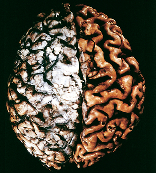
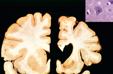
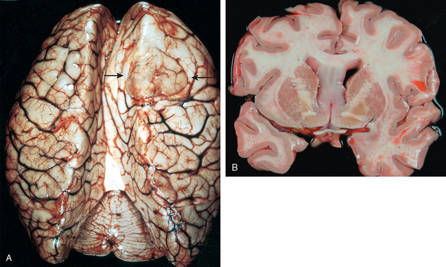
Morphology. MS is a white matter disease that is best appreciated in sections of the brain and spinal cord. Lesions appear as multiple, well-circumscribed, somewhat depressed, glassy, graytan, irregularly shaped plaques (Fig. 28-33). In the fresh state these are firmer than the surrounding white matter (sclerosis). Plaques can be found throughout the white matter and also extend into gray matter, since these have myelinated fibers running through them. The size of lesions varies considerably, from small foci that are only recognizable microscopically to confluent plaques that involve large portions of the centrum semiovale. The lesions often have sharply defined borders (Fig. 28-34). Plaques commonly occur adjacent to the lateral ventricles. They are also frequent in the optic nerves and chiasm, brainstem, ascending and descending fiber tracts, cerebellum, and spinal cord.
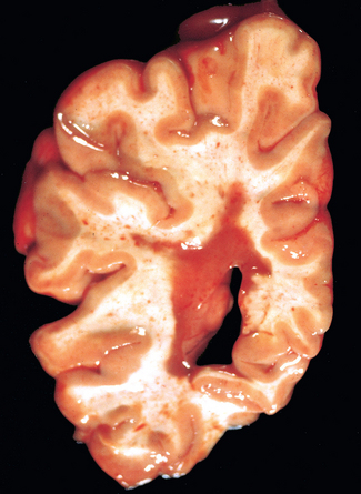
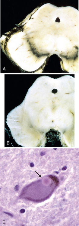
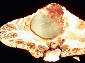
FIGURE 28-33 Multiple sclerosis. Section of fresh brain showing brown plaque around occipital horn of the lateral ventricle.
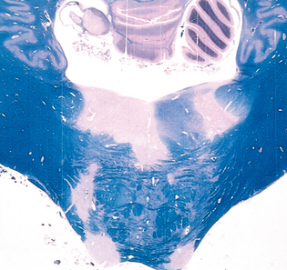
FIGURE 28-34 Multiple sclerosis (MS). Unstained regions of demyelination (MS plaques) around the fourth ventricle (Luxol fast blue PAS stain for myelin).
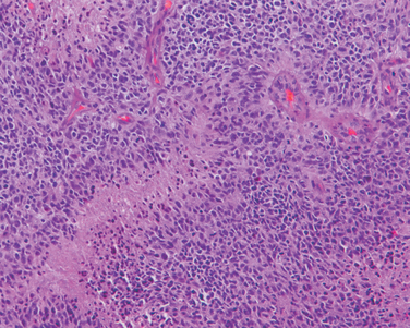
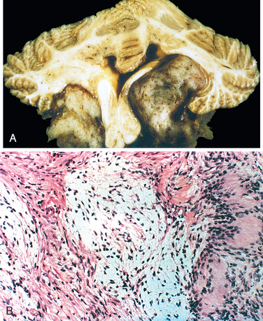
Microscopically, in an active plaque there is evidence of ongoing myelin breakdown with abundantmacrophages containing lipid-rich, PAS-positive debris. Inflammatory cells, including both lymphocytes and monocytes, are present, mostly as perivascular cuffs, especially at the outer edge of the lesion (Fig. 28-35A). Active lesions are often centered on small veins. Within a plaque there is relative preservation of axons (Fig. 28-35B) and depletion of oligodendrocytes. In time, astrocytes undergo reactive changes. As lesions become quiescent, the inflammatory cells slowly disappear. Within inactive plaques, little to no myelin is found, and there is a reduction in the number of oligodendrocyte nuclei; instead, astrocytic proliferation and gliosis are prominent. Axons in old gliotic plaques show severe depletion of myelin and are also greatly diminished in number.
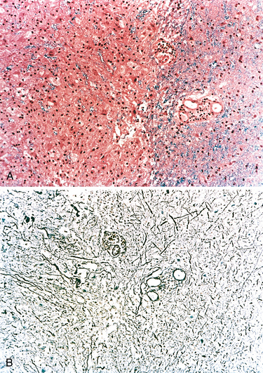
FIGURE 28-35 Multiple sclerosis. A, Myelin-stained section shows the sharp edge of a demyelinated plaque and perivascular lymphocytic cuffs. B, The same lesion stained for axons shows relative preservation.
Active plaques can also be grouped into four basic patterns: those that are sharply demarcated and centered on blood vessels, either with (pattern I) or without (pattern II) deposition of immunoglobulin and complement, and those that are less well demarcated and are not centered on vessels (patterns III and IV). These latter two are distinguished by the distribution of oligodendrocyte apoptosis (III, widespread; IV, central only). It has been observed that only one pair of patterns (I/II or III/IV) may be present in a given individual, suggesting that these may reflect distinct mechanisms rather than different stages of lesion.
In some MS plaques (shadow plaques) the border between normal and affected white matter is not sharply circumscribed. In this type of lesion some abnormally thinned-out myelin sheaths can be demonstrated, especially at the outer edges. This phenomenon is most commonly interpreted as evidence of partial and incomplete remyelination by surviving oligodendrocytes. Abnormally myelinated fibers have also been observed at the edges of typical plaques. Although these histologic findings suggest a limited potential for remyelination in the CNS, the remaining axons within most MS plaques remain unmyelinated; studies aimed at promoting remyelination are an important focus of research.33
Although MS lesions can occur anywhere in the CNS and consequently may induce a wide range of clinical manifestations, certain patterns of neurologic symptoms and signs are commonly observed. Unilateral visual impairment, due to involvement of the optic nerve (optic neuritis, retrobulbar neuritis), is a frequent initial manifestation of MS. However, only some affected individuals (10% to 50%, depending on the population studied) with optic neuritis go on to develop MS. Involvement of the brainstem produces cranial nerve signs, ataxia, nystagmus, and internuclear ophthalmoplegia from interruption of the fibers of the medial longitudinal fasciculus. Spinal cord lesions give rise to motor and sensory impairment of trunk and limbs, spasticity, and difficulties with the voluntary control of bladder function. Examination of the CSF in individuals with MS shows a mildly elevated protein level, and in one third of cases, there is moderate pleocytosis. IgG levels in the CSF are increased and oligoclonal IgG bands are usually observed on immunoelectrophoresis; these are indicative of the presence of a small number of activated B cell clones, postulated to be self-reactive, in the CNS. Radiologic studies using magnetic resonance imaging, typically based on identifying gadolinium-enhancing lesions, have taken on a prominent role in assessing disease progression; these studies, when correlated with autopsy studies as well as clinical findings, have indicated that some plaques may be clinically silent even in otherwise symptomatic patients.
The development of synchronous (or near synchronous) bilateral optic neuritis and spinal cord demyelination is referred to as neuromyelitis optica or Devic disease. White cells are common in the CSF, often including neutrophils. Within the damaged areas of white matter, there is typically necrosis, an inflammatory infiltrate including neutrophils, and vascular deposition of immunoglobulin and complement. These lesions have been suggested to be mediated by humoral immune mechanisms.34 Many affected individuals show antibodies to aquaporins, which are in part responsible for maintenance of astrocytic foot process and thus the integrity of the blood-brain barrier.35,36
Acute disseminated encephalomyelitis (ADEM, perivenous encephalomyelitis) is a diffuse, monophasic demyelinating disease that follows either a viral infection or, rarely, a viral immunization. Symptoms typically develop a week or two after the antecedent infection and include headache, lethargy, and coma rather than focal findings, as seen in MS. The clinical course is rapid, and as many as 20% of those affected die; the remaining patients recover completely.
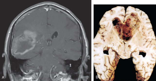
Acute necrotizing hemorrhagic encephalomyelitis (ANHE, acute hemorrhagic leukoencephalitis of Weston Hurst) is a fulminant syndrome of CNS demyelination, typically affecting young adults and children. The illness is almost invariably preceded by a recent episode of upper respiratory infection, most often of unknown cause. The disease is fatal in many patients, with significant deficits present in most survivors.
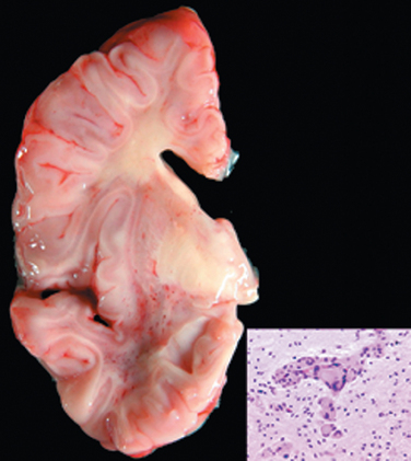
Morphology. In ADEM, macroscopic examination of the brain shows only grayish discoloration around white-matter vessels. On microscopic examination, myelin loss with relative preservation of axons can be found throughout the white matter. In the early stages, polymorphonuclear leukocytes can be found within the lesions; later, mononuclear infiltrates predominate. The breakdown of myelin is associated with the accumulation of lipid-laden macrophages. In contrast with MS, all lesions appear similar, consistent with the clinically monophasic nature of the disorder.
ANHE shows histologic similarities with ADEM, including a perivenular distribution of demyelination and widespread dissemination throughout the CNS (sometimes with extensive confluence of lesions). However, the lesions are much more severe than those of ADEM and include destruction of small blood vessels, disseminated necrosis of white and gray matter with acute hemorrhage, fibrin deposition, and abundant neutrophils. Scattered lymphocytes are seen in foci of demyelination.
The lesions of ADEM are similar to those induced by immunization of animals with myelin components or with early rabies vaccines that had been prepared from brains of infected animals. This has suggested that ADEM may represent an acute autoimmune reaction to myelin and that ANHE may represent a hyperacute variant, although no inciting antigens have been identified.
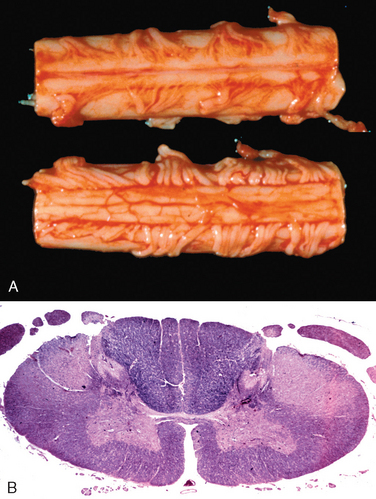
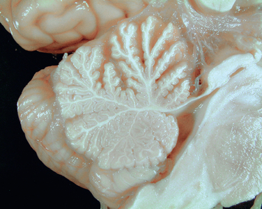
Central pontine myelinolysis is characterized by loss of myelin (with relative preservation of axons and neuronal cell bodies) in a roughly symmetric pattern involving the basis pontis and portions of the pontine tegmentum but sparing the periventricular and subpial regions.37 Lesions may be found more rostrally; it is extremely rare for the process to extend below the pontomedullary junction. Extra-pontine lesions occur in the supratentorial compartment, with similar appearance and apparent etiology. The condition is most commonly associated with rapid correction of hyponatremia, although it can be associated with other severe electrolyte or osmolar imbalance, as well as orthotopic liver transplantation. The clinical presentation of central pontine myelinolysis is that of a rapidly evolving quadriplegia; radiologic imaging studies localize the lesion to the basis pontis. Morphologically there is myelin loss without evidence of inflammation; neurons and axons are well preserved. Again, because of the monophasic nature of the disease all lesions appear to be at the same stage of myelin loss and reaction.
These are diseases of gray matter characterized by the progressive loss of neurons with associated secondary changes in white matter tracts. The pattern of neuronal loss is selective, affecting one or more groups of neurons while leaving others, sometimes immediately adjacent, intact. As genetic and molecular studies of these diseases have progressed certain shared features have emerged. A common theme among the neurodegenerative disorders is the presence of protein aggregates that are resistant to degradation through the ubiquitin-proteasome system. These aggregates are recognized histologically as inclusions, which often form the diagnostic hallmarks of these different diseases. The basis for aggregation varies across diseases. It may be directly related to an intrinsic feature of a mutated protein (e.g., expanded polyglutamine repeats in Huntington disease), a feature of a peptide derived from a larger precursor protein (e.g., Aβ in Alzheimer disease), or an unexplained alteration of a normal cellular protein (e.g., α-synuclein in sporadic Parkinson disease).
Degenerative diseases differ in terms of the distribution of disease burden and in the specific neuropathologic findings (e.g., tangles, plaques, Lewy bodies). They can be grouped using two approaches:
The discussion that follows is primarily based on the first approach (diseases of cortex, basal ganglia, etc.) with a few exceptions (all of the tau diseases and the synuclein diseases are considered together).
The major cortical degenerative disease is Alzheimer disease, and its principal clinical manifestation is dementia, that is, progressive loss of cognitive function independent of the state of attention. There are many other causes of dementia, including the various forms of frontotemporal dementia, vascular disease, dementia with Lewy bodies (considered later in the context of Parkinson disease, the other Lewy body disorder), CJD, and neurosyphilis (both considered earlier). These diseases also involve subcortical structures, but many of the clinical symptoms are related to the changes in the cerebral cortex. Regardless of etiology, dementia is not part of normal aging and always represents a pathologic process.
Alzheimer disease (AD) is the most common cause of dementia in the elderly. The disease usually becomes clinically apparent as insidious impairment of higher intellectual function, with alterations in mood and behavior. Later, progressive disorientation, memory loss, and aphasia become manifest, indicating severe cortical dysfunction. Eventually, in 5 to 10 years, the affected individual becomes profoundly disabled, mute, and immobile. Patients rarely become symptomatic before 50 years of age, but the incidence of the disease rises with age, and the prevalence roughly doubles every 5 years, starting from a level of 1% for the 60- to 64-year-old population and reaching 40% or more for the 85- to 89-year-old cohort. This progressive increase in the incidence of the disease with age has given rise to major medical, social, and economic problems in countries with a growing number of elderly individuals. Most cases are sporadic, and although 5% to 10% are familial, the study of such familial cases has provided important insight into the pathogenesis of the more common sporadic form. While pathologic examination of brain tissue remains necessary for the definitive diagnosis of Alzheimer disease, the combination of clinical assessment and modern radiologic methods allows accurate diagnosis in 80% to 90% of cases.
Morphology. Grossly, the brain shows a variable degree of cortical atrophy marked by widening of the cerebral sulci that is most pronounced in the frontal, temporal, and parietal lobes (Fig. 28-36). With significant atrophy, there is compensatory ventricular enlargement (hydrocephalus ex vacuo) secondary to loss of parenchyma and reduced brain volume. Structures of the medial temporal lobe, including hippocampus, entorhinal cortex and amygdala, are involved early in the course and are usually severely atrophied in the later stages. The major microscopic abnormalities of AD, which form the basis of the histologic diagnosis, are neuritic (senile) plaques and neurofibrillary tangles. There is progressive and eventually severe neuronal loss and reactive gliosis in the same regions that bear the burden of plaques and tangles.
FIGURE 28-36 Alzheimer disease with cortical atrophy most evident on the right, where meninges have been removed.
(Courtesy of the late Dr. E.P. Richardson, Jr., Massachusetts General Hospital, Boston, MA.)
Neuritic plaques are focal, spherical collections of dilated, tortuous, neuritic processes (dystrophic neurites) often around a central amyloid core, which may be surrounded by clear halo (Fig. 28-37A). Neuritic plaques range in size from 20 to 200 μm in diameter; microglial cells and reactive astrocytes are present at their periphery. Plaques are found in the hippocampus, amygdala, and neocortex, although there is usually relative sparing of primary motor and sensory cortices (this also applies to neurofibrillary tangles). The amyloid core, which can be stained by Congo Red, contains several abnormal proteins. The dominant component of the amyloid plaque core is Aβ, a peptide derived through specific processing events from a larger molecule, amyloid precursor protein (APP) (Figs. 28-37 and 28-38). The two dominant species of Aβ, called Aβ40 and Aβ42, share an Nterminus and differ in length by two amino acids at the C-terminus. Other proteins are present in plaques in lesser abundance, including components of the complement cascade, pro-inflammatory cytokines, α1-antichymotrypsin, and apolipoproteins. In some cases, there is deposition of Aβ peptides with staining characteristics of amyloid in the absence of the surrounding neuritic reaction. These lesions, termed diffuse plaques, are found in superficial portions of cerebral cortex as well as in basal ganglia and cerebellar cortex. Diffuse plaques appear to represent an early stage of plaque development. This conclusion is based primarily on studies of brains from individuals with trisomy 21. Recall that in patients with trisomy 21 (Down syndrome), early onset of Alzheimer disease is common (Chapter 5). In some brain regions (cerebellar cortex and striatum) these diffuse plaques represent a major manifestation of the disease, with other clear-cut findings of Alzheimer disease, or in isolation. While neuritic plaques contain both Aβ40 and Aβ42, diffuse plaques are predominantly made up of Aβ42.
FIGURE 28-37 Alzheimer disease. A, Plaques with dystrophic neurites surrounding amyloid cores are visible (arrows). B, Plaque core and surrounding neuropil are immunoreactive for Aβ. C, Neurofibrillary tangle is present within one neuron, and several extracellular tangles are also present (arrows). D, Silver stain showing a neurofibrillary tangle within the neuronal cytoplasm. E, Tangle (upper left) and neurites around a plaque (lower right) contain tau, demonstrated by immunohistochemistry.
FIGURE 28-38 Mechanisms of processing of amyloid precursor protein (APP). APP can be processed by two pathways; sequential cleavage by β-secretase and γ-secretase is the pathway that results in the generation of Aβ and the formation of amyloid fibrils.
Neurofibrillary tangles are bundles of filaments in the cytoplasm of the neurons that displace or encircle the nucleus. In pyramidal neurons, they often have an elongated “flame” shape; in rounder cells, the basket weave of fibers around the nucleus takes on a rounded contour (“globose” tangles). Neurofibrillary tangles are visible as basophilic fibrillary structures with H&E staining (Fig. 28-37C) but are dramatically demonstrated by silver (Bielschowsky) staining (Fig. 28-37D). They are commonly found in cortical neurons, especially in the entorhinal cortex, as well as in other sites such as pyramidal cells of the hippocampus, the amygdala, the basal forebrain, and the raphe nuclei. Neurofibrillary tangles are insoluble and apparently resistant to clearance in vivo, thus remaining visible in tissue sections as “ghost” or “tombstone” tangles long after the death of the parent neuron. Ultrastructurally, neurofibrillary tangles are composed predominantly of paired helical filaments along with some straight filaments that appear to have a comparable composition. A major component of paired helical filaments is abnormally hyperphosphorylated forms of the protein tau, an axonal microtubule-associated protein that enhances microtubule assembly (Fig. 28-37E). Other components include MAP2 (another microtubule-associated protein) and ubiquitin. Paired helical filaments are also found in the dystrophic neurites that form the outer portions of neuritic plaques and in axons coursing through the affected gray matter as neuropil threads. Tangles are not specific to AD, being found in other diseases as well.
In addition to the diagnostic features of plaques and tangles, several other pathologic findings are seen in the setting of AD. Cerebral amyloid angiopathy (CAA) is an almost invariable accompaniment of Alzheimer disease; however, it can also be found in brains of individuals without AD (see Fig. 28-18B). Vascular amyloid is predominantly Aβ40, as is also the case when CAA occurs without AD. Granulovacuolar degeneration is the formation of small (∼5 μm in diameter), clear intraneuronal cytoplasmic vacuoles, each of which contains an argyrophilic granule. While it occurs with normal aging, it is most commonly found in great abundance in hippocampus and olfactory bulb in AD. Hirano bodies, found especially in AD, are elongated, glassy, eosinophilic bodies consisting of paracrystalline arrays of beaded filaments, with actin as their major component. They are found most commonly within hippocampal pyramidal cells.
Since both plaques and tangles may be present in low abundance in nondemented individuals, the diagnosis of Alzheimer disease is based on a combination of clinical and pathologic features. The progression of changes is fairly constant. Pathologic changes (specifically plaques, tangles, and the associated neuronal loss and glial reaction) are evident earliest in the entorhinal cortex, then spread through the hippocampal formation and isocortex, and then extend into the neocortex. Plaques are assessed semiquantitatively (absent, sparse, moderate, abundant) in each cortical area, while tangles are assessed based on how widespread they are in the brain.38,39 These assessments are combined in the current NIA-Reagan criteria to provide an estimate of the likelihood that AD pathology caused a particular patient’s dementia.40,41
The fundamental abnormality in AD is the deposition of Aβ peptides, which are derived through processing of APP (Fig. 28-38). APP is a cell surface protein with a single transmembrane domain that may function as a receptor, although ligands have remained elusive. The Aβ portion of the protein extends from the extracellular region into the transmembrane domain. Processing of APP begins with cleavage in the extracellular domain, followed by an intramembranous cleavage. There are two potential pathways, determined by the type of initial proteolytic event. If the first cut occurs at the α-secretase site within the Aβ sequence, then Aβ is not generated (the nonamyloidogenic pathway). This mostly occurs at the cell surface, since the various enzymes with α-secretase activity are involved in the shedding of surface proteins. Surface APP can also be endocytosed and may undergo cleavage by β-secretase, which cuts at the N-terminal region of the Aβ sequence (the amyloidogenic pathway). Following cleavage of APP at either of these sites, the γ-secretase complex performs an intramembranous cleavage. When paired with a first cut by α-secretase, it will produce a soluble fragment, but when paired with β-secretase cleavage, it generates Aβ. The variation in peptide length (Aβ40 vs Aβ42) arises from alterations in the exact location of the γ-secretase cleavage. The γ-secretase complex—containing presenilin, nicastrin, pen-2, and aph-1—is also responsible for processing of Notch, a cell fate-determining molecule, as well as many other membrane proteins.42 Once generated, Aβ is highly prone to aggregation—first into small oligomers (which may be the toxic form responsible for neuronal dysfunction), and eventually into large aggregates and fibrils.
Familial forms of AD have provided support for the central role of Aβ generation as a critical step for at least initiation of AD pathogenesis. The gene encoding APP, on chromosome 21, lies in the Down syndrome region; AD pathology is an eventual feature of the cognitive impairment of these individuals. Histologic findings appear in the second and third decades followed by neurologic decline about 20 years later. A similar gene dosage effect is produced by localized chromosome 21 duplications that span the APP locus in some patients with familial AD.44 Point mutations in APP are another cause of familial AD. Some mutations lie near the β-secretase and γ-secretase cleavage sites, and others sit in the Aβ sequence and increase its propensity to aggregate. The two loci identified as causes of the majority of early-onset familial AD encode the two presenilins (PS1 on chromosome 14 and PS2 on chromosome 1). These mutations lead to a gain of function, such that the γ-secretase complex generates increased amounts of Aβ, particularly Aβ42. Thus, the genetic evidence strongly supports the notion that the underlying pathogenetic event in AD is the accumulation of Aβ.
The Aβ peptides readily aggregate, and can be directly neurotoxic. There are various lines of evidence indicating that the small aggregates of Aβ can result in synaptic dysfunction, such as blocking of long-term potentiation and changes in other membrane properties.4 While aggregates are difficult to degrade, monomeric Aβ can be degraded by a variety of proteases. Both small aggregates and larger deposits elicit an inflammatory response from microglia and astrocytes. This response probably assists in the clearance of the aggregated peptide, but may also stimulate the secretion of mediators that cause damage.43 Additional consequences of the activation of these inflammatory cascades may include alterations in tau phophosrylation, along with oxidative injury to the neurons.
The genetic locus on chromosome 19 that encodes apolipoprotein E (ApoE) has a strong influence on the risk of developing AD. Three alleles exist (ε2, ε3, and ε4) based on two amino acid polymorphisms. The dosage of the ε4 allele increases the risk of AD and lowers the age of onset of the disease, such that individuals with the ε4 allele are over-represented in populations of patients with AD. This ApoE isoform promotes Aβ generation and deposition, although the mechanisms have not been established. Overall, this locus has been estimated to convey about a quarter of the risk for development of sporadic AD. It is likely that other risk factor alleles will have much smaller population effects.45 The newly evolving approach of genome-wide association screening may help locate these loci with weaker effects.46
Because neurofibrillary tangles contain the tau protein, there has been much interest in the role of this protein in AD. Tau is a microtubule-associated protein present in axons in association with the microtubular network. With the development of tangles in AD, it shifts to a somatic-dendritic distribution, becomes hyperphosphorylated, and loses the ability to bind to microtubules. It is, however, thought that the primary abnormality in AD is in Aβ and not in tau, because mutations affecting Aβ lead to the formation of tangles and AD but mutations in the gene encoding tau, MAPT, cause one of the frontotemporal demential (see below) but neither Aβ deposition nor AD. The mechanism of tangle injury to neurons remains poorly understood.
While there remains disagreement regarding the best correlate of dementia in individuals with AD, it is clear that the presence of a large burden of plaques and tangles is highly associated with severe cognitive dysfunction. The number of neurofibrillary tangles correlates better with the degree of dementia than does the number of neuritic plaques. Biochemical markers that have been correlated with the degree of dementia include loss of choline acetyltransferase, synaptophysin immunoreactivity, and amyloid burden.
The progression of AD is slow but relentless, with a symptomatic course often running more than 10 years. Initial symptoms are forgetfulness and other memory disturbances; with progression of the disease other symptoms emerge, including language deficits, loss of mathematical skills, and loss of learned motor skills. In the final stages of AD, affected individuals may become incontinent, mute, and unable to walk. Intercurrent disease, often pneumonia, is usually the terminal event in these individuals. Discovery of biomarkers for AD is an area of continuing interest; the amyloid-binding positron emission tomography imaging agent PiB is beginning to be used for this purpose.47,48
Frontotemporal dementias (FTDs) are a group of disorders that were first classified together because they shared clinical features (progressive deterioration of language and changes in personality) corresponding to degeneration and atrophy of temporal and frontal lobes.49 These entities have recently been better understood through a combination of immunohistochemical and biochemical, and genetic approaches. Several of the disorders to be considered share the accumulation of tau-containing deposits as their characteristic finding, giving rise to the term tauopathy.
This is a genetically determined disorder in which the clinical syndrome of a FTD is often accompanied by parkinsonian symptoms.
The study of families with FTD led to the recognition that in some, but not all, pedigrees there are mutations in the MAPT gene encoding tau. The mutations fall into several broad categories: coding-region mutations and intronic mutations. The tau protein has six splice forms. When exon 10 is present, the protein contains four microtubule-binding domains (called 4R), and in its absence there are three such domains (3R). Some of the intronic mutations influence the inclusion of this exon and thus determine the form of the protein that is produced. The ratio of the 4R to 3R form varies in different diseases but the basis of this effect is unknown and both forms can produce tangles. Coding-region mutations seem to have several different consequences, including alterations in the interaction of tau with microtubules and in the intrinsic tendency of tau to aggregate.
Morphology. There is evidence of atrophy of frontal and temporal lobes in various combinations and to various degrees. The pattern of atrophy can often be predicted in part by the clinical symptomatology. The atrophic regions of cortex are marked by neuronal loss, gliosis, and the presence of tau-containing neurofibrillary tangles. These tangles may contain either 4R tau or a mixture of 3R and 4R tau. Nigral degeneration may also occur. Inclusions can also be found in glial cells in some forms of the disease.
Pick disease (lobar atrophy) is a rare, distinct, progressive dementia characterized clinically by early onset of behavioral changes together with alterations in personality (frontal lobe signs) and language disturbances (temporal lobe signs). While most cases of Pick disease are sporadic, there have been some familial forms identified and linked to mutated tau protein.
Morphology. The brain invariably shows a pronounced, frequently asymmetric, atrophy of the frontal and temporal lobes with conspicuous sparing of the posterior two thirds of the superior temporal gyrus and only rare involvement of either the parietal or occipital lobe. The atrophy can be severe, reducing the gyri to a wafer-thin (“knife-edge”) appearance. This pattern of lobar atrophy is often prominent enough to distinguish Pick disease from AD on gross examination. In addition to the localized cortical atrophy there may also be bilateral atrophy of the caudate nucleus and putamen.
Microscopically, neuronal loss is most severe in the outer three layers of the cortex. Some of the surviving neurons show a characteristic swelling (Pick cells), while others contain Pick bodies, which are cytoplasmic, round to oval, filamentous inclusions that are only weakly basophilic but stain strongly with silver methods (Fig. 28-39). Ultrastructurally, these are composed of straight filaments, vesiculated endoplasmic reticulum, and paired helical filaments that are immunocytochemically similar to those found in AD, and contain 3R tau. Unlike the neurofibrillary tangles of AD, Pick bodies do not survive the death of their host neuron and do not remain as markers of the disease.
Progressive supranuclear palsy is an illness characterized clinically by truncal rigidity with dysequilibrium and nuchal dystonia; pseudobulbar palsy and abnormal speech; ocular disturbances, including vertical gaze palsy progressing to difficulty with all eye movements; and mild progressive dementia in most affected individuals. The onset of the disease is usually between the fifth and seventh decades, and males are affected approximately twice as frequently as are females. The disease is often fatal within 5 to 7 years of onset.
Morphology. There is widespread neuronal loss in the globus pallidus, subthalamic nucleus, substantia nigra, colliculi, periaqueductal gray matter, and dentate nucleus of the cerebellum. Globose neurofibrillary tangles are found in these affected regions, in neurons as well as in glia. Ultrastructural analysis reveals 15-nm straight filaments that are composed of 4R tau.
Mutations in the MAPT gene have not been found in progressive supranuclear palsy. However, MAPT contains a series of polymorphisms in linkage dysequilibrium that fall into two haplotypes, one of which is highly over-represented in individuals with progressive supranuclear palsy. How this haplotype influences the risk of the disease is unknown.
This is a disease of the elderly, with considerable clinical and neuropathologicheterogeneity. Because of the extrapyramidal signs and symptoms in this disorder, it can also be grouped with syndromes of basal ganglia dysfunction.
Morphology. On macroscopic examination there is cortical atrophy, mainly of the motor, premotor, and anterior parietal lobes. These regions of cortex show severe loss of neurons, gliosis, and “ballooned” neurons (neuronal achromasia) that can be highlighted with immunocytochemical methods for phosphorylated neurofilaments. Tau immunoreactivity has been found in astrocytes (“tufted astrocytes”), oligodendrocytes (“coiled bodies”), basal ganglionic neurons, and, variably, cortical neurons. Clusters of tau-positive processes around an astrocyte (“astrocytic plaques”) and the presence of tau-positive threads in gray and white matter may be the most specific pathologic findings of corticobasal degeneration. The substantia nigra and locus ceruleus show loss of pigmented neurons, neuronal achromasia, and tangles. Similar to progressive supranuclear palsy, the tau deposits in corticobasal degeneration contain predominantly 4R tau.
The disease is characterized by extrapyramidal rigidity, asymmetric motor disturbances (jerking movements of limbs), and sensory cortical dysfunction (apraxias, disorders of language); cognitive decline occurs, and may be prominent in some cases. The same MAPT haplotype linked to progressive supranuclear palsy is also highly associated with corticobasal degeneration.
Some cases with clinical and pathologic findings involving the frontal and temporal lobes lack tau deposition; instead, usually tau-negative, ubiquitin-containing inclusions are found in superficial cortical layers in temporal and frontal lobes and in the dentate gyrus (giving rise to the term FTD-U for ubiquitin). Some of these cases are familial and show linkage to chromosome 17 but are caused by mutations in the gene for progranulin (an inflammatory modulator protein), which is close to the MAPT locus.50 Similar pathology is seen accompanying the cognitive impairment that sometimes occurs with amyotrophic lateral sclerosis.51
While some individuals with cognitive decline due to vasculitis show improvement with treatment, there is also an irreversible and progressive cognitive disorder associated with vascular injury to the brain.52 Various etiologies include widespread areas of infarction (abundant cortical microinfarcts, multiple lacunar infarcts, cortical laminar necrosis associated with reduced perfusion/oxygenation), and diffuse white-matter injury (hypertension, cerebral autosomal dominant arteriopathy with subcortical infarcts and leukoencephalopathy). Additionally, dementia has been associated with so-called strategic infarcts, which are usually embolic and involve brain regions such as the hippocampus, dorsomedial thalamus, or the cingulate gyrus of the frontal cortex. Many individuals, in fact, demonstrate a combination of pathologic changes. There is also a relationship between vascular injury and other dementing disorders, such as AD. It has been found that individuals with vascular changes above a certain threshold have a lower burden of plaques and tangles for their level of cognitive impairment than do those without vascular-based cerebral pathology.
Diseases affecting these regions of the brain are frequently associated with movement disorders, including rigidity, abnormal posturing, and chorea. In general, they can be categorized as manifesting either a reduction of voluntary movement or an abundance of involuntary movement. The basal ganglia, especially the nigrostriatal pathway, play an important role in the system of positive and negative regulatory synaptic pathways that serve to modulate feedback from the thalamus to the motor cortex. The most important disorders in this group are those associated with parkinsonism and Huntington disease.
Parkinsonism is a clinical syndrome characterized by diminished facial expression, stooped posture, slowness of voluntary movement, festinating gait (progressively shortened, accelerated steps), rigidity, and a “pill-rolling” tremor. This type of motor disturbance is seen in a number of conditions that have in common damage to the nigrostriatal dopaminergic system. Parkinsonism may also be induced by drugs that affect this system, particularly dopamine antagonists and toxins. The principal diseases that involve the nigrostriatal system are as follows:
This diagnosis is made in individuals with progressive L-DOPA-responsive signs of parkinsonism (tremor, rigidity, and bradykinesia) in the absence of a toxic or other known underlying etiology. Familial forms of PD with autosomal dominant or autosomal recessive inheritance exist. Although these make up a limited number of cases, they have contributed to our understanding of the pathogenesis of the disease.
Morphology. The typical macroscopic findings are pallor of the substantia nigra (compare Fig. 28-40A and B) and locus ceruleus. On microscopic examination, there is loss of the pigmented, catecholaminergic neurons in these regions, associated with gliosis. Lewy bodies (Fig. 28-40C) may be found in some of the remaining neurons. These are single or multiple, cytoplasmic, eosinophilic, round to elongated inclusions that often have a dense core surrounded by a pale halo. Ultrastructurally, Lewy bodies are composed of fine filaments, densely packed in the core but loose at the rim; these filaments are composed of α-synuclein. Lewy bodies may also be found in the cholinergic cells of the basal nucleus of Meynert, which is depleted of neurons (particularly in patients with abnormal mental function), as well as in other brainstem nuclei including the locus ceruleus and the dorsal motor nucleus of the vagus.
More than a dozen genetic loci for PD have been identified through linkage studies. The five genes currently known to be clearly associated with the disease point to a complex set of possible disease mechanisms.53,54 The first gene to be identified as a cause of autosomal dominant PD encodes α-synuclein, an abundant lipid-binding protein normally associated with synapses that is also a major component of the Lewy body. Mutations in α-synuclein are rare; they take the form of point mutations and amplifications of the region of chromosome 4q21 that contains the gene. The occurrence of disease caused by changes in gene copy number implies a gene dosage effect, similar to what has been observed with APP in AD, and suggests that polymorphisms in the α-synuclein promoter that alter its expression may influence the risk of PD. Mutations in the gene encoding LRRK2 (leucine-rich repeat kinase 2) are a more common cause of autosomal dominant PD and are found in some sporadic cases of the disease. Several of these pathogenic mutations increase the kinase activity of LRRK2, suggesting that gains in LRRK2 function contribute to the development of PD.
A juvenile autosomal recessive form of PD is caused by loss of function mutations in the gene encoding parkin, an E3 ubiquitin ligase with a wide range of substrates. The pathology of parkin-linked PD is similar to that of α-synuclein–linked or sporadic PD except that Lewy bodies are absent in most cases. Other cases of autosomal recessive PD are the result of mutations in the gene encoding DJ-1, a protein involved in regulating redox responses to stress; or the gene encoding the kinase PINK1, which appears to regulate normal mitochondrial function.
No unifying pathogenic mechanism has emerged yet from these diverse genetic and biochemical clues, and many possibilities have been suggested, including a misfolded protein/stress response triggered by α-synuclein aggregation; defective proteosomal function due to the loss of the E3 ubiquitin ligase parkin; and altered mitochondrial function caused by the loss of DJ-1 and PINK1. Intriguingly, other lines of evidence also point to a role for mitochondrial dysfunction; for example, levels of mitochondrial complex I, a component of the oxidative phosphorylation cascade, are reduced in the brains of patients with sporadic PD, and some models of experimental PD are produced by the administration of mitochondrial inhibitors.
The dopaminergic neurons of the substantia nigra project to the striatum, and their degeneration in PD is associated with a reduction in the striatal dopamine content. The severity of the motor syndrome is proportional to the dopamine deficiency, which can, at least in part, be corrected by replacement therapy with L-DOPA (the immediate precursor of dopa mine). Treatment does not, however, reverse the morphologic changes or arrest the progress of the disease; moreover, with progression, drug therapy tends to become less effective and symptoms become more difficult to manage. An acute parkinsonian syndrome and destruction of neurons in the substantia nigra follows exposure to MPTP (1-methyl-4-phenyl-1,2,3,6-tetrahydropyridine), discovered as a contaminant in the illicit synthesis of psychoactive meperidine analogues. The use of this toxin in experimental animals has proved highly useful in studies of therapeutic interventions for PD, including transplantation. Epidemiologic evidence has also suggested pesticide exposure as a risk factor for PD, while caffeine and nicotine may be protective.
In addition to the signs of parkinsonism, autonomic dysfunction is common, as is some impairment of cognitive function. Parkinson disease is sometimes accompanied by a dementia, either early in the course of the illness or as a late additional morbidity. While L-DOPA therapy is often extremely effective in symptomatic treatment, it does not significantly alter the intrinsically progressive nature of the disease. Over time, L-DOPA becomes less able to help the patient through symptomatic relief and begins to lead to fluctuations in motor function on its own. Given the wellcharacterized biochemical defect in PD, it has been the focus of early therapeutic trials for neural transplantation and gene therapy.55 Other current neurosurgical approaches to this disease include the strategic placement of lesions elsewhere in the extrapyramidal system to compensate for the loss of nigrostriatal function and placement of stimulating electrodes (deep brain stimulation).56
About 10% to 15% of individuals with PD develop dementia, with increasing incidence with advancing age. Characteristic features of this disorder include a fluctuating course, hallucinations, and prominent frontal signs. While some affected individuals have pathologic evidence of AD (or, less frequently, other degenerative diseases associated with cognitive changes) in combination with the findings of PD, in others the most prominent histologic correlate appears to be the presence of Lewy bodies in a wide range of cortical locations.57,58 These inclusions are less distinct than those observed in the brainstem but similarly contain predominantly α-synuclein. Immunohistochemical staining for α-synuclein also reveals the presence of abnormal neurites, which contain aggregated protein—called Lewy neurites even though he never saw them! In this setting, the gross pathologic findings typically include depigmentation of the substantia nigra and locus ceruleus, paired with relative preservation of the cortex, hippocampus, and amygdala. The burden of cortical Lewy bodies is usually extremely low, and the mechanism by which this disease wreaks havoc on cognitive functioning is not clear. It has been suggested that Lewy body diseases represent a continuum; there is evidence that Lewy bodies and Lewy neurites are found first in the medulla, progress over time to reach the midbrain (when it becomes manifest as PD), and can eventually progress across the nervous system to reach the cortex (and manifest as dementia with Lewy bodies).59
The designation multiple system atrophy (MSA) describes a group of disorders characterized by the presence of glial cytoplasmic inclusions, typically within the cytoplasm of oligodendrocytes, that can have different patterns of clinical presentation.60 The dominant symptoms can be parkinsonism (MSA-P, historically known as striatonigral degeneration), or cerebellar dysfunction (MSA-C, previously known as olivopontocerebellar atrophy), or autonomic dysfunction (MSA-A, once known as Shy-Drager syndrome). Of these, MSA-C is the least frequently observed pure syndrome. These variants appear to stem from a single disease mechanism, and many affected individuals develop symptoms during the course of the illness that fall into more than one clinical pattern.
Morphology. The gross pathology matches the clinical presentation. In cerebellar forms there is typically atrophy of the cerebellum, including the cerebellar peduncles, pons (especially the basis pontis), and medulla (especially the inferior olive), while in parkinsonian forms the atrophy involves both the substantia nigra and striatum (especially putamen). Since autonomic symptoms are related to cell loss from the catecholaminergic nuclei of the medulla and the intermediolateral cell column of the spinal cord, there are usually no specific gross findings. Atrophic brain regions show evidence of neuronal loss as well as variable numbers of neuronal cytoplasmic and nuclear inclusions.
The diagnostic glial cytoplasmic inclusions were originally demonstrated in oligodendrocytes with silver impregnation methods and contain α-synuclein as well as ubiquitin and αB-crystallin. The inclusions are ultrastructurally distinct from those found in other neurodegenerative diseases and are composed primarily of 20- to 40-nm tubules. Similar inclusions may also be found in the cytoplasm of neurons, sometimes in neuronal and glial nuclei, and in axons.
As in PD, α-synuclein is the major component of the inclusions, but unlike PD, no mutations in the gene encoding this protein have been found in patients with MSA. Furthermore, unlike in PD, α-synuclein–containing inclusions are found in glial cells, notably oligodendrocytes. The relationship between glial cytoplasmic inclusions and disease is supported by the observation that the inclusions are present in low numbers at earliest stages of MSA and increase in abundance as the disease progresses, although they eventually disappear as cells die in the final stages. It appears that glial cytoplasmic inclusions can occur in the absence of neuronal loss, suggesting that they may represent a primary pathologic event; for example, glial cytoplasmic inclusions are consistently observed in the white matter projecting to and from the motor cortex. The origin of the α-synuclein in oligodendrocytes remains perplexing, since this is a neuronal protein associated with synaptic vesicles. Several studies have shown that there is no up-regulation of α-synuclein expression in white matter or in oligodendrocytes in MSA, suggesting that the protein may be acquired secondarily by oligodendrocytes from injured or dying neurons. It may be that the less conspicuous neuronal inclusions of α-synuclein, which are also present in MSA, are more closely linked to the disease process.61
Huntington disease (HD) is an autosomal dominant disease characterized clinically by progressive movement disorders and dementia, and histologically by degeneration of striatal neurons. Jerky, hyperkinetic, sometimes dystonic movements involving all parts of the body (chorea) are characteristic; affected individuals may later develop parkinsonism with bradykinesia and rigidity. The disease is relentlessly progressive, with an average course of about 15 years to death.
Huntington disease is the prototype of the polyglutamine trinucleotide repeat expansion diseases (see Chapter 5).62,63 The HD gene, located on chromosome 4p16.3, encodes a 348-kD protein known as huntingtin. In the first exon of the gene there is a stretch of CAG repeats, which encodes a polyglutamine region near the N terminus of the protein. Normal HD genes contain 6 to 35 copies of the repeat; when the number of repeats is increased beyond this level it is associated with disease. There is an inverse relationship between repeat number and age of onset, such that longer repeats are associated with earlier onset. Because other factors modify the effect of the repeats, determination of repeat length is not an accurate predictor of age of onset. Repeat expansions occur during spermatogenesis, so that paternal transmission is associated with early onset in the next generation, the phenomenon of anticipation. Newly occurring mutations are uncommon; most apparently sporadic cases can be related to non-paternity, the death of a parent before expression of the disease, or a father as yet unaffected but with a mild repeat expansion that further enlarged during spermatogenesis.
Morphology. Macroscopically, the brain is small and shows striking atrophy of the caudate nucleus and, less markedly at early stages, the putamen (Fig. 28-41). The globus pallidus may be atrophied secondarily, and the lateral and third ventricles are dilated. Atrophy is frequently also seen in the frontal lobe, less often in the parietal lobe, and occasional in the entire cortex. On microscopic examination, there is severe loss of striatal neurons; the most marked changes are found in the caudate nucleus, especially in the tail and portions nearer the ventricle. The putamen is involved in the later stages of disease. Pathologic changes develop in a medial-to-lateral direction in the caudate and from dorsal to ventral in the putamen. The nucleus accumbens is the best preserved portion of the striatum. Both the large and small neurons are affected, but loss of the small neurons generally occurs first. The medium-sized, spiny neurons that use γ-aminobutyric acid as theirneurotransmitter, along with enkephalin, dynorphin, and substance P, are especially affected. Two populations of neurons are relatively spared, the diaphorasepositive neurons that contain nitric oxide synthase and the large cholinesterase-positive neurons; both appear to serve as local interneurons. There is also fibrillary gliosis that is more extensive than in the usual reaction to neuronal loss. There is a direct relationship between the degree of degeneration in the striatum and the severity of clinical symptoms. Protein aggregates containing huntingtin can be found in neurons in the striatum and cerebral cortex (Fig. 28-41, inset).
FIGURE 28-41 Huntington disease. Normal hemisphere on the left, compared with the hemisphere with Huntington disease on the right showing atrophy of the striatum and ventricular dilation.Inset, Intranuclear inclusions in neurons are highlighted by immunohistochemistry against ubiquitin.
(Courtesy of Dr. J.-P. Vonsattel, Columbia University, New York, NY.)
The loss of medium spiny striatal neurons leads to dysregulation of the basal ganglia circuitry that modulates motor output. These neurons normally function to dampen motor activity; thus, their degeneration in HD results in increased motor output, often manifested as choreoathetosis. The cognitive changes associated with the disease are probably related to the neuronal loss from cerebral cortex.
The biologic function of normal huntingtin remains unknown, but there is little evidence to suggest that the disease is caused by haploinsufficiency related to a mutated allele. Rather, the expansion of the polyglutamine region seems to bestow a toxic gain of function on huntingtin. While the expanded polyglutamine repeat results in protein aggregation and formation of intranuclear inclusions as described above, it is not established that this is a direct pathway to cellular injury. Transcriptional dysregulation has been implicated in HD, with mutant forms of huntingtin binding important transcriptional regulators such as Sp1 and CBP (cyclic adenosine monophosphate response-element binding protein). A proposed consequence of this sequestration of critical transcription factors is the down-regulation of PGC-1α, itself a transcription factor involved in mitochondrial biogenesis and protection against oxidative injury.
The age at onset is most commonly in the fourth and fifth decades and is related to the length of the CAG repeat in the HD gene. Motor symptoms often precede the cognitive impairment. The movement disorder of HD is choreiform, with increased and involuntary jerky movements of all parts of the body; writhing movements of the extremities are typical. Early symptoms of higher cortical dysfunction include forgetfulness and thought and affective disorders, but there is progression to a severe dementia. Although individuals with HD have an increased risk of suicide, intercurrent infection is the most common natural cause of death. Given the ability to screen for disease-causing mutations and the devastating nature of the disease, HD is often the focal point of discussion of ethical issues in genetic diagnosis.
This group of diseases affects, to a variable extent, the cerebellar cortex, spinal cord, peripheral nerves, and other regions of the neuraxis. The clinical spectrum includes cerebellar and sensory ataxia, spasticity, and sensorimotor peripheral neuropathy. This is a clinically and genetically heterogeneous group of illnesses, with differences in patterns of inheritance, age at onset, and signs and symptoms. Degeneration of neurons, sometimes without other distinctive histopathologic changes, occurs in the affected areas and is associated with gliosis. Genetic analysis continues to redefine and subclassify these illnesses.
This is a group of genetically distinct diseases characterized by signs and symptoms referable to the cerebellum (progressive ataxia), brainstem, spinal cord, and peripheral nerves, as well as other brain regions in different subtypes. Pathologically they are characterized by neuronal loss from the affected areas and secondary degeneration of white-matter tracts.
The list of spinocerebellar ataxias (SCAs) has expanded to reach 29 distinct entities at this time. Three distinct types of mutations have been recognized: polyglutamine diseases linked to expansion of a CAG repeat, similar to HD; expansion of non–coding region repeats, similar to myotonic dystrophy; and other types of mutations.64 Expanded polyglutamine repeats affecting different proteins underlie six forms of SCA (SCA1, 2, 3 [also known as Machado-Joseph disease], 6, 7, and 17). Intranuclear inclusions can be found in these forms of SCA, but just as in HD it remains unclear whether this contributes to or protects against neuronal injury. Putative disease mechanisms include sequestration and depletion of chaperone proteins by the formation of abnormal aggregates driven by the polyglutamine tracts as well as transcriptional dysregulation. In general, these forms of SCA show anticipation. The basis for the targeting of the cerebellar system for specific injury remains unknown.
In the diseases caused by non–coding region repeat expansions (SCA8, 10, and 12), the underlying mechanisms are even more obscure. Point mutations have been found in βIII spectrin (SCA5), a voltage-gated potassium channel (Kv3.3 in SCA13), protein kinase Cγ (SCA14), and fibroblast growth factor 14 (SCA27). These have been linked to a wide range of potential disease mechanisms without any obvious shared pathways of neuronal injury.
Friedreich ataxia, a distinctive spinocerebellar degeneration, is an autosomal recessive progressive illness, generally beginning in the first decade of life with gait ataxia, followed by hand clumsiness and dysarthria. Deep tendon reflexes are depressed or absent, but an extensor plantar reflex is typically present. Joint position and vibratory sense are impaired, and there is sometimes loss of pain and temperature sensation and light touch. Most affected individuals develop pes cavus and kyphoscoliosis. There is a high incidence of cardiac arrhythmias and congestive heart failure. Concomitant diabetes is found in about 10% of patients. Most patients become wheelchair-bound within about 5 years of onset; the cause of death is intercurrent pulmonary infections and cardiac disease.
Friedreich ataxia is caused by expansion of a GAA trinucleotide-repeat in the first intron of a gene on chromosome 9q13 that encodes a protein called frataxin.65 Affected individuals have extremely low levels of the protein, which normally localizes to the inner mitochondrial membrane, where it may have a role in regulation of iron levels. Because iron is an essential component of many of the complexes of the oxidative phosphorylation chain, mutations in frataxin have been suggested to result in generalized mitochondrial dysfunction. Thus, Friedreich ataxia shares biologic features with other SCAs (anatomic distribution of pathology and trinucleotide-repeat expansion) as well as the mitochondrial encephalopathies.
Morphology. The spinal cord shows loss of axons and gliosis in the posterior columns, the distal portions of corticospinal tracts, and the spinocerebellar tracts. There is degeneration of neurons in the spinal cord (Clarke column), the brainstem (cranial nerve nuclei VIII, X, and XII), the cerebellum (dentate nucleus and the Purkinje cells of the superior vermis), and the Betz cells of the motor cortex. Large dorsal root ganglion neurons are also decreased in number; their large myelinated axons, traveling both in the dorsal roots and in dorsal columns, undergo secondary degeneration. The heart is enlarged and may have pericardial adhesions. Multifocal destruction of myocardial fibers with inflammation and fibrosis is detectable in about half the affected individuals who come to autopsy.
Ataxia-telangiectasia (Chapter 7) is an autosomal recessive disorder characterized by an ataxic-dyskinetic syndrome beginning in early childhood, with the subsequent development of telangiectasias in the conjunctiva and skin; and immunodeficiency. The ataxia-telangiectasia mutated (ATM) gene on chromosome 11q22–q23 encodes a kinase with a critical role in orchestrating the cellular response to double-stranded DNA breaks (Chapter 7). Cells from individuals with the disease show increased sensitivity to x-ray-induced chromosome abnormalities; these cells continue to replicate damaged DNA rather than stopping to allow repair or undergoing apoptosis. The carrier frequency of ataxia-telangiectasia has been estimated at 1%; in these individuals the mutated ATM allele may underlie an increased risk of cancer, specifically breast cancer. The link between DNA repair mechanisms and neurodegenerative disease is harder to understand than the connection to neoplasia. It has been suggested that mutations in ATM result in failure to remove cells with DNA damage from the developing nervous system, predisposing it to degeneration.66
Morphology. The abnormalities are predominantly in the cerebellum, with loss of Purkinje and granule cells; there is also degeneration of the dorsal columns, spinocerebellar tracts, and anterior horn cells, and a peripheral neuropathy. Telangiectatic lesions have been reported in the CNS as well as in the conjunctiva and skin of the face, neck, and arms. Cells in many organs (e.g., Schwann cells in dorsal root ganglia and peripheral nerves, endothelial cells, pituicytes) show a bizarre enlargement of the nucleus to two to five times normal size and are referred to as amphicytes. The lymph nodes, thymus, and gonads are hypoplastic.
The disease is relentlessly progressive, with death early in the second decade. Affected individuals first come to medical attention because of recurrent sinopulmonary infections and unsteadiness in walking. Later on, speech is noted to become dysarthric, and eye movement abnormalities develop. Many affected individuals develop lymphoid neoplasms, often T-cell leukemias; gliomas and carcinomas have been reported in some.
These are a group of inherited or sporadic diseases that affect both lower motor neurons in the anterior horns of the spinal cord and brainstem motor nuclei and upper motor neurons in the motor cortex (also known as Betz cells).
These diseases occur in several age groups, and the course of the illnesses range from slowly progressive or nonprogressive to rapidly progressive and fatal in a period of months or a few years. Denervation of muscles from loss of lower motor neurons and their axons results in muscular atrophy, weakness, and fasciculations; the corresponding histologic changes in nerve and muscle are discussed in Chapter 27. The clinical manifestations of upper motor neuron loss include paresis, hyperreflexia, spasticity, and extensor plantar responses (Babinski sign). Sensory systems are unaffected, but some of these diseases may be associated with manifestations of cortical dysfunction, such as behavioral abnormalities and dementia.
ALS is characterized by loss of lower motor neurons in spinal cord and brainstem and upper motor neurons that project in corticospinal tracts. This relatively rare disease (incidence of about 2 cases per 100,000 population) affects men slightly more frequently than women and becomes clinically manifest in the fifth decade or later. Five to 10% of cases are familial (fALS), mostly with autosomal dominant inheritance.
Close to a quarter of familial cases of ALS are caused by mutations in the gene encoding copper-zinc superoxide dismutase (SOD1) on chromosome 21.67 A wide variety of mutations, nearly all missense mutations, have been identified throughout the gene; ALS seems to be caused by an adverse gain-of-function phenotype associated with mutant SOD1. A mutation resulting in an alanine to valine substitution in residue 4 is the most common in the United States; it is associated with a rapid course, and rarely has upper motor neuron signs. Other loci for ALS have been mapped, although none appears in linkage with as large a fraction of the patient population as SOD1. These mendelian loci include the genes encoding dynactin (a protein involved in retrograde axonal transport), VAMP-associated protein B (involved in regulation of vesicle transport), and alsin (containing guanine nucleotide exchange factor domains and associated with regulation of endosomal trafficking through interaction with Rab5b).
The pathogenesis of ALS is still not understood despite the identification of numerous genetic associations. The discovery of SOD1 mutations initially suggested that a reduced capacity to detoxify free radicals (the physiologic function of SOD) may account for neuronal death in ALS. However, this hypothesis has not been proved, and currently a more accepted idea is that the mutated SOD1 protein is misfolded and triggers an injurious unfolded protein response.68 Mutated SOD1 in non-neuronal (glial and smooth muscle) cells may also contribute to the disease.69 Alterations in axonal transport, neurofilament abnormalities, toxicity mediated by increased levels of the neurotransmitter glutamate, and aggregation of other proteins (such as one called TDP-43 that is sometimes found in cytoplasmic inclusions in neurons in ALS)70, have all been suggested as mechanisms contributing to the progressive loss of motor neurons.
Morphology. On gross examination, the anterior roots of the spinal cord are thin (Fig. 28-42A). The precentral gyrus may be atrophic in especially severe cases. Microscopic examination demonstrates a reduction in the number of anterior-horn neurons throughout the length of the spinal cord with associated reactive gliosis and loss of anterior-root myelinated fibers. Similar findings are seen in the hypoglossal, ambiguus, and motor trigeminal cranial nerve nuclei. Remaining neurons often contain PAS-positive cytoplasmic inclusions, called Bunina bodies, that appear to be remnants of autophagic vacuoles. Skeletal muscles innervated by the degenerated lower motor neurons show neurogenic atrophy. Loss of the upper motor neurons leads to degeneration of the corticospinal tracts, resulting in volume loss and absence of myelinated fibers, which may be particularly evident at the lower segmental levels (Fig. 28-42B).
FIGURE 28-42 Amyotrophic lateral sclerosis. A, Segment of spinal cord viewed from anterior (upper) and posterior (lower) surfaces showing attenuation of anterior (motor) roots compared to posterior (sensory) roots. B, Spinal cord showing loss of myelinated fibers (lack of stain) in corticospinal tracts as well as degeneration of anterior roots.
Early symptoms include asymmetric weakness of the hands, manifested as dropping objects and difficulty in performing fine motor tasks, and cramping and spasticity of the arms and legs. As the disease progresses, muscle strength and bulk diminish, and involuntary contractions of individual motor units, termed fasciculations, occur. The disease eventually involves the respiratory muscles, leading to recurrent bouts of pulmonary infection. The severity of involvement of the upper and lower motor neurons is variable; the term progressive muscular atrophy applies to those relatively uncommon cases in which lower motor neuron involvement predominates. In some affected individuals, degeneration of the lower brainstem cranial motor nuclei occurs early and progresses rapidly, a pattern referred to as progressive bulbar palsy or bulbar ALS. In these individuals, abnormalities of deglutition and phonation dominate, and the clinical course is inexorable during a 1- or 2-year period; when bulbar involvement is less severe, about half of affected individuals are alive 2 years after diagnosis. Although it has been suggested that the motor neurons innervating extra-ocular muscles were spared in ALS, it is now clear that these cells are susceptible to the disease process when individuals survive longer. Familial cases develop symptoms earlier than most sporadic cases, but the clinical course is comparable.
This X-linked adult-onset disease is characterized by distal limb amyotrophy and bulbar signs such as atrophy and fasciculations of the tongue and dysphagia. Affected individuals manifest androgen insensitivity, gynecomastia, testicular atrophy, and oligospermia. On microscopic examination there is degeneration of lower motor neurons in the spinal cord and brainstem. The gene defect is expansion of a CAG/polyglutamine repeat in the androgen receptor (40 to 60 for affected males as opposed to 11 to 33 for normals); nuclear inclusions containing aggregated androgen receptor can be found, although it remains unclear whether these inclusions are critical to cellular injury.71
This group of diseases affects mainly the lower motor neurons in children. As in ALS, there is a selective loss of anterior-horn cells and atrophy of anterior spinal roots. It includes several entities with distinct clinical courses (Chapter 27).
A subset of genetic metabolic diseases affects the nervous system preferentially and will be discussed here; other metabolic diseases are covered elsewhere in this book. Many of these disorders express themselves in children who are normal at birth but who begin to miss developmental milestones during infancy and childhood.
These are a set of inherited lysosomal storage diseases that are grouped because they share the accumulation of lipofuscin—an autofluorescent substance with a variety of ultrastructural appearances—in neurons. Neuronal dysfunction typically leads to a combination of blindness, mental and motor deterioration, and seizures. These disorders are classified based on age of onset into infantile (INCL), late infantile (LINCL), juvenile (JNCL), and adult neuronal ceroid lipofuscinoses (ANCL; Kuf disease), or on the pattern of inclusions by electron microscopy. Genetic studies indicate that there are likely eight causative loci, which encode a variety of proteins involved in the modification and degradation of proteins.72 The CLN1 locus, a common cause of INCL, encodes palmitoyl protein thioesterase 1 (PPT1), which removes palmitate residues from proteins; this is similar to CLN3, implicated in most cases of JNCL (also known as Batten disease), which encodes palmitoyl-protein Δ-9 desaturase, another enzyme related to regulation of membrane-associated palmitoylated proteins. How the abnormalities in protein modification lead to the accumulation of lipofuscin or the neuronal dysfunction is not understood.
This disease begins in early infancy with developmental delay, followed by paralysis and loss of neurologic function, and death after several years. It is discussed in Chapter 5 along with other lysosomal storage diseases.
This disease is an autosomal recessive leukodystrophy resulting from a deficiency of galactocerebroside β-galactosidase (galactosylceramidase), the enzyme required for the catabolism of galactocerebroside to ceramide and galactose. More than 40 different mutations have been found in the gene encoding this enzyme, which is located on chromosome 14q31. While accumulation of galactocerebroside occurs, this is not the direct toxic agent in this disease. Instead, it seems that an alternative catabolic pathway removes a fatty acid from this molecule, generating galactosylsphingosine, which is a cytotoxic compound that may cause oligodendrocyte injury.
The clinical course is rapidly progressive, with onset of symptoms often between the ages of 3 and 6 months. Survival beyond 2 years of age is uncommon. The clinical symptoms are dominated by motor signs, including stiffness and weakness, with gradually worsening difficulties in feeding. The brain shows loss of myelin and oligodendrocytes in the CNS and a similar process in peripheral nerves (Fig. 28-43). Neurons and axons are relatively spared. A unique and diagnostic feature of Krabbe disease is the aggregation of engorgedmacrophages (globoid cells) in the parenchyma and around blood vessels (Fig. 28-43, inset). The potential exists for treatment with cord blood transplantation in the pre-symptomatic state.73
This disorder is transmitted in an autosomal recessive pattern and results from a deficiency of the lysosomal enzyme arylsulfatase A. This enzyme, present in a variety of tissues, cleaves the sulfate from sulfate-containing lipids (sulfatides), the first step in their degradation. Enzyme deficiency leads to an accumulation of the sulfatides, especially cerebroside sulfate; how this leads to myelin breakdown is not known, although sulfatides are reported to inhibit differentiation of oligodendrocytes. The gene encoding arylsulfatase A has been localized to the distal end of chromosome 22q, and a wide range of mutations have been described. Recognized clinical subtypes of the disorder include a late infantile form (the most common), a juvenile form, and an adult form. The two forms with childhood onset often present with motor symptoms and progress gradually, leading to death in 5 to 10 years. In the adult form psychiatric or cognitive symptoms are the usual initial complaint, with motor symptoms coming later. Approaches using various types of bone marrow stem cell transplantation have been shown to provide benefit, most commonly when performed before the neurologic deficits appear.74
The most striking histologic finding is demyelination with resulting gliosis. Macrophages with vacuolated cytoplasm are scattered throughout the white matter. The membrane-bound vacuoles contain complex crystalloid structures composed of sulfatides; when bound to certain dyes such as toluidine blue, sulfatides shift the absorbance spectrum of the dye, a property called metachromasia. Similar changes in peripheral nerves are observed. The detection of metachromatic material in the urine is also a sensitive method of establishing the diagnosis.
This disorder, which has several clinically and genetically distinct forms, is a progressive disease with symptoms referable to myelin loss from the CNS and peripheral nerves as well as adrenal insufficiency. In general, forms with earlier onset have a more rapid course. The X-linked form usually presents in the early school years with neurologic symptoms and adrenal insufficiency and is rapidly progressive and fatal. In individuals with later onset the course is more protracted; when it develops in adults it is usually a slowly progressive disorder with predominantly peripheral nerve involvement developing over a period of decades. The disease is associated with mutations in the ALD gene on chromosome Xq28, which encodes a member of the ATP-binding cassette transporter family of proteins, ABCD1. However, there is little correlation between clinical course and the underlying mutations. The disease is characterized by the inability to properly catabolize very-long-chain fatty acids (VLCFAs) within peroxisomes, with elevated levels of VLCFAs in serum. There is loss of myelin, accompanied by gliosis and extensive lymphocytic infiltration. Atrophy of the adrenal cortex is present, and VLCFA accumulation can be seen in remaining cells.
This is an X-linked, invariably fatal leukodystrophy beginning either in early childhood or just after birth, and characterized by slowly progressive signs and symptoms resulting from widespread white-matter dysfunction. Affected individuals present with pendular eye movements, hypotonia, choreoathetosis, and pyramidal signs early in the disease, followed later by spasticity, dementia, and ataxia. Although myelin is nearly completely lost in the cerebral hemispheres, patches may remain, giving a “tigroid” appearance to tissue sections stained for myelin. The disease has been shown to arise in most cases from mutations in a gene on the X chromosome that encodes two distinct myelin proteins, distinguished by alternative splicing: proteolipid protein (PLP) and DM20. Gene duplications are the most common mutation observed, although point mutations giving rise to null alleles are also observed; it remains unclear how these distinct mutations cause the disease.75 This same genetic locus is also the site of one form of spastic paraplegia (SPG2).
This disease is characterized by megalocephaly, severe mental deficits, blindness, and signs and symptoms of white matter injury beginning in early infancy and relentlessly progressing to death within a few years of onset. Autopsy studies show spongy degeneration of the white matter. There is accumulation of N-acetylaspartic acid in this disorder as a consequence of loss-of-function mutations in the gene encoding the deactylating enzyme aspartoacylase, located on chromosome 17. The mechanisms of myelin injury remain uncertain.76
This disease is characterized by megalencephaly, seizures, and progressive psychomotor retardation. There is white-matter loss, typically with a frontal-to-occipital gradient. The characteristic pathologic finding is the exuberant accumulation of Rosenthal fibers around blood vessels, in the subpial and subependymal zones and in the brain parenchyma. Even though Rosenthal fibers are primarily composed of various heat-shock proteins, including αB-crystallin, the basis of Alexander disease lies in mutations in the gene encoding glial fibrillary acid protein (GFAP).77 The disease is believed to be caused by dominant gain-of-function mutations associated with decreased capacity to form filaments as well as induction of stress responses.
Vanishing-white-matter leukodystrophy, named for the characteristic progression of the disorder as revealed by imaging studies, is associated with mutations in the genes encoding any of the five subunits of eukaryotic initiation factor 2B (eIF2B).78,79 The disease usually begins insidiously during the first few years of life, with ataxia and seizures as common symptoms. With a relentless downward course, sometimes exacerbated by intercurrent illnesses, affected individuals typically survive for a few years after onset of symptoms. Levels of eIF2B are reduced throughout the body; the basis for selective injury to the brain, with the primary burden falling on white matter, is not known.
While many of the inherited disorders of mitochondrial oxidative phosphorylation present as muscle diseases (Chapter 27), the CNS is the second most commonly affected tissue.80,81 In addition to the diseases discussed below, Friedreich ataxia (considered above) is also recognized as a mitochondrial disorder associated with mutations in the frataxin gene.
Mitochondrial encephalomyopathy, lactic acidosis, and strokelike episodes (MELAS) is the most common neurologic syndrome caused by mitochondrial abnormalities. The syndrome is characterized by recurrent episodes of acute neurologic dysfunction, cognitive changes, and evidence of muscle involvement with weakness and lactic acidosis. The stroke-like episodes are often associated with reversible deficits that do not correspond well to specific vascular territories. Pathologically, areas of infarction are observed, often with vascular proliferation and focal calcification. The most prevalent mutations associated with MELAS occurs in tRNAs; coding-gene mutations have also been reported.
Myoclonic epilepsy and ragged red fibers (MERRF) is a maternally transmitted disease in which affected individuals have myoclonus, a seizure disorder, and evidence of a myopathy. Ataxia, associated with neuronal loss from the cerebellar system (including the inferior olive in the medulla, cerebellar cortex, and deep nuclei), is also a common component. Most cases of MERRF are associated with mutations in tRNA that are distinct from those in MELAS. In some affected individuals there seems to be an overlap between MERRF and MELAS.
This disease of early childhood is characterized by lactic acidemia, arrest of psychomotor development, feeding problems, seizures, extra-ocular palsies, and weakness with hypotonia. Death usually occurs within 1 to 2 years. On histologic examination there are multifocal, moderately symmetric regions of destruction of brain tissue with a spongiform appearance and proliferation of blood vessels. The areas that are most commonly affected include the periventricular gray matter of the midbrain, the tegmentum of the pons, and the periventricular regions of the thalamus and hypothalamus. A wide spectrum of mutations has been identified as causing Leigh syndrome, including both nuclear and mitochondrial DNA mutations. Diverse mutations affecting mitochondrial genome–encoded components of oxidative phosphorylation complexes, as well as mitochondrial tRNA mutations, have been found; there does not seem to be a genotype-phenotype relationship. Interestingly, a point mutation in the mitochondrial gene for an ATPase subunit of complex V causes a maternally inherited form of Leigh syndrome when the cells contain a large proportion of the mutated mitochondrial DNA. However, when there is a higher fraction of normal mitochondria, the disease takes on a different clinical and pathologic appearance, as neuropathy, ataxia, and retinitis pigmentosa (NARP). Such unequal distribution of mitochondrial DNA among cells is called heteroplasmy (Chapter 5).
Kearns-Sayre syndrome (“ophthalmoplegia plus”) is a sporadic disorder most often associated with a large mitochondrial DNA deletion/rearrangement. The disorder presents with cerebellar ataxia, progressive external ophthalmoplegia, pigmentary retinopathy, and cardiac conduction defects. Pathologically, there is spongiform change in gray and white matter, with neuronal loss most evident in the cerebellum. The basis of the disease is large defects in the mitochondrial genome.
This disorder combines neurologic symptoms with evidence of hepatic dysfunction and pathologic findings including hepatitis and bile duct proliferation.82 Alpers disease typically begins in the first few years of life with severe intractable seizures, followed by developmental delay, hypotonia, ataxia, and cortical blindness. There is neuronal loss in cerebral cortex and throughout deeper structures, and spongiform degeneration of gray matter. Mutations in the nuclear gene encoding DNA polymerase γ—the isoform of DNA polymerase responsible for replication of the mitochondrial genome—have been identified in Alpers disease.83
Toxic and acquired metabolic diseases are relatively common causes of neurologic illnesses. These diseases were discussed in Chapter 9; only aspects that are relevant to CNS pathology are presented below.
As was discussed in Chapter 9, thiamine deficiency may result in the slowly evolving clinical disorder beriberi, which is associated with cardiac failure. In certain affected individuals, thiamine deficiency may also lead to the development of psychotic symptoms or ophthalmoplegia that begin abruptly, a syndrome termed Wernicke encephalopathy. The acute stages, if unrecognized and untreated, may be followed by a prolonged and largely irreversible condition, Korsakoff syndrome, characterized clinically by memory disturbances and confabulation. Because the two syndromes are closely linked, the term Wernicke-Korsakoff syndrome is often applied. The syndrome is particularly common in the setting of chronic alcoholism, but it may also be encountered in individuals with thiamine deficiency resulting from gastric disorders, including carcinoma, chronic gastritis, or persistent vomiting. Treatment with thiamine may reverse the manifestations of Wernicke syndrome.
Morphology. Wernicke encephalopathy is characterized by foci of hemorrhage and necrosis in the mamillary bodies and the walls of the third and fourth ventricles. Early lesions show dilated capillaries with prominent endothelial cells. Subsequently, the capillaries become leaky, producing hemorrhagic areas. With time, there is infiltration of macrophages and development of a cystic space with hemosiderin-laden macrophages. These chronic lesions predominate in individuals with Korsakoff syndrome. Lesions in the dorsomedial nucleus of the thalamus seem to be the best correlate of the memory disturbance and confabulation.
Deficiency of vitamin B12 often causes anemia (Chapter 14) but can also have severe and potentially irreversible effects on the nervous system. The neurologic symptoms may present in the course of a few weeks, initially with numbness, tingling, and slight ataxia in the lower extremities, but may progress rapidly to include spastic weakness of the lower extremities. Complete paraplegia may occur, usually only later in the course. With prompt vitamin replacement therapy, clinical improvement occurs; however, if complete paraplegia has developed, recovery is poor. On microscopic examination vitamin B12 deficiency is associated with a swelling of myelin layers, producing vacuoles. This begins segmentally at the midthoracic level of the spinal cord in the early stages. With time, axons in both the ascending tracts of the posterior columns and the descending pyramidal tracts degenerate. While isolated involvement of descending or ascending tracts may be observed in a variety of spinal cord diseases, the combined degeneration of both ascending and descending tracts of the spinal cord is characteristic of vitamin B12 deficiency and has led to the designation subacute combined degeneration of the spinal cord.
Since the brain requires glucose and oxygen for its energy production, the cellular effects of diminished glucose resemble those of oxygen deprivation, as described earlier. Some regions of the brain are more sensitive to hypoglycemia than are others. Glucose deprivation initially leads to selective injury to large pyramidal neurons of the cerebral cortex, which, if severe, may result in pseudolaminar necrosis of the cortex, predominantly involving deep layers. The hippocampus is also vulnerable to glucose depletion and may show a marked loss of pyramidal neurons in Sommer sector (area CA1 of the hippocampus). Purkinje cells of the cerebellum are also sensitive to hypoglycemia, although to a lesser extent than to hypoxia. If the level and duration of hypoglycemia are of sufficient severity, there may be widespread injury to many areas of the brain.
Hyperglycemia is most commonly found in the setting of inadequately controlled diabetes mellitus and can be associated with either ketoacidosis or hyperosmolar coma. The affected individual becomes dehydrated and develops confusion, stupor, and eventually coma. The fluid depletion must be corrected gradually; otherwise, severe cerebral edema may follow.
The pathogenesis of hepatic encephalopathy is discussed in Chapter 18. The cellular response in the CNS is predominantly glial. Alzheimer type II cells are evident in the cortex and basal ganglia and other subcortical gray matter regions.
Cellular and tissue injury from toxic agents is discussed in Chapter 1. Aspects of several important toxic disorders that are of unique neurologic importance are discussed here.
Many of the pathologic findings that follow acute carbon monoxide exposure are the result of hypoxia from altered oxygen-carrying capacity of hemoglobin. Selective injury of the neurons of layers III and V of the cerebral cortex, Sommer sector of the hippocampus, and Purkinje cells is characteristic. Bilateral necrosis of the globus pallidus may also occur; it is more common in carbon monoxide–induced hypoxia than in hypoxia from other causes. Demyelination of white matter tracts may be a later event.
Methanol toxicity preferentially affects the retina, where degeneration of retinal ganglion cells may cause blindness. Selective bilateral putamenal necrosis and focal white-matter necrosis also occur when the exposure is severe. Formate, a major metabolite of methanol, may have a role in the retinal toxicity.
Experience tells us that the effects of acute ethanol intoxication are reversible, but chronic alcohol abuse is associated with a variety of neurologic sequelae, including Wernicke-Korsakoff syndrome, considered above. The toxic effects of chronic alcohol intake may be either direct or secondary to nutritional deficits. Cerebellar dysfunction occurs in about 1% of chronic alcoholics, associated with a clinical syndrome of truncal ataxia, unsteady gait, and nystagmus. The histologic changes are atrophy and loss of granule cells predominantly in the anterior vermis (Fig. 28-44). In advanced cases there is loss of Purkinje cells and proliferation of the adjacent astrocytes (Bergmann gliosis) between the depleted granular cell layer and the molecular layer of the cerebellum. The fetal alcohol syndrome is discussed in Chapter 10.
As discussed in Chapter 9, exposure to very high doses of radiation (>1000 rems) can cause intractable nausea, confusion, convulsions, and rapid onset of coma, followed by death. Delayed effects of radiation can also present with rapidly evolving symptoms, including headaches, nausea, vomiting, and papilledema that may appear months to years after irradiation. The pathologic findings consist of large areas of coagulative necrosis and adjacent edema. The typical lesion is restricted to white matter, and all elements within the area undergo necrosis, including astrocytes, axons, oligodendrocytes, and blood vessels. Adjacent to the area of coagulative necrosis, proteinaceous spheroids may be identified, and blood vessels show thickened walls with intramural fibrin-like material. Radiation can also induce tumors, which usually develop years after radiation therapy and include sarcomas, gliomas, and meningiomas.
Methotrexate toxicity most commonly develops when the drug has been administered in association with radiation therapy, either together or at separate times. The interval between the inciting events and the onset of symptoms varies considerably but may be as long as months. Symptoms often begin with drowsiness, ataxia, and confusion, and may progress rapidly. While some affected individuals recover function, others may become comatose; rarely, methotrexate neurotoxicity may be responsible for the patient’s death. The mechanisms of these delayed effects of methotrexate are unclear.
The pathologic basis of the symptoms are focal areas of coagulative necrosis within white matter, often adjacent to the lateral ventricles but at times distributed throughout the white matter or in the brainstem. Surrounding axons are often dilated and form axonal spheroids. Axons and cell bodies in the vicinity of the lesions undergo dystrophic mineralization, and there is adjacent gliosis.
The annual incidence of tumors of the CNS ranges from 10 to 17 per 100,000 persons for intra-cranial tumors and 1 to 2 per 100,000 persons for intra-spinal tumors; about half to three quarters are primary tumors, and the rest are metastatic. Tumors of the CNS account for 20% of all cancers of childhood. Seventy percent of childhood CNS tumors arise in the posterior fossa; a comparable number of tumors in adults arise within the cerebral hemispheres above the tentorium. There is great interest in identifying tumor initiating (stem) cells that maintain tumor growth and, therefore, may be key targets of new therapies.84,85
While pathologists have developed classification schemes that distinguish between benign and malignant lesions on histologic grounds, the clinical course of disease is also influenced by relatively unique features of brain tumors. Thus, some glial tumors with benign histologic features (low mitotic rate, cellular uniformity, and slow growth) may infiltrate large regions of the brain and lead to serious clinical deficits and poor prognosis. Because of their infitrative behavior, it is often not feasible to resect glial neoplasms completely without compromising neurologic function. Also, any neoplasm can have lethal consequences if it is located in a critical region, as when a benign meningioma, by compressing the medulla, causes cardiorespiratory arrest. Even the most highly malignant gliomas rarely metastasize outside the CNS. The subarachnoid space provides a pathway for spread, so seeding along the brain and spinal cord can occur in highly anaplastic as well as in well-differentiated neoplasms that extend into the CSF.
Classification of tumors is one of the arts of pathology, drawing on emerging molecular methods and the traditional recognition of histologic and biologic patterns.86,87 Treatment protocols and experimental trials of glial tumors are usually based on the World Health Organization (WHO) grading scheme, which segregates tumors into one of four grades according to their biologic behavior, ranging from grade I to grade IV. Under the current classification scheme, lesions of different grade are always given distinct names. When tumors recur, they often show progression to a higher histologic grade and, thus, acquire a different name; this actually represents progression along a classification scheme rather than a new disease.
The major classes of primary brain tumors to be considered here include gliomas, neuronal tumors, poorly differentiated tumors, as well as a small collection of other tumors. In addition, we will discuss tumors of the meninges as well as tumors of peripheral nerves.
Gliomas, the most common group of primary brain tumors, include astrocytomas, oligodendrogliomas, and ependymomas.
The two major categories of astrocytic tumors are infiltrating astrocytomas and non-infiltrating neoplasms, of which the most common are the pilocytic astrocytomas. These tumor types have characteristic histologic features, distribution within the brain, age groups typically affected, and clinical course.
These account for about 80% of adult primary brain tumors in adults. Usually found in the cerebral hemispheres, they may also occur in the cerebellum, brainstem, or spinal cord, most often in the fourth through sixth decades. The most common presenting signs and symptoms are seizures, headaches, and focal neurologic deficits related to the anatomic site of involvement. Infiltrating astrocytomas show a spectrum of histologic differentiation that correlates well with clinical course and outcome; within this spectrum, tumors range from diffuse astrocytoma (grade II/IV) through anaplastic astrocytoma (grade III/IV) to glioblastoma (grade IV/IV). (The grade I/IV category is limited to pilocytic astrocytoma.)
Morphology. The gross appearance of diffuse astrocytoma is that of a poorly defined, gray, infiltrative tumor that expands and distorts the invaded brain (Fig. 28-45). These tumors range in size from a few centimeters to enormous lesions that replace an entire hemisphere. The cut surface of the tumor is either firm or soft and gelatinous; cystic degeneration may be seen. The tumor may appear well demarcated from the surrounding brain tissue, but infiltration beyond the outer margins is always present.
FIGURE 28-45 Diffuse astrocytoma. A, The right frontal tumor has expanded gyri, which led to flattening (arrows). B, There is bilateral expansion of the septum pellucidum by gray, glassy tumor.
On microscopic examination, diffuse astrocytomas are characterized by a mild to moderate increase in glial cellularity, variable nuclear pleomorphism, and an intervening feltwork of fine, GFAP-positive astrocytic processes that give the background a fibrillary appearance. The transition between neoplastic and normal tissue is indistinct, and tumor cells can be seen infiltrating normal tissue at some distance from the main lesion.
Anaplastic astrocytomas show regions that are more densely cellular and have greater nuclear pleomorphism; mitotic figures are often observed. The term gemistocytic astrocytoma is used for tumors in which the predominant neoplastic astrocyte shows a brightly eosinophilic cell body from which emanate abundant, stout processes.
In glioblastoma (previously called glioblastoma multiforme) variation in the gross appearance of the tumor from region to region is characteristic (Fig. 28-46). Some areas are firm and white, others are soft and yellow due to necrosis, and yet others show regions of cystic degeneration and hemorrhage. The histologic appearance of glioblastoma is similar to anaplastic astrocytoma with the additional features of necrosis and vascular or endothelial cell proliferation. Necrosis in glioblastoma often occurs in a serpentine pattern in areas of hypercellularity. Tumor cells collect along the edges of the necrotic regions, producing a histologic pattern referred to as pseudo-palisading (Fig. 28-47). Vascular cell proliferation is characterized by tufts of piled-up cells that bulge into the lumen; the minimal criterion for this feature is a double layer of endothelial cells. With marked vascular cell proliferation the tuft forms a ball-like structure, the glomeruloid body (see Fig. 28-47). VEGF, produced by malignant astrocytes in response to hypoxia, contributes to this distinctive vascular change. Since histologic features can be extremely variable from one region of the neoplasm to another, a single small biopsy specimen might not be representative of the worst aspects of a tumor.
FIGURE 28-46 A, Post-contrast T1-weighted coronal MRI shows a large mass in the right parietal lobe with “ring” enhancement. B, Glioblastoma appearing as a necrotic, hemorrhagic, infiltrating mass.
FIGURE 28-47 Glioblastoma. Foci of necrosis with pseudopalisading of malignant nuclei and endothelial cell proliferation.
In the condition called gliomatosis cerebri, multiple regions of the brain, in some cases the entire brain, are infiltrated by neoplastic astrocytes. Because of the widespread infiltration, this process follows an aggressive course and is considered to be a grade III/IV lesion—independent of the appearance of the individual tumor cells.
Certain genetic alterations correlate with the progression of infiltrating astrocytomas from low to high grade, which is part of the natural course of the disease in many patients.86,88 Among the alterations that are most common in the low-grade astrocytomas are mutations affecting p53 and overexpression of platelet-derived growth factor α (PDGF-A) and its receptor. The transition to higher grade astrocytoma is associated with disruption of two well-known tumor suppressor genes, RB and p16/CDKNaA, and an unknown putative tumor suppressor on chromosome 19q.
It was recognized well before advances in genetic analyses that glioblastoma tends to occur in one of two clinical settings: most commonly as new onset disease, typically in older individuals (primary glioblastoma), and in younger patients with a past history of lower-grade astrocytoma (secondary glioblastoma). While primary and secondary glioblastomas show some molecular distinctions, the molecular lesions found in the two types of glioblastoma tend to impinge on the same pathways. For example, whereas secondary glioblastomas usually have p53 mutations, primary astrocytomas more commonly have amplification of MDM2, a gene that encodes an inhibitor of p53. Similarly, while secondary glioblastomas have increased signaling through the PDGF-A receptor, primary glioblastomas often have amplified, mutated epidermal growth factor receptor (EGFR) genes, which encode aberrant forms of EGFR known as EGFRvIII. Both types of mutations lead to increased receptor tyrosine kinase activity and the activation of the RAS and PI-3 kinase pathways, whichstimulate the growth and survival of tumor cells (Chapter 7). Based on whole genome sequencing, it is estimated that combinations of mutations that activate RAS and PI-3 kinase and inactivate p53 and RB are present in 80% to 90% of primary glioblastomas.90
The presenting symptoms of infiltrating astrocytomas depend, in part, on the location and growth rate of the tumor. Well-differentiated diffuse astrocytomas may remain static or progress only slowly over a number of years; the mean survival is more than 5 years. Eventually, however, clinical deterioration occurs that is usually due to the appearance of a more rapidly growing tumor of higher histologic grade. Radiologic studies show mass effect as well as changes in the brain adjacent to the tumor, such as edema. High-grade astrocytomas have abnormal vessels that are “leaky” and therefore demonstrate contrast enhancement on imaging studies. The prognosis for individuals with glioblastoma is very poor, although the use of newer chemotherapeutic agents has provided some benefit.92 Methylation of the promoter for the gene encoding the DNA repair enzyme MGMT predicts responsiveness to DNA alkylating drugs—as would be expected since MGMT is critical for the repair of the chemotherapeutically induced DNA modification.91 Treatment of primary glioblastoma patients with tyrosine kinase inhibitors that target EGFR has produced some encouraging results.89 With current optimal treatment, consisting of resection followed by radiation therapy and chemotherapy, the mean length of survival after diagnosis has increased to 15 months; 25% of such patients are alive after 2 years. Survival is substantially shorter in older patients, for those with lower performance status, and for large unresectable lesions.
Pilocytic astrocytomas (grade I/IV) are distinguished from the other types by their pathologic appearance and relatively benign behavior. They typically occur in children and young adults, and are usually located in the cerebellum but may also appear in the floor and walls of the third ventricle, the optic nerves, and occasionally the cerebral hemispheres.
Morphology. On macroscopic examination, a pilocytic astrocytoma is often cystic (Fig. 28-48); if solid, it may be well circumscribed or, less frequently, infiltrative. On microscopic examination the tumor is composed of bipolar cells with long, thin “hairlike” processes that are GFAP-positive and form dense fibrillary meshworks; Rosenthal fibers and eosinophilic granular bodies, are often present. Tumors are often biphasic with a loose microcystic pattern in addition to the fibrillary areas. An increase in the number of blood vessels, often with thickened walls or vascular cell proliferation, is seen but does not imply an unfavorable prognosis; necrosis and mitoses are uncommon. Unlike diffuse fibrillary astrocytomas of any grade, pilocytic astrocytomas have a narrow infiltrative border with the surrounding brain.
These tumors grow very slowly, and, in the cerebellum particularly, may be treated by resection. Symptomatic recurrence of incompletely resected lesions is often associated with cyst enlargement rather than growth of the solid component. Tumors that extend into the hypothalamic region from the optic tract can have a more ominous clinical course because of their location. The histologic separation of these tumors from other astrocytomas is supported by the rarity of p53 mutations or other genetic changes that are found in infiltrating astrocytomas. Those pilocytic astrocytomas that occur in the setting of NF1 show functional loss of neurofibromin; this genetic alteration is not observed in sporadic forms.
This is a tumor that occurs most often in the temporal lobe of children and young adults, usually with a history of seizures. The tumor consists of neoplastic, occasionally bizarre, astrocytes, which are sometimes lipidized; these cells often express neuronal and glial markers. The degree of nuclear atypia can be extreme and may suggest a high-grade astrocytoma, but the presence of abundant reticulin deposits, relative circumscription, and chronic inflammatory cell infiltrates, along with the absence of necrosis and mitotic activity, redirects the pathologist toward the diagnosis. This is usually a low-grade tumor (WHO grade II/IV), with a 5 year survival rate estimated at 80%. Necrosis and mitotic activity are indicative of a higher grade tumor and predict a more aggressive course.
A clinical subgroup of astrocytomas, brainstem gliomas occur mostly in the first two decades of life and make up about 20% of primary brain tumors in this age group. Several distinct anatomic patterns have been defined in the pediatric age group, each differing in clinical course: intrinsic pontine gliomas (the most common, with an aggressive course and short survival); tumors, often exophytic, arising in the cervicomedullary junction region (with a less aggressive course); and tectal gliomas (with an even more benign course). Among the rarer brainstem gliomas affecting adults, most are intrinsic pontine gliomas. These can be separated into low-grade diffuse fibrillary astrocytomas and glioblastoma, with the expected differences in clinical course and survival.
These tumors constitute 5% to 15% of gliomas and are most common in the fourth and fifth decades. Patients may have had several years of neurologic complaints, often including seizures. The lesions are found mostly in the cerebral hemispheres, with a predilection for white matter.
Morphology. On gross examination, oligodendrogliomas are well-circumscribed, gelatinous, gray masses, often with cysts, focal hemorrhage, and calcification. On microscopic examination, the tumors are composed of sheets of regular cells with spherical nuclei containing finely granular chromatin (similar to normal oligodendrocytes) surrounded by a clear halo of cytoplasm (Fig. 28-49). The tumor typically contains a delicate network of anastomosing capillaries. Calcification, present in as many as 90% of these tumors, ranges from microscopic foci to massive depositions. As the tumor cells infiltrate cerebral cortex, there is often formation of secondary structures, often with tumor cells arrayed around neurons (perineuronal satellitosis). Mitotic activity is usually very difficult to detect, and proliferation indices are low. Oligodendrogliomas are considered to be WHO grade II/IV lesions.
FIGURE 28-49 Oligodendroglioma. Tumor nuclei are round, with cleared cytoplasm forming “halos” and vasculature composed of thin-walled capillaries.
Anaplastic oligodendrogliomas (WHO grade III/IV) are characterized by increased cell density, nuclear anaplasia, increased mitotic activity, and necrosis. These changes can often be found in nodules within an otherwise grade II/IV oligodendroglioma. Also often present in these higher grade lesions are discrete round cells with cytoplasmic GFAP and nuclei that resemble the other elements of the tumor. These microgemistocytes differ from gemistocytic astro cytes in that they lack abundant processes; the intermediate filaments are restricted to a small lump of cytoplasm. Some of these high-grade oligodendroglial tumors also show patterns that are indistinguishable from glioblastoma. Because several studies have shown that such appearance correlates with worse behavior, these tumors are grouped with glioblastoma.
The underlying molecular abnormalities, along with histologic appearance, distinguish oligodendrogliomas from astrocytic tumors. The most common genetic alterations in oligodendrogliomas are loss of heterozygosity for chromosomes 1p and 19q, seen in up to 80% of cases (depending on the level of histologic stringency in the study). The specific tumor suppressor loci that are involved in the generation of these tumors remain unknown. Additional genetic alterations tend to accumulate with progression to anaplastic oligodendroglioma. The more common of these include loss of 9p, loss of 10q, and mutation in CDKN2A. In contrast to high-grade astrocytic tumors, EGFR gene amplification is not seen in these tumors, but a significant proportion do show increased EGFR protein levels.
In addition to having implications for the biology of the tumors, the molecular alterations in anaplastic oligodendrogliomas impact treatment. Tumors with loss of 1p and 19q but without other alterations have consistent, long-lasting responses to chemotherapy and radiation.91 Those with additional genetic changes have shorter-lived responses, and those without loss of 1p and 19q seem to be resistant to therapy.
In general, individuals with oligodendrogliomas have a better prognosis than do those with astrocytomas. Current treatment with surgery, chemotherapy, and radiation therapy has yielded an average survival of 5 to 10 years. Individuals with anaplastic oligodendroglioma have an overall worse prognosis. Progression from low- to higher-grade lesions occurs, typically over about 6 years.
The terms oligoastrocytoma and anaplastic oligoastrocytoma refer to neoplasms consisting of distinct regions of oligodendroglioma and astrocytoma. The diagnostic criteria for these entities remain controversial. Despite their biphasic nature, these tumors are monoclonal, and show either 1p/19q deletion or p53 mutations.
Ependymomas, as would be expected, most often arise next to the ependyma-lined ventricular system, including the oft-obliterated central canal of the spinal cord. In the first two decades of life they typically occur near the fourth ventricle and constitute 5% to 10% of the primary brain tumors in this age group. In adults the spinal cord is the most common location; tumors in this site are particularly frequent in the setting of neurofibromatosis type 2 (NF2).
Morphology.In the fourth ventricle, ependymomas are typically solid or papillary masses extending from the floor of the ventricle (Fig. 28-50A). Although ependymomas are moderately well demarcated from adjacent brain, the proximity of vital pontine and medullary nuclei usually makes complete extirpation impossible. In the intra-spinal tumors this sharp demarcation sometimes makes total removal feasible. On microscopic examination ependymomas are composed of cells with regular, round to oval nuclei and abundant granular chromatin. Between the nuclei there is a variably dense fibrillary background. Tumor cells may form glandlike round or elongated structures (rosettes, canals) that resemble the embryologic ependymal canal, with long, delicate processes extending into a lumen (Fig. 28-50B); more frequently present are perivascular pseudorosettes (Fig. 28-50B), in which tumor cells are arranged around vessels with an intervening zone consisting of thin ependymal processes directed toward the wall of the vessel. GFAP expression is found in most ependymomas. While most ependymomas are well differentiated and behave as WHO grade II/IV lesions, anaplastic ependymomas (WHO grade III/IV) reveal increased cell density, high mitotic rates, areas of necrosis, and less evident ependymal differentiation.
FIGURE 28-50 Ependymoma. A, Tumor growing into the fourth ventricle, distorting, compressing, and infiltrating surrounding structures. B, Microscopic appearance of ependymoma.
Myxopapillary ependymomas are distinct but related lesions that occur in the filum terminale of the spinal cord and contain papillary elements in a myxoid background, admixed with ependymoma-like cells. Cuboidal cells, sometimes with clear cytoplasm, are arranged around papillary cores containing connective tissue and blood vessels. The myxoid areas contain neutral and acidic mucopolysaccharides. Prognosis depends on completeness of surgical resection; if the tumor has extended into the subarachnoid space and surrounded the roots of the cauda equina, recurrence is likely.
Given the association of spinal ependymomas with NF2, it is not surprising that the NF2 gene on chromosome 22 is commonly mutated in ependymomas in the spinal cord but not at other sites. Supratentorial lesions are more likely to show alterations in chromosome 9. Ependymomas do not seem to share the genetic alterations that are found in other gliomas, such as mutations in p53.
Posterior fossa ependymomas often manifest with hydrocephalus secondary to progressive obstruction of the fourth ventricle rather than invasion of the pons or medulla. Because of the relationship of ependymomas to the ventricular system, CSF dissemination is a common occurrence and portends a poor prognosis. Posterior fossa lesions have the worst overall outcome, particularly in younger children, with a 5-year survival of roughly 50%. The clinical outcome for completely resected supratentorial and spinal ependymomas is better.
Several other tumors occur either immediately below the ependymal lining of the ventricle or in association with the choroid plexus, which sits in continuity with the ependyma. With the exception of the rare choroid plexus carcinoma, these are benign to low-grade lesions; however, their location may cause clinical problems.
The most common CNS tumor containing mature-appearing neurons (ganglion cells) is ganglioglioma, since there is usually an admixed glial neoplasm. Most of these tumors are slow growing, but the glial component occasionally becomes frankly anaplastic, and the disease then progresses rapidly. Lesions that contain mixtures of neuronal and glial elements often present as a seizure disorder; surgical resection of the tumor is usually effective in controlling the seizures.
Morphology. Gangliogliomas are most commonly found in the temporal lobe and often have a cystic component. The neoplastic ganglion cells are irregularly clustered and have apparently random orientation of neurites. Binucleate forms are frequent. The glial component of these lesions usually resembles a low-grade astrocytoma, lacking mitotic activity and necrosis.
Dysembryoplastic neuroepithelial tumor is a rare, lowgrade tumor of childhood that often presents as a seizure disorder, and has a relatively good prognosis after surgical resection. These lesions are typically located in the superficial temporal lobe, although other cortical sites are seen. There is often attenuation of the overlying skull, suggesting that the lesion has been present for a long time.
Morphology. These lesions typically form multiple discrete intracortical nodules of small, round cells, arranged in columns around central cores of processes, and are associated with a myxoid background, known as the “specific glioneuronal element.” There are well-differentiated “floating neurons” that sit in the pools of mucopolysaccharide-rich fluid of the myxoid background. The larger neurons and the small, round cells of the specific element express neuronal markers. Surrounding the nodules, there may be focal cortical dysplasia and sometimes low-grade astrocytoma. Lesions that show both the specific element and a glial component are termed complex.
Central neurocytoma typically is a low-grade neuronal neoplasm found within the ventricular system (most commonly the lateral or third ventricles), characterized by evenly spaced, round, uniform nuclei and often islands of neuropil. Although in pattern and shape the cells resemble oligodendroglioma, ultrastructural and immunohistochemical studies reveal the neuronal lineage of the tumor cells.
Some tumors, though of neuroectodermal origin, express few if any of the phenotypic markers of mature cells of the nervous system and are described as poorly differentiated, or embryonal, meaning that they retain cellular features of primitive, undifferentiated cells. The most common is the medulloblastoma, which accounts for 20% of the brain tumors in children.
This tumor occurs predominantly in children and exclusively in the cerebellum (by definition). Neuronal and glial markers may be expressed, but the tumor is often largely undifferentiated.
Morphology. In children, medulloblastomas are located in the midline of the cerebellum, but lateral locations are more often found in adults. Rapid growth may occlude the flow of CSF, leading to hydrocephalus. The tumor is often well circumscribed, gray, and friable, and may be seen extending to the surface of the cerebellar folia and involving the leptomeninges (Fig. 28-51A). On microscopic examination medulloblastoma is extremely cellular, with sheets of anaplastic cells (Fig. 28-51B). Individual tumor cells are small, with scant cytoplasm and hyperchromatic nuclei that are frequently elongated or crescent shaped. Mitoses are abundant, and markers of cellular proliferation, such as Ki-67, are detected in a high percentage of the cells. The tumor may express neuronal (neurosecretory granules or Homer Wright rosettes, as occur in neuroblastoma; Chapter 10) and glial (GFAP+) phenotypes. The desmoplastic variant is characterized by areas of stromal response, marked by collagen and reticulin deposition and nodules of cells forming “pale islands” that have more neuropil and show greater expression of neuronal markers.
FIGURE 28-51 Medulloblastoma. A, Sagittal section of brain showing medulloblastoma destroying the superior midline cerebellum. B, Microscopic appearance of medulloblastoma.
At the edges of the main tumor mass, medulloblastoma cells have a propensity to form linear chains of cells infiltrating through cerebellar cortex to aggregate beneath the pia, penetrate the pia, and seed into the subarachnoid space. Dissemination through the CSF is a common complication, presenting as nodular masses elsewhere in the CNS, including metastases to the cauda equina that are sometimes termed drop metastases.
The most common genetic alteration is loss of material from 17p, with an abnormal chromosome derived from duplication of this chromosome’s long arm (isochromosome 17q or i(17q)). Loss of 17p signals a poor pro gnosis. MYC amplification may also be found and is also associated with a more aggressive clinical course. Several other signaling pathways involved in normal cerebellar development are altered in subsets of medulloblastoma. These include the sonic hedgehog–patched pathway (involved in control of normal proliferation of cerebellar granule cells), the WNT signaling pathway (including APC and β-catenin), and Notch signaling pathway. Tumors that have increased levels of neurotrophin receptor TRKC have a better clinical outcome, as do those that show nuclear accumulation of β-catenin.
The tumor is highly malignant, and the prognosis for untreated patients is dismal; however, it is exquisitely radiosensitive. With total excision and irradiation, the 5-year survival rate may be as high as 75%.
Tumors of similar histology and poor degree of differentiation, resembling medulloblastomas, can be found in the cerebral hemispheres. These lesions are known as CNS supratentorial primitive neuroectodermal tumors (CNS PNET). This term can lead to confusion with the peripheral lesion (peripheral neuroectodermal tumor), that shares a genetic alteration with Ewing sarcoma. In the CNS, PNET is genetically distinct from medulloblastoma and from the peripheral tumor.
This highly malignant tumor of young children occurs in the posterior fossa and supratentorial compartments in nearly equal proportions. The presence of rhabdoid cells, resembling those of a rhabdomyosarcoma, is the defining characteristic of the lesion.
Morphology. Atypical teratoid/rhabdoid tumors tend to be large, with a soft consistency, and spread along the surface of the brain. The rhabdoid cells have eosinophilic cytoplasm, sharp cell borders and eccentrically located nuclei. When these cells are smaller the cytoplasm can take on an elongated appearance that mimics a rhabdomyosarcoma cell. The cytoplasm of the rhabdoid cell contains intermediate filaments and is immunoreactive for epithelial membrane antigen and vimentin. Some other markers that may be positive include smooth muscle actin and keratins. Other muscle markers such as desmin and myoglobin are not present. Rhabdoid cells are rarely a majority of the tumor; instead, islands of tumor with this pattern of differentiation are mixed with a small-cell component, as well as other histologic patterns (including mesenchymal and epithelial). Mitotic activity is extremely prominent.
Consistent genetic alterations in chromosome 22 (>90% of cases) are a hallmark of rhabdoid tumor. The relevant gene is hSNF5/INI1, which encodes a protein that is part of a large complex involved in chromatin remodeling; functional deletions of the locus and loss of nuclear staining for INI1 protein are seen in the majority of tumors.
Primary CNS lymphoma accounts for 2% of extra-nodal lymphomas and 1% of intra-cranial tumors. It is the most common CNS neoplasm in immunosuppressed individuals, including those with AIDS and immunosuppression after transplantation. In non-immunosuppressed populations, the age spectrum is relatively wide, and the frequency increases after 60 years of age.
The term primary emphasizes the distinction between these lesions and secondary involvement of the CNS by lymphoma arising elsewhere in the body (Chapter 13). Primary brain lymphoma is often multifocal within the brain parenchyma, yet nodal, bone marrow, or extra-nodal involvement outside of the CNS is a rare and late complication. Conversely, lymphoma arising outside the CNS rarely involves the brain parenchyma; involvement of the nervous system, when it occurs in lymphoma, is usually manifested by the presence of malignant cells within the CSF and around intradural nerve roots, and occasionally by the infiltration of superficial areas of the cerebrum or spinal cord by malignant cells.
Most primary brain lymphomas are of B-cell origin. In the setting of immunosuppression, the cells in nearly all such tumors are latently infected by Epstein-Barr virus. Overall, primary lymphomas of the CNS are aggressive, with relatively poor response to chemotherapy compared with peripheral lymphomas.
Morphology. Lesions are frequently multiple and often involve deep gray matter as well as white matter and cortex. Periventricular spread is common. The tumors are relatively well defined in comparison with glial neoplasms but are not as discrete as metastases and often show extensive areas of central necrosis. Diffuse large-cell B-cell lymphomas are the most common histologic group. Within the tumor malignant cells infiltrate the parenchyma of the brain and accumulate around blood vessels. Reticulin stains demonstrate that the infiltrating cells are separated from one another by silver-staining material; this pattern, referred to as “hooping,” is characteristic of primary brain lymphoma. In addition to expressing B-cell markers, most of the cells also express BCL-6; when tumors arise in the setting of immunosuppression, various markers of Epstein-Barr viral infection can be used as an aid in diagnosis.
Intravascular lymphoma, an unusual lymphoid malignancy in which the tumor cells grow intraluminally within small vessels, often involves the brain along with other regions of the body.93 Instead of presenting as a mass lesion, the occlusion of vessels by malignant cells can result in widespread microscopic infarcts. Affected individuals present with evidence of multifocal lesions, with the differential diagnosis usually including processes such as vasculitis and showers of emboli.
Primary brain germ cell tumors occur along the midline, most commonly in the pineal and the suprasellar regions. They account for 0.2% to 1% of brain tumors in people of European descent but up to 10% in Japanese people. They are tumors of the young, with 90% occurring during the first two decades. Germ cell tumors, particularly teratomas, are among the more common congenital tumors. Germ cell tumors in the pineal region show a strong male predominance, which is not seen in suprasellar lesions.
The source of germ cells in the CNS is not clear; they may be “rests” that remain in the CNS or perhaps migrate there from other sites late in development. Germ cell tumors share many features with their counterparts in the gonads. In contrast to lymphomas, however, metastasis of a gonadal germ cell tumor to the CNS is not uncommon; thus, the presence of a non-CNS primary tumor must be excluded before a diagnosis of primary germ cell tumor is made. The histologic classification of brain germ cell tumors is similar to that used in the testis (Chapter 21), but the tumor that is histologically similar to the seminoma in the testis is referred to as germinoma in the CNS. The responses to radiation therapy and chemotherapy roughly parallel those of similar histologic lesions at other sites. As in the periphery, CSF levels of tumor markers including α-fetoprotein and β-human chorionic gonadotropin can be used to aid in diagnosis and track response to therapy.
These lesions arise from specialized cells of the pineal gland (pineocytes) that have features of neuronal differentiation. The tumors range from well-differentiated lesions (pineocytomas, with areas of neuropil, cells with small, round nuclei, and no evidence of mitoses or necrosis) to high-grade tumors (pineoblastomas, with little evidence of neuronal differentiation, densely packed small cells with necrosis, and frequent mitotic figures). High-grade pineal tumors tend to affect children, while lower-grade lesions are found more often in adults. The highly aggressive pineoblastoma commonly spreads throughout the CSF space. It occurs with increased frequency in individuals with germline mutations in RB (so-called trilateral retinoblastoma). Gliomas are also found in the pineal region, arising from the glial stroma of the gland. Often low grade, these gliomas can be difficult to distinguish from the glial reaction that can accompany non-neoplastic pineal region cysts.
Meningiomas are predominantly benign tumors of adults, usually attached to the dura, that arise from the meningothelial cell of the arachnoid. Meningiomas may be found along any of the external surfaces of the brain as well as within the ventricular system, where they arise from the stromal arachnoid cells of the choroid plexus. Prior radiation therapy, typically decades earlier, is a risk factor for development of meningiomas. Other tumors such as metastases, solitary fibrous tumors, and a range of poorly differentiated sarcomas may also grow as dural-based masses.
Morphology. Meningiomas are usually rounded masses with well-defined dural bases that compress underlying brain but are easily separated from it (Fig. 28-52A). Extension into the overlying bone may be present. The surface of the mass is usually encapsulated with thin, fibrous tissue and may have a bosselated or polypoid appearance. They may also grow en plaque, in which the tumor spreads in a sheetlike fashion along the surface of the dura. This form is commonly associated with hyperostotic reactive changes in the overlying bone. The lesions range from firm and fibrous to finely gritty, or they may contain numerous calcified psammoma bodies. Gross evidence of necrosis or extensive hemorrhage is not present.
FIGURE 28-52 A, Parasagittal multilobular meningioma attached to the dura with compression of underlying brain. B, Meningioma with a whorled pattern of cell growth and psammoma bodies.
Most meningiomas have a relatively low risk of recurrence or aggressive growth, and so are considered WHO grade I/IV. Various histologic patterns are observed, with no prognostic significance. These include syncytial (“meningothelial”), appropriately named for the whorled clusters of cells that sit in tight groups without visible cell membranes; fibroblastic, with elongated cells and abundant collagen deposition between them; transitional, which share features of the syncytial and fibroblastic types; psammomatous, with psammoma bodies, apparently formed from calcification of the syncytial nests of meningothelial cells (Fig. 28-52B); secretory, with PAS-positive intracytoplasmic droplets and intracellular lumens by electron microscopy; and microcystic, with a loose, spongy appearance. Xanthomatous degeneration, metaplasia (often osseous), and moderate nuclear pleomorphism are common in meningiomas. Among these lesions, proliferation index has been shown to be a predictor of biologic behavior.
Atypical meningiomas (WHO grade II/IV) are lesions with a higher rate of recurrence and more aggressive local growth, and may require radiation therapy in addition to surgery. They are distinguished from lower grade meningiomas by the presence of either a mitotic index of four or more mitoses per 10 high power fields or at least three atypical features (increased cellularity, small cells with a high nuclear-to-cytoplasmic ratio, prominent nucleoli, patternless growth, or necrosis). Certain histologic patterns (clear cell and chordoid) are also considered to be grade II/IV because of their more aggressive behavior.
Anaplastic (malignant) meningioma (WHO grade III/IV) is a highly aggressive tumor with the appearance of a high-grade sarcoma, but retaining some histologic evidence of meningothelial origin. Mitotic rates are often extremely high (>20 mitoses per 10 high power fields). Papillary meningioma (with pleomorphic cells arranged around fibrovascular cores) and rhabdoid meningioma (with sheets of tumor cells with hyaline eosinophilic cytoplasm containing intermediate filaments) both have such a high propensity to recur that they are also considered to be WHO grade III/IV tumors.
While most meningiomas are easily separable from the brain even if they displace it, some tumors infiltrate the brain. This can occur with broad, pushing edges or as single cells. The presence of brain invasion is associated with increased risk of recurrence but does not alter the histologic grade of the lesion. Meningiomas are commonly immunoreactive for epithelial membrane antigen, in contrast to other tumors arising in this region, although with a higher grade, this may be less prominent. Keratin is restricted to lesions with the secretory pattern, and these tumors are also positive for carcinoembryonic antigen.
The most common cytogenetic abnormality is loss of chromosome 22, especially the long arm (22q). The deletions include the region of 22q12 that harbors the NF2 gene, which encodes the protein merlin; as expected, meningiomas are a common lesion in the setting of NF2 (see later). Of sporadic fibroblastic, transitional and psammomatous meningiomas, 50% to 60% harbor mutations in the NF2 gene; most of these mutations are predicted to result in absence of functional merlin protein. Higher grade meningiomas often accumulate other genetic alterations as well; several studies have also supported the existence of a locus on chromosome 22 distinct from NF2 as contributing to meningiomas.
Meningiomas are usually slow-growing lesions that present either with vague nonlocalizing symptoms or with focal findings referable to compression of underlying brain. Common sites of involvement include the parasagittal aspect of the brain convexity, dura over the lateral convexity, wing of the sphenoid, olfactory groove, sella turcica, and foramen magnum. They are uncommon in children and generally show a moderate (3 : 2) female predominance, although the ratio is 10 : 1 for spinal meningiomas, which are also commonly psammomatous. Lesions are usually solitary, but when present at multiple sites, especially in association with acoustic neuromas or glial tumors, a diagnosis of NF2 should be considered. Clonality studies indicate that multiple lesions are much more likely to represent dissemination from a single tumor rather than distinct tumors. Meningiomas often express progesterone receptors and may grow more rapidly during pregnancy.
Metastatic lesions, mostly carcinomas, account for approximately a quarter to half of intra-cranial tumors in hospitalized patients. The five most common primary sites are lung, breast, skin (melanoma), kidney, and gastrointestinal tract, accounting for about 80% of all metastases. Some rare tumors (e.g., choriocarcinoma) have a high likelihood of metastasizing to the brain, whereas some more common tumors (e.g., prostatic carcinoma) almost never grow in the brain, even when they are metastatic to adjacent bone and dura. The meninges are also a frequent site of involvement by metastatic disease. Metastatic tumors present clinically as mass lesions and may occasionally be the first manifestation of the cancer. In general, there is a benefit to quality of life from localized treatment of solitary brain metastases. Metastases to the epidural or subdural space can cause spinal cord compression, which requires emergency treatment.
Morphology. Intraparenchymal metastases form sharply demarcated masses, often at the junction of gray matter and white matter, usually surrounded by a zone of edema. The boundary between tumor and brain parenchyma is well defined microscopically as well, although melanoma is one tumor that does not always follow this rule. Nodules of tumor, often with central areas of necrosis, are surrounded by reactive gliosis. Meningeal carcinomatosis, with tumor nodules studding the surface of the brain, spinal cord, and intradural nerve roots, is associated particularly with carcinoma of the lung and the breast.
In addition to the direct and localized effects produced by metastases, paraneoplastic syndromes may involve the peripheral and central nervous systems, sometimes even preceding the clinical recognition of the malignant neoplasm.94,95 A variety of specific clinical syndromes have been described.96 The major underlying mechanism of paraneoplastic syndromes appears to be the development of an immune response against tumor antigens that cross-react with antigens in the central or peripheral nervous systems. The relationship among the underlying malignant process, the clinical features, and the target antigens is unclear. Some tumor types are associated with multiple types of autoantibodies, and the same antibodies can be present in different clinical syndromes. Among the well-recognized syndromes are various patterns of encephalomyelitis:
The peripheral nervous system can also be affected:
For some disorders, such as limbic encephalitis associated with antibodies against voltage-gated potassium channels, there is evidence that therapies aimed at reducing the antibody titer result in clinical improvement. In other settings there remain questions as to how an immune response to intracellular proteins elicits disease, whether the antibodies are directly pathogenic or merely a marker of the disease, and even which components of the immune system are critically involved.
These tumors arise from cells of the peripheral nerve, including Schwann cells, perineurial cells, and fibroblasts. Many express Schwann cell characteristics, including the presence of S-100 antigen as well as the potential for melanocytic differentiation. There is a transition between myelination by oligodendrocytes (central myelin) and myelination by Schwann cells (peripheral myelin) that occurs within several millimeters of the substance of the brain. Thus, peripheral nerve tumors can arise within the dura, as well as along the peripheral course of nerves. The various forms of peripheral nerve sheath tumors are also associated with the two forms of neurofibromatosis (discussed below with “Familial Tumor Syndromes”).
These benign tumors arise from the neural crest–derived Schwann cell and cause symptoms by local compression of the involved nerve or adjacent structures (such as brainstem or spinal cord). Schwannomas are a component of NF2, and even sporadic schwannomas are commonly associated with inactivating mutations in the NF2 gene on chromosome 22. Loss of expression of the NF2 gene product, merlin, is a consistent finding in all schwannomas. Merlin normally restricts the cell-surface expression of growth factor receptors, such as EGFR, through interactions involving the actin cytoskeleton; in its absence, cells hyperproliferate in response to growth factors.
Morphology. Schwannomas are well-circumscribed, encapsulated masses that are attached to the nerve but can be separated from it (Fig. 28-53A). Tumors form firm, gray masses that may have areas of cystic and xanthomatous change. On microscopic examination tumors show a mixture of two growth patterns (Fig. 28-53B). In the Antoni A pattern of growth, elongated cells with cytoplasmic processes are arranged in fascicles in areas of moderate to high cellularity and scant stromal matrix; the “nuclear-free zones” of processes that lie between the regions of nuclear palisading are termed Verocay bodies. In the Antoni B pattern of growth, the tumor is less densely cellular and consists of a loose meshwork of cells, microcysts and myxoid stroma. In both areas the individual cells have an elongated shape and regular oval nuclei. Electron microscopy shows basement membrane deposits encasing single cells and collagen fibers. Because the lesion displaces the nerve of origin as it grows, silver stains or immunostains for neurofilament proteins demonstrate that axons are largely excluded from the tumor, although they may become entrapped in the capsule. The Schwann cell origin of these tumors is borne out by their S-100 immunoreactivity. A variety of degenerative changes may be found in schwannomas, including nuclear pleomorphism, xanthomatous change, and vascular hyalinization. Malignant change is extremely rare, but local recurrence can follow incomplete resection.
Within the cranial vault most schwannomas occur at the cerebellopontine angle, where they are attached to the vestibular branch of the eighth nerve. Affected individuals often present with tinnitus and hearing loss; the tumor is often referred to as an “acoustic neuroma,” although it actually is a vestibular schwannoma. Elsewhere within the dura, sensory nerves are preferentially involved, including branches of the trigeminal nerve and dorsal roots. When extradural, schwannomas are most commonly found in association with large nerve trunks, where motor and sensory modalities are intermixed.
Neurofibromas can present as discrete localized masses—most commonly as a cutaneous neurofibroma or in peripheral nerve as a solitary neurofibroma—or as an infiltrative lesion growing within and expanding a peripheral nerve (plexiform neurofibroma). The presence of either multiple neurofibromas or plexiform neurofibromas strongly suggests the diagnosis of neurofibromatosis type 1 (NF1). Skin lesions grow as nodules, sometimes with overlying hyperpigmentation; they may become large and pedunculated. The risk of malignant transformation of these tumors is extremely small, and they are mostly of cosmetic concern. In contrast, plexiform tumors may result in significant neurologic deficits when they involve major nerve trunks, are difficult to remove because of their intraneural spread, and have a significant potential for malignant transformation.
Cutaneous Neurofibroma. Present in the dermis and subcutaneous fat, these well-delineated but unencapsulated masses are composed of spindle cells. Although they are not invasive, the adnexal structures are sometimes enwrapped by the edges of the lesion. The stroma of these tumors is highly collagenized and contains little myxoid material. Lesions within peripheral nerves are of identical histologic appearance.
Plexiform Neurofibroma. These tumors may arise anywhere along a nerve, although the large nerve trunks are most commonly involved. They are frequently multiple. The affected nerves are irregularly expanded, as each of the fascicles is infiltrated by the neoplasm. Unlike schwannomas, it is not possible to separate the lesion from the nerve. The proximal and distal extremes of the tumor may have poorly defined margins, as fingers of tumor and individual neoplastic cells insert themselves between the nerve fibers. On microscopic examination, the lesion has a loose, myxoid background with a low cellularity. Several cell types are present, including Schwann cells with typical elongated nuclei and extensions of pink cytoplasm, larger multipolar fibroblastic cells, and a sprinkling of inflammatory cells, including mast cells. Although the myxoid stroma dominates, there are often areas of collagen bundles, which have a “shredded carrot” appearance. In contrast to schwannomas, axons can be demonstrated within the tumor.
Alterations in both copies of the NF1 gene have been consistently observed in the Schwann cell components of plexiform neurofibromas, supporting a critical role for loss of NF1 function in the genesis of this tumor. The product of the NF1 gene (neurofibromin) stimulates the activity of a GTPase that inhibits RAS activity (recall that RAS is active only when bound to GTP).
Malignant peripheral nerve sheath tumors are highly malignant tumors that are locally invasive, frequently with multiple recurrences and eventual metastatic spread. They are most commonly associated with medium to large nerves, rather than either cranial nerves or distal small nerves. While many occur sporadically, close to 50% of cases arise in the setting of NF1—either from transformation of a plexiform neurofibroma or following radiation therapy. NF1 function is lost at an early stage of development of malignant peripheral nerve sheath tumors; subsequent alterations often disrupt both the p53- and RB-dependent pathways for regulation of cell proliferation.
Morphology. The lesions are poorly defined tumor masses that frequently infiltrate along the axis of the parent nerve and invade adjacent soft tissues. On microscopic examination a wide range of histologic findings can be encountered. Patterns reminiscent of fibrosarcoma or pleomorphic sarcoma may be found. In other areas the tumor cells resemble Schwann cells, with elongated nuclei and prominent bipolar processes. Fascicle formation may be present. Mitoses, necrosis, and extreme nuclear anaplasia are common. Some but not all tumors are immunoreactive for S-100 protein. In addition, a wide variety of “divergent” histologic patterns may be admixed, including epithelial structures, rhabdomyoblastic differentiation (termed Triton tumors), cartilage, and even bone. Epithelioid malignant schwannomas are aggressive variants with tumor cells that have visible cell borders and grow in nests. They are immunoreactive for S-100 but not for keratin, allowing distinction from true epithelial tumors.
A variety of inherited diseases are associated with the occurrence of tumors. In most, the pattern of inheritance is autosomal dominant, with involvement of tumor suppressor genes.97 In several of these syndromes, tumors of the nervous system are a prominent aspect of the disease and are discussed below. Other syndromes include tumors of the CNS as part of their spectrum, but the bulk of disease burden lies elsewhere. These include:
This autosomal dominant disorder, one of the more common genetic disorders, having a frequency of 1 in 3000, is characterized by neurofibromas (plexiform and solitary), gliomas of the optic nerve, pigmented nodules of the iris (Lisch nodules), and cutaneous hyperpigmented macules (café au lait spots). In individuals with NF1 there is a propensity for the neurofibromas, particularly plexiform neurofibromas, to undergo malignant degeneration at a higher rate than that observed for comparable tumors in the general population. As described earlier under “Neurofibroma”, the NF1 gene, located at 17q11.2, encodes neurofibromin—a large protein with a GTPase-activating domain that inhibits RAS. The tumor cells in NF1-related tumors lack NF1 expression due to biallelic inactivation of the gene.
The course of the disease is highly variable; some individuals who carry a mutated gene have no symptoms, while others develop progressive disease with spinal deformities, disfiguring lesions, and compression of vital structures, including the spinal cord.
This is an autosomal dominant disorder resulting in a range of tumors, most commonly bilateral eighth-nerve schwannomas and multiple meningiomas. Gliomas, typically ependymomas of the spinal cord, also occur in these patients. Many individuals with NF2 also have non-neoplastic lesions, which include nodular ingrowth of Schwann cells into the spinal cord (schwannosis), meningioangiomatosis (a proliferation of meningeal cells and blood vessels that grows into the brain), and glial hamartia (microscopic nodular collections of glial cells at abnormal locations, often in the superficial and deep layers of cerebral cortex). This disorder is much less common than NF1, having a frequency of 1 in 40,000 to 50,000.
The NF2 gene is located on chromosome 22q12, and the gene product, merlin, shows structural similarity to a series of cytoskeletal proteins; the NF2 gene is also commonly mutated in sporadic meningiomas and schwannomas. The protein is believed to regulate membrane receptor signaling, including contact growth inhibition.98 There is some correlation between the type of mutation and clinical symptoms, with nonsense and frameshift mutations causing a more severe phenotype than missense mutations.
Tuberous sclerosis is an autosomal dominant syndrome, occurring at a frequency of approximately 1 in 6000 births. It is characterized by the development of hamartomas and benign neoplasms involving the brain and other tissues. Hamartomas within the CNS take the form of cortical tubers and subependymal nodules; subependymal giant-cell astrocytomas are low grade neoplasms that appear to develop from the hamartomatous nodules in the same location. Cortical tubers are often epileptogenic, and surgical resection can be beneficial when medical management of the seizures is difficult. Elsewhere in the body, lesions include renal angiomyolipomas, retinal glial hamartomas, pulmonary lymphangioleiomyomatosis and cardiac rhabdomyomas. Cysts may be found at various sites, including the liver, kidneys, and pancreas. Cutaneous lesions include angiofibromas, localized leathery thickenings (shagreen patches), hypopigmented areas (ash-leaf patches), and subungual fibromas.
One tuberous sclerosis locus (TSC1) is found on chromosome 9q34, and it encodes a protein known as hamartin; the more commonly mutated tuberous sclerosis locus (TSC2) is at 16p13.3 and encodes tuberin. These two proteins bind, forming a complex that inhibits the kinase mTOR, which is a key regulator of protein synthesis and other aspects of anabolic metabolism. Of note, mTOR is well-known to control cell size, and the tumors associated with tuberous sclerosis are remarkable for having voluminous amounts of cytoplasm, particularly giant-cell astrocytomas in the CNS, and cardiac rhabdomyomas. Cortical and subependymal tubers are associated with an intact copy of the wild-type allele, while in subependymal giant-cell astrocytomas there is biallelic loss. Treatment is symptomatic, including anticonvulsant therapy for control of seizures.
Morphology. Cortical hamartomas of tuberous sclerosis are firm areas of the cortex that, in contrast to the softer adjacent cortex, have been likened to potatoes, hence the appellation “tubers.” These hamartomas are composed of haphazardly arranged neurons that lack the normal laminar organization of neocortex. In addition, some large cells have appearances intermediate between glia and neurons (large vesicular nuclei with nucleoli, resembling neurons, and abundant eosinophilic cytoplasm like gemistocytic astrocytes) and often express intermediate filaments of both neuronal (neurofilament) and glial (GFAP) types. Consistent with the preservation of the wild-type allele, these cells usually stain for both tuberin and hamartin. Similar hamartomatous features are present in the subependymal nodules, where the large astrocyte-like cells cluster beneath the ventricular surface. These multiple droplike masses that bulge into the ventricular system gave rise to the term candle-guttering. In subependymal areas a tumor unique to tuberous sclerosis, subependymal giant-cell astrocytoma, occurs, which is marked by having very large amounts of eosinophilic cytoplasm.
This is an autosomal dominant disease in which affected individuals develop hemangioblastomas and cysts involving the pancreas, liver, and kidneys, and have a propensity to develop renal cell carcinoma and pheochromocytoma. Hemangioblastomas are most common in the cerebellum and retina. The disease frequency is 1 in 30,000 to 40,000.
The gene associated with von Hippel–Lindau disease (VHL), a tumor suppressor gene, is located on chromosome 3p25–p26 and encodes a protein (pVHL) that, among its other functions, is a component of a ubiquitin ligase complex that down-regulates hypoxia-induced factor 1 (HIF-1), a transcription factor involved in regulating expression of vascular endothelial growth factor, erythropoietin, and other growth factors.99 It is the dysregulation of erythropoietin that is responsible for the polycythemia observed in association with hemangioblastomas in about 10% of cases. There may be other targets of this ubiquitin ligase complex whose normal degradation is disrupted by loss of pVHL, explaining the other tumors associated with this syndrome. Missense mutations in VHL, but not other types of mutations, are highly likely to be associated with pheochromocytomas.
Morphology. Hemangioblastomas are highly vascular neoplasms that occur as a mural nodule associated with a large fluid-filled cyst. On microscopic examination, the lesion consists of a mixture of variable proportions of capillary-size or somewhat larger thin-walled vessels with intervening stromal cells of uncertain histogenesis characterized by vacuolated, lightly PAS-positive, lipid-rich cytoplasm and an indefinite immunohistochemical phenotype; nonetheless, studies have shown that these cells are the neoplastic element of the hemangioblastoma based on the presence of a second “hit” in the previously normal VHL allele.
Therapy is directed at the symptomatic neoplasms, including resection of the cerebellar hemangioblastomas and laser therapy for retinal hemangioblastomas.
1 Alvarez-Buylla A, Garcia-Verdugo JM. Neurogenesis in adult subventricular zone. J Neurosci. 2002;22:629.
2 Love S, et al, editors. Greenfield’s Neuropathology, 8th ed., London: Hodder Arnold, 2008.
3 Roper A, Samuels MA. Adams and Victor’s Principles of Neurology, 9th ed., Philadelphia: McGraw-Hill, 2009.
4 Haass C, Selkoe DJ. Soluble protein oligomers in neurodegeneration: lessons from the Alzheimer’s amyloid beta-peptide. Nat Rev Mol Cell Biol. 2007;8:101.
5 Golden J, Harding B, editors. Pathology and Genetics. Developmental Neuropathology. Basel: ISN Neuropath Press, 2004.
6 Copp A, Harding B. Neural tube defects. In: Golden J, Harding B, editors. Pathology and Genetics. Developmental Neuropathology. Basel: ISN Neuropath Press; 2004:2.
7 Caviness VS, et al. Cell output, cell cycle duration and neuronal specification: a model of integrated mechanisms of the neocortical proliferative process. Cereb Cortex. 2003;13:592.
8 Ayala R, et al. Trekking across the brain: the journey of neuronal migration. Cell. 2007;128:29.
9 Bystron I, et al. Development of the human cerebral cortex: Boulder Committee revisited. Nat Rev Neurosci. 2008;9:110.
10 Guerrini R, Marini C. Genetic malformations of cortical development. Exp Brain Res. 2006;173:322.
11 Jansen A, Andermann E. Genetics of the polymicrogyria syndromes. J Med Genet. 2005;42:369.
12 Lian G, Sheen V. Cerebral developmental disorders. Curr Opin Pediatr. 2006;18:614.
13 Ming J, Golden J. Midline patterning defect. In: Golden J, Harding B, editors. Pathology and Genetics. Developmental Neuropathology. Basel: ISN Neuropath Press; 2004:14.
14 Lo EH, et al. Exciting, radical, suicidal: how brain cells die after stroke. Stroke. 2005;36:189.
15 Mehta SL, et al. Molecular targets in cerebral ischemia for developing novel therapeutics. Brain Res Rev. 2007;54:34.
16 Lo EH, et al. Mechanisms, challenges and opportunities in stroke. Nat Rev Neurosci. 2003;4:399.
17 Monet M, et al. The archetypal R90C CADASIL-notch3 mutation retains Notch3 function in vivo. Hum Mol Genet. 2007;16:982.
18 Revencu N, Vikkula M. Cerebral cavernous malformation: new molecular and clinical insights. J Med Genet. 2006;43:716.
19 Debiasi RL, Tyler KL. Molecular methods for diagnosis of viral encephalitis. Clin Microbiol Rev. 2004;17:903.
20 Bell JE. An update on the neuropathology of HIV in the HAART era. Histopathology. 2004;45:549.
21 Vago L, et al. Pathological findings in the central nervous system of AIDS patients on assumed antiretroviral therapeutic regimens: retrospective study of 1597 autopsies. AIDS. 2002;16:1925.
22 Kramer-Hammerle S, et al. Cells of the central nervous system as targets and reservoirs of the human immunodeficiency virus. Virus Res. 2005;111:194.
23 Collinge J. Molecular neurology of prion disease. J Neurol Neurosurg Psychiatry. 2005;76:906.
24 Wadsworth JD, Collinge J. Update on human prion disease. Biochim Biophys Acta. 2007;1772:598.
25 Gambetti P, et al. Hereditary Creutzfeld-Jakob disease and fatal familial insomnia. Clin Lab Med. 2003;23:43.
26 Mead S, et al. Creutzfeldt-Jakob disease, prion protein gene codon 129VV, and a novel PrPSc type in a young British woman. Arch Neurol. 2007;64:1780.
27 Frohman EM, et al. Multiple sclerosis—the plague and its pathogenesis. N Engl J Med. 2006;354:942.
28 Lassmann H, et al. The immunopathology of multiple sclerosis: an overview. Brain Pathol. 2007;17:210.
29 Hafler DA, et al. Risk alleles for multiple sclerosis identified by a genomewide study. N Engl J Med. 2007;357:851.
30 Baxter AG. The origin and application of experimental autoimmune encephalomyelitis. Nat Rev Immunol. 2007;7:904.
31 Owens GP, et al. The B cell response in multiple sclerosis. Neurol Res. 2006;28:236.
32 Hauser SL, et al. B-cell depletion with rituximab in relapsing-remitting multiple sclerosis. N Engl J Med. 2008;358:676.
33 Miller RH, Mi S. Dissecting demyelination. Nat Neurosci. 2007;10:1351.
34 Lucchinetti CF, et al. A role for humoral mechanisms in the pathogenesis of Devic’s neuromyelitis optica. Brain. 2002;125:1450.
35 Misu T, et al. Loss of aquaporin 4 in lesions of neuromyelitis optica: distinction from multiple sclerosis. Brain. 2007;130:1224.
36 Roemer SF, et al. Pattern-specific loss of aquaporin-4 immunoreactivity distinguishes neuromyelitis optica from multiple sclerosis. Brain. 2007;130:1194.
37 Love S. Demyelinating diseases. J Clin Pathol. 2006;59:1151.
38 Mirra SS, et al. The Consortium to Establish a Registry for Alzheimer’s Disease (CERAD). Part II. Standardization of the neuropathologic assessment of Alzheimer’s disease. Neurology. 1991;41:479.
39 Braak H, Braak E. Neuropathological staging of Alzheimer-related changes. Acta Neuropathol. 1991;82:239.
40 Newell KL, et al. Application of the National Institute on Aging (NIA)–Reagan Institute criteria for the neuropathological diagnosis of Alzheimer disease. J Neuropathol Exp Neurol. 1999;58:1147.
41 Hyman BT, Trojanowski JQ. Consensus recommendations for the postmortem diagnosis of Alzheimer disease from the National Institute on Aging and the Reagan Institute Working Group on diagnostic criteria for the neuropathological assessment of Alzheimer disease. J Neuropathol Exp Neurol. 1997;56:1095.
42 Selkoe DJ, Wolfe MS. Presenilin: running with scissors in the membrane. Cell. 2007;131:215.
43 Heneka MT, O’Banion MK. Inflammatory processes in Alzheimer’s disease. J Neuroimmunol. 2007;184:69.
44 Rovelet-Lecrux A, et al. APP locus duplication causes autosomal dominant early-onset Alzheimer disease with cerebral amyloid angiopathy. Nat Genet. 2006;38:24.
45 Bertram L, et al. Systematic meta-analyses of Alzheimer disease genetic association studies: the AlzGene database. Nat Genet. 2007;39:17.
46 Reiman EM, et al. GAB2 alleles modify Alzheimer’s risk in APOE epsilon4 carriers. Neuron. 2007;54:713.
47 Klunk WE, et al. Imaging brain amyloid in Alzheimer’s disease with Pittsburgh Compound-B. Ann Neurol. 2004;55:306.
48 Pike KE, et al. Beta-amyloid imaging and memory in non-demented individuals: evidence for preclinical Alzheimer’s disease. Brain. 2007;130:2837.
49 Cairns NJ, et al. Neuropathologic diagnostic and nosologic criteria for frontotemporal lobar degeneration: consensus of the Consortium for Frontotemporal Lobar Degeneration. Acta Neuropathol. 2007;114:5.
50 Eriksen JL, Mackenzie IR. Progranulin: normal function and role in neurodegeneration. J Neurochem. 2008;104:287.
51 Neumann M, et al. Ubiquitinated TDP-43 in frontotemporal lobar degeneration and amyotrophic lateral sclerosis. Science. 2006;314:130.
52 Hachinski V, et al. National Institute of Neurological Disorders and Stroke–Canadian Stroke Network vascular cognitive impairment harmonization standards. Stroke. 2006;37:2220.
53 Thomas B, Beal MF. Parkinson’s disease. Hum Mol Genet. 2007;16(Spec No. 2):R183.
54 Tan EK, Skipper LM. Pathogenic mutations in Parkinson disease. Hum Mutat. 2007;28:641.
55 Freed CR, et al. Transplantation of embryonic dopamine neurons for severe Parkinson’s disease. N Engl J Med. 2001;344:710.
56 Perlmutter JS, Mink JW. Deep brain stimulation. Annu Rev Neurosci. 2006;29:229.
57 Bonanni L, et al. Diagnosis and management of dementia with Lewy bodies: third report of the DLB Consortium. Neurology. 2006;66:1455. author reply 1455,
58 McKeith IG. Consensus guidelines for the clinical and pathologic diagnosis of dementia with Lewy bodies (DLB): report of the Consortium on DLB International Workshop. J Alzheimers Dis. 2006;9:417.
59 Del Tredici K, et al. Where does Parkinson disease pathology begin in the brain? J Neuropathol Exp Neurol. 2002;61:413.
60 Yoshida M. Multiple system atrophy: alpha-synuclein and neuronal degeneration. Neuropathology. 2007;27:484.
61 Wakabayashi K, Takahashi H. Cellular pathology in multiple system atrophy. Neuropathology. 2006;26:338.
62 Greenamyre JT. Huntington’s disease—making connections. N Engl J Med. 2007;356:518.
63 Orr HT, Zoghbi HY. Trinucleotide repeat disorders. Annu Rev Neurosci. 2007;30:575.
64 Soong BW, Paulson HL. Spinocerebellar ataxias: an update. Curr Opin Neurol. 2007;20:438.
65 Babady NE, et al. Advancements in the pathophysiology of Friedreich’s ataxia and new prospects for treatments. Mol Genet Metab. 2007;92:23.
66 Rass U, et al. Defective DNA repair and neurodegenerative disease. Cell. 2007;130:991.
67 Pasinelli P, Brown RH. Molecular biology of amyotrophic lateral sclerosis: insights from genetics. Nat Rev Neurosci. 2006;7:710.
68 Kabashi E, et al. Oxidized/misfolded superoxide dismutase-1: the cause of all amyotrophic lateral sclerosis? Ann Neurol. 2007;62:553.
69 Lobsiger CS, Cleveland DW. Glial cells as intrinsic components of non-cell-autonomous neurodegenerative disease. Nat Neurosci. 2007;10:1355.
70 Kwong LK, et al. TDP-43 Proteinopathesies: neurodegenerative protein misfolding diseases without amyloidosis. Neurosignals. 2008;16:41.
71 Adachi H, et al. Pathogenesis and molecular targeted therapy of spinal and bulbar muscular atrophy. Neuropathol Appl Neurobiol. 2007;33:135.
72 Haltia M. The neuronal ceroid-lipofuscinoses: from past to present. Biochim Biophys Acta. 2006;1762:850.
73 Escolar ML, et al. Transplantation of umbilical-cord blood in babies with infantile Krabbe’s disease. N Engl J Med. 2005;352:2069.
74 Koc ON, et al. Allogeneic mesenchymal stem cell infusion for treatment of metachromatic leukodystrophy (MLD) and Hurler syndrome (MPS-IH). Bone Marrow Transplant. 2002;30:215.
75 Inoue K. PLP1-related inherited dysmyelinating disorders: Pelizaeus-Merzbacher disease and spastic paraplegia type 2. Neurogenetics. 2005;6:1.
76 Moffett JR, et al. N-Acetylaspartate in the CNS: from neurodiagnostics to neurobiology. Prog Neurobiol. 2007;81:89.
77 Quinlan RA, et al. GFAP and its role in Alexander disease. Exp Cell Res. 2007;313:2077.
78 Pronk JC, et al. Vanishing white matter disease: a review with focus on its genetics. Ment Retard Dev Disabil Res Rev. 2006;12:123.
79 van der Knaap MS, et al. Vanishing white matter disease. Lancet Neurol. 2006;5:413.
80 Finsterer J. Central nervous system manifestations of mitochondrial disorders. Acta Neurol Scand. 2006;114:217.
81 Wong LJ. Pathogenic mitochondrial DNA mutations in protein-coding genes. Muscle Nerve. 2007;36:279.
82 Gordon N. Alpers syndrome: progressive neuronal degeneration of children with liver disease. Dev Med Child Neurol. 2006;48:1001.
83 Hudson G, Chinnery PF. Mitochondrial DNA polymerase-gamma and human disease. Hum Mol Genet. 2006;15(Spec No 2):R244.
84 Bao S, et al. Glioma stem cells promote radioresistance by preferential activation of the DNA damage response. Nature. 2006;444:756.
85 Singh SK, et al. Identification of a cancer stem cell in human brain tumors. Cancer Res. 2003;63:5821.
86 Louis DN, et al, editors. WHO Classification of Tumours of the Central Nervous System. Lyon: International Agency for Research on Cancer, 2007.
87 Louis DN, et al. The 2007 WHO classification of tumours of the central nervous system. Acta Neuropathol. 2007;114:97.
88 Louis DN. Molecular pathology of malignant gliomas. Annu Rev Pathol. 2006;1:97.
89 Mellinghoff IK, et al. Molecular determinants of the response of glioblastomas to EGFR kinase inhibitors. N Engl J Med. 2005;353:2012.
90 The Cancer Genome Atlas Research Network. Comprehensive genomic characterization defines human glioblastoma genes and core pathways. Nature. 2008;455:1061.
91 Yip S, et al. Molecular diagnostic testing in malignant gliomas: a practical update on predictive markers. J Neuropathol Exp Neurol. 2008;67:1.
92 Stupp R, et al. Radiotherapy plus concomitant and adjuvant temozolomide for glioblastoma. N Engl J Med. 2005;352:987.
93 Ponzoni M, Ferreri AJ. Intravascular lymphoma: a neoplasm of “homeless” lymphocytes? Hematol Oncol. 2006;24:105.
94 Bataller L, Dalmau J. Paraneoplastic neurologic syndromes. Neurol Clin. 2003;21:221.
95 Graus F, Dalmau J. Paraneoplastic neurological syndromes: diagnosis and treatment. Curr Opin Neurol. 2007;20:732.
96 Graus F, et al. Recommended diagnostic criteria for paraneoplastic neurological syndromes. J Neurol Neurosurg Psychiatry. 2004;75:1135.
97 Farrell CJ, Plotkin SR. Genetic causes of brain tumors: neurofibromatosis, tuberous sclerosis, von Hippel–Lindau, and other syndromes. Neurol Clin. 2007;25:925.
98 Curto M, McClatchey AI. Nf2/Merlin: a coordinator of receptor signalling and intercellular contact. Br J Cancer. 2008;98:256.
99 Kaelin WG. Von Hippel–Lindau disease. Annu Rev Pathol. 2007;2:145.