Chapter 7 Environmental and Nutritional Diseases
See Targeted Therapy available online at studentconsult.com
Many diseases are caused or influenced by environmental factors. Broadly defined, the term environment encompasses the various outdoor, indoor, and occupational settings in which humans live and work. In each of these settings, the air people breathe, the food and water they consume, and the toxic agents they are exposed to are major determinants of health. Other environmental factors pertain to the individual (“personal environment”) and include tobacco use, alcohol ingestion, therapeutic and “recreational” drug consumption, diet, and the like. Factors in the personal environment generally have a larger effect on human health than that of the ambient environment, but new threats related to global warming (described later on) may change this equation.
The term environmental disease refers to disorders caused by exposure to chemical or physical agents in the ambient, workplace, and personal environments, including diseases of nutritional origin. Environmental diseases are surprisingly common. The International Labor Organization has estimated that work-related injuries and illnesses kill 1.1 million people per year globally—more deaths than are caused by road accidents and wars combined. Most of these work-related problems are caused by illnesses rather than accidents. The burden of disease in the general population created by nonoccupational exposures to toxic agents is much more difficult to estimate, mostly because of the diversity of agents and the difficulties in measuring the dose and duration of exposures. Whatever the precise numbers, environmental diseases are major causes of disability and suffering and constitute a heavy financial burden, particularly in developing countries.
Environmental diseases are sometimes the consequence of major disasters, such as the methyl mercury contamination of Minamata Bay in Japan in the 1960s, the leakage of methyl isocyanate gas in Bhopal, India, in 1984, the Chernobyl nuclear accident in 1986, and the intentional contamination of Tokyo subways by the organophosphate pesticide sarin in 1995. Fortunately, these are unusual and infrequent occurrences. Less dramatic, but much more common, are diseases and injury produced by chronic exposure to relatively low levels of contaminants. Several agencies in the United States set permissible levels of exposure to known environmental hazards (e.g., the maximum level of carbon monoxide [CO] in air that is noninjurious or the level of radiation exposure that is harmless or “safe”). But a host of factors, including complex interactions between pollutants producing multiplicative effects, as well as the age, genetic predisposition, and different tissue sensitivities of exposed persons, create wide variations in individual sensitivity. Nevertheless, such “safe” levels are useful for comparative studies of the effects of harmful agents between populations, and for estimating disease risk in heavily exposed persons. From this brief overview of the nature and magnitude of the problem, we turn to a consideration of mechanisms of toxicity and then some of the more important environmental hazards.
Health Effects of Climate Change
Temperature measurements show that the earth has warmed at an accelerating pace over the last 50 years, perhaps at a rate greater than in any period during the preceding 1000 years. Since 1960 the global average temperature has increased by 0.6°C, with the greatest increases seen over land areas between 40°N and 70°N. These changes have been accompanied by the rapid loss of glacial and sea ice, leading to predictions that the glaciers of Glacier National Park in Montana and Mt. Kilimanjaro in Kenya will disappear by the year 2025, and that the Arctic Ocean will be completely ice-free in summer by no later than the year 2040.
Although politicians quibble, among scientists there is a general acceptance that climate change is, at least in part, man-made. The culprit is the rising atmospheric level of greenhouse gases, particularly carbon dioxide (CO2) released through the burning of fossil fuels (Fig. 7–1, A), as well as ozone (an important air pollutant, discussed later) and methane. These gases, along with water vapor, produce the so-called greenhouse effect by absorbing energy radiated from Earth’s surface that otherwise would be lost into space. The annual average level of atmospheric CO2 (about 387 ppm) in 2009 was higher than at any point in approximately 650,000 years and, without changes in human behavior, is expected to increase to 500 to 1200 ppm by the end of this century—levels not experienced for tens of millions of years. This increase stems not only from increased CO2 production but also from deforestation and the attendant decrease in carbon fixation by plants. Depending on which computer model is used, increased levels of greenhouse gases are projected to cause the global temperature to rise by 2°C to 5°C by the year 2100 (Fig. 7–1, B). Part of the uncertainty about the extent of the temperature increase stems from questions about the degree to which positive-feedback loops will exacerbate factors driving the process. Examples of such self-reinforcing loops are increases in heat absorption due to loss of reflective ice and snow; increases in water vapor due to greater evaporation from rivers, lakes, and oceans; large releases of CO2 and methane from organic matter in thawing Arctic “permafrost” and submarine methane hydrates; and decreased sequestration of CO2 in oceans due to reduced growth of organisms, such as diatoms, that serve as carbon sinks.
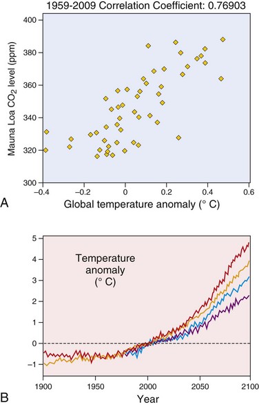
Figure 7–1 Climate change, past and future.
A, Correlation of CO2 levels measured at the Mauna Loa Observatory in Hawaii with average global temperature trends over the past 50 years. “Global temperature” in any given year was deduced at the Hadley Center (United Kingdom) from measurements taken at over 3000 weather stations located around the globe. B, Predicted temperature increases during the 21st century. Different computer models plot anticipated rises in global temperatures of 2°C to 5°C by the year 2100.
(A, Courtesy of Dr. Richard Aster, Department of Earth and Environmental Science, New Mexico Institute of Mining and Technology, Socorro, New Mexico.)
The health consequences of climate change will depend on its extent and rapidity, the severity of the ensuing consequences, and humankind’s ability to mitigate the damaging effects. Even in the best-case scenario, however, climate change is expected to have a serious negative impact on human health by increasing the incidence of a number of diseases, including
• Cardiovascular, cerebrovascular, and respiratory diseases, all of which will be exacerbated by heat waves and air pollution.
• Gastroenteritis, cholera, and other food- and water-borne infectious diseases, caused by contamination as a consequence of floods and disruption of clean water supplies and sewage treatment, after heavy rains and other environmental disasters
• Vector-borne infectious diseases, such as malaria and dengue fever, due to changes in vector number and geographic distribution related to increased temperatures, crop failures and more extreme weather variation (e.g., more frequent and severe El Niño events)
• Malnutrition, caused by changes in local climate that disrupt crop production. Such changes are anticipated to be most severe in tropical locations, in which average temperatures may already be near or above crop tolerance levels; it is estimated that by 2080, agricultural productivity may decline by 10% to 25% in some developing countries as a consequence of climate change.
Beyond these disease-specific effects, it is estimated that melting of glacial ice, particularly in Greenland and other parts of the Northern Hemisphere, combined with the thermal expansion of warming oceans, will raise sea levels by 2 to 6 feet by 2100. Approximately 10% of the world’s population—roughly 600 million people—live in low-lying areas that are at risk for flooding even if the rise in ocean levels is at the low end of these estimates. The resulting displacement of people will disrupt lives and commerce, creating conditions ripe for political unrest, war, and poverty, the “vectors” of malnutrition, sickness, and death.
Both developed and developing countries will suffer the consequences of climate change, but the burden will be greatest in developing countries, which are least culpable for increases in greenhouse gases to date. This picture is changing rapidly, however, owing to the growth of the economies of India and China, which has recently surpassed the United States to become the largest producer of CO2 in the world. The urgent challenge is to develop new renewable energy resources that stem the production of greenhouse gases. Without immediate action, climate change stands to become the preeminent global cause of environmental disease in the 21st century and beyond.
Toxicity of Chemical and Physical Agents
Toxicology is defined as the science of poisons. It studies the distribution, effects, and mechanisms of action of toxic agents. More broadly, it also includes the study of the effects of physical agents such as radiation and heat. Approximately 4 billion pounds of toxic chemicals, including 72 million pounds of known carcinogens, are produced each year in the United States. In general, however, little is known about the potential health effects of chemicals. Of the approximately 100,000 chemicals in use in the United States, less than 1% have been tested experimentally for health effects. In Europe the number of available chemicals is less than one-half that in the United States, but many of these chemicals are released into the environment as industrial products or discharged as human and animal wastes.
We now consider some basic principles regarding the toxicity of exogenous chemicals and drugs.
• The definition of a poison is not straightforward. It is basically a quantitative concept strictly dependent on dosage. The quote from Paracelsus in the 16th century that “all substances are poisons; the right dosage differentiates a poison from a remedy” is perhaps even more valid today, in view of the proliferation of therapeutic drugs with potentially harmful effects.
• Xenobiotics are exogenous chemicals in the environment that may be absorbed by the body through inhalation, ingestion, or skin contact (Fig. 7–2).
• Chemicals may be excreted in urine or feces or eliminated in expired air, or they may accumulate in bone, fat, brain, or other tissues.
• Chemicals may act at the site of entry, or they may be transported to other sites. Some agents are not modified upon entry in the body, but most solvents and drugs are metabolized to form water-soluble products (detoxification) or are activated to form toxic metabolites.
• Most solvents and drugs are lipophilic, which facilitates their transport in the blood by lipoproteins and penetration through lipid components of cell membranes.
• The reactions that metabolize xenobiotics into nontoxic products, or activate xenobiotics to generate toxic compounds (Fig. 7–3; see also Fig. 7–2), occur in two phases. In phase I reactions, chemicals can undergo hydrolysis, oxidation, or reduction. Products of phase I reactions often are metabolized into water-soluble compounds through phase II reactions of glucuronidation, sulfation, methylation, and conjugation with glutathione (GSH). Water-soluble compounds are readily excreted.
• The most important cellular enzyme system involved in phase I reactions is the cytochrome P-450 system, located primarily in the endoplasmic reticulum (ER) of the liver but also present in skin, lungs, and gastrointestinal (GI) mucosa and in practically every organ. The system catalyzes reactions that either detoxify xenobiotics or activate xenobiotics into active compounds that cause cellular injury. Both types of reactions may produce, as a byproduct, reactive oxygen species (ROS), which can cause cellular damage (discussed in Chapter 1). Examples of metabolic activation of chemicals through the P-450 system are the conversion of carbon tetrachloride to the toxic trichloromethyl free radical and the generation of a DNA-binding metabolite from benzo[a]pyrene (BaP), a carcinogen present in cigarette smoke. The cytochrome P-450 system also participates in the metabolism of a large number of common therapeutic drugs such as acetaminophen, barbiturates, and anticonvulsants, and in alcohol metabolism (discussed later).
• P-450 enzymes vary widely in activity among different people, owing to both polymorphisms in the genes encoding the enzymes and interactions with drugs that are metabolized through the system. The activity of the enzymes also may be decreased by fasting or starvation, and increased by alcohol consumption and smoking.
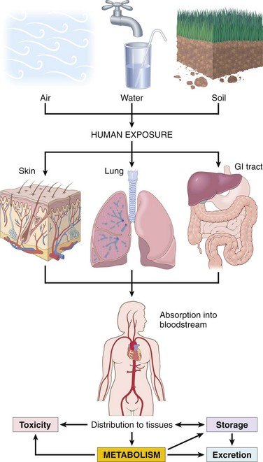
Figure 7–2 Human exposure to pollutants.
Pollutants contained in air, water, and soil are absorbed through the lungs, gastrointestinal tract, and skin. In the body, they may act at the site of absorption, but they generally are transported through the bloodstream to various organs, where they may be stored or metabolized. Metabolism of xenobiotics may result in the formation of water-soluble compounds, which are excreted, or in activation of the agent, creating a toxic metabolite.
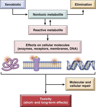
Figure 7–3 Xenobiotic metabolism.
Xenobiotics can be metabolized to nontoxic metabolites and eliminated from the body (detoxification). However, their metabolism also may result in activation of the chemical, leading to formation of a reactive metabolite that is toxic to cellular components. If repair is not effective, short- and long-term effects develop.
(Modified from Hodgson E: A Textbook of Modern Toxicology, 3rd ed, and Fig. 1–1. Hoboken, NJ, John Wiley & Sons, 2004.)
Environmental Pollution
Air Pollution
The life-giving air that we breathe is also often laden with many potential causes of disease. Airborne microorganisms have long been major causes of morbidity and death. More widespread are the chemical and particulate pollutants found in the air, both in so-called “developed” and “underdeveloped” countries. Specific hazards have been recognized for both outdoor and indoor air.
Outdoor Air Pollution
The ambient air in industrialized nations is contaminated with an unsavory mixture of gaseous and particulate pollutants, more heavily in cities and in proximity to heavy industry. In the United States, the Environmental Protection Agency (EPA) monitors and sets allowable upper limits for six pollutants: sulfur dioxide, CO, ozone, nitrogen dioxide, lead, and particulate matter. Together, some of these agents produce the well-known smog that sometimes stifles large cities such as Cairo, Los Angeles, Houston, Mexico City, and São Paulo. It may seem that air pollution is a modern phenomenon. This is not the case; Seneca wrote in ad 61 that he felt an alteration of his disposition as soon as he left the “pestilential vapors, soot, and heavy air of Rome.” The first environmental control law was proclaimed by Edward I in 1306 and was straightforward in its simplicity: “Whoever should be found guilty of burning coal shall suffer the loss of his head.” What has changed in modern times is the nature and sources of air pollutants, and the types of regulations that control their emission. It could be argued that modern man has lost his head to drown himself in pollution!
The lungs bear the brunt of the adverse consequences of air pollution, but air pollutants can affect many organ systems (as with the effects of lead poisoning and CO, discussed later). Except for some comments on smoking later in this chapter, pollutant-caused lung diseases are discussed in Chapter 12. Discussed here are the major health effects of ozone, sulfur dioxide, particulates, and CO (Table 7–1).
Table 7–1 Health Effects of Outdoor Air Pollutants
| Pollutant | Populations at Risk | Effect(s) |
|---|---|---|
| Ozone | Healthy adults and children | Decreased lung function |
| Increased airway reactivity | ||
| Lung inflammation | ||
| Athletes, outdoor workers | Decreased exercise capacity | |
| Asthmatics | Increased hospitalizations | |
| Nitrogen dioxide | Healthy adults | Increased airway reactivity |
| Asthmatics | Decreased lung function | |
| Children | Increased respiratory infections | |
| Sulfur dioxide | Healthy adults | Increased respiratory symptoms |
| Patients with chronic lung disease | Increased mortality | |
| Asthmatics | Increased hospitalization | |
| Decreased lung function | ||
| Acid aerosols | Healthy adults | Altered mucociliary clearance |
| Children | Increased respiratory infections | |
| Asthmatics | Decreased lung function | |
| Increased hospitalizations | ||
| Particulates | Children | Increased respiratory infections |
| Decreased lung function | ||
| Patients with chronic lung or heart disease | Excess mortality | |
| Asthmatics | Increased attacks |
Data from Health effects of outdoor air pollution. Part 2. Committee of the Environmental and Occupational Health Assembly of the American Thoracic Society. Am J Respir Crit Care Med 153:477, 1996.
Ozone is one of the most pervasive air pollutants, with levels in many cities exceeding EPA standards. It is a gas formed by sunlight-driven reactions involving nitrogen oxides, which are released mostly by automobile exhaust. Together with oxides and fine particulate matter, ozone forms the familiar smog (from smoke and fog). Its toxicity stems from its participation in chemical reactions that generate free radicals, which injure the lining cells of the respiratory tract and the alveoli. Low levels of ozone may be tolerated by healthy persons but are detrimental to lung function, especially in those with asthma or emphysema, and when present along with particulate pollution. Unfortunately, pollutants rarely occur singly but combine to create a veritable “witches’ brew.”
Sulfur dioxide, particles, and acid aerosols are emitted by coal- and oil-fired power plants and industrial processes burning these fuels. Of these, particles (although not well characterized chemically or physically) appear to be the main cause of morbidity and death. Particles less than 10 µm in diameter are particularly harmful, since when inhaled they are carried by the airstream all the way to the alveoli. Here, they are phagocytosed by macrophages and neutrophils, causing the release of mediators and inciting an inflammatory reaction. By contrast, larger particles are removed in the nose or are trapped by the mucociliary “escalator” and as a result are less dangerous.
Carbon monoxide (CO) is a nonirritating, colorless, tasteless, odorless gas. It is produced by the incomplete oxidation of carbonaceous materials. Its sources include automotive engines, industries using fossil fuels, home oil burners, and cigarette smoke. The low levels often found in ambient air may contribute to impaired respiratory function but usually are not life-threatening. However, persons working in confined environments with high exposure to fumes, such as tunnel and underground garage workers, may develop chronic poisoning. CO is included here as an air pollutant, but it also is an important cause of accidental and suicidal death. In a small, closed garage, exhaust from a running car engine can induce lethal coma within 5 minutes. CO is a systemic asphyxiant that kills by binding to hemoglobin and preventing oxygen transport. Hemoglobin has a 200-fold greater affinity for CO than for O2. The resultant compound, carboxyhemoglobin, is incapable of carrying oxygen. Hypoxia leads to central nervous system (CNS) depression, which develops so insidiously that victims may not be aware of their plight and indeed may be unable to help themselves. Systemic hypoxia appears when the hemoglobin is 20% to 30% saturated with CO, and unconsciousness and death are probable with 60% to 70% saturation.
 Morphology
Chronic poisoning by CO develops because carboxyhemoglobin, once formed, is remarkably stable. As a result, with low-level persistent exposure to CO, carboxyhemoglobin may accumulate to a life-threatening concentration in the blood. The slowly developing hypoxia can insidiously evoke widespread ischemic changes in the brain; these changes are particularly marked in the basal ganglia and lenticular nuclei. With cessation of exposure to CO, the patient usually recovers, but there may be permanent neurologic damage. The diagnosis of CO poisoning is based on detection of high levels of carboxyhemoglobin in the blood.
Acute poisoning by CO generally is a consequence of accidental exposure or suicide attempt. In light-skinned people, it is marked by a characteristic generalized cherry-red color of the skin and mucous membranes, a color imparted by carboxyhemoglobin. If death occurs rapidly, morphologic changes may not be present; with longer survival, the brain may be slightly edematous and exhibit punctate hemorrhages and hypoxia-induced neuronal changes. These changes are not specific; they simply imply systemic hypoxia. In victims who survive CO poisoning, complete recovery is possible; however, sometimes impairments of memory, vision, hearing, and speech may remain.
Indoor Air Pollution
As modern homes are increasingly “buttoned up” to exclude the environment, the potential for pollution of the indoor air increases. The commonest pollutant is tobacco smoke (discussed later), but additional offenders are CO, nitrogen dioxide (already mentioned as outdoor pollutants), and asbestos (discussed in Chapter 12). A few comments about some other agents are presented here.
Wood smoke, containing various oxides of nitrogen and carbon particulates, is an irritant that predisposes exposed persons to lung infections and may contain carcinogenic polycyclic hydrocarbons. Radon, a radioactive gas derived from uranium, is widely present in soil and in homes. Although radon exposure can cause lung cancer in uranium miners (particularly in those who smoke), it does not appear that low-level chronic exposures in the home increase lung cancer risk, at least for nonsmokers. Bioaerosols may contain pathogenic microbiologic agents, such as those that can cause Legionnaires’ disease, viral pneumonia, and the common cold, as well as allergens derived from pet dander, dust mites, and fungi and molds, which can cause rhinitis, eye irritation, and even asthma.
 Summary
Environmental Diseases and Environmental Pollution
• Environmental diseases are conditions caused by exposure to chemical or physical agents in the ambient, workplace, and personal environments.
• Exogenous chemicals known as xenobiotics enter the body through inhalation, ingestion, and skin contact, and can either be eliminated or accumulate in fat, bone, brain, and other tissues.
• Xenobiotics can be converted into nontoxic products, or activated to generate toxic compounds, through a two-phase reaction process that involves the cytochrome P-450 system.
• The most common air pollutants are ozone (which in combination with oxides and particulate matter forms smog), sulfur dioxide, acid aerosols, and particles less than 10 µm in diameter.
• Carbon monoxide is an air pollutant and important cause of death from accidents and suicide; it binds hemoglobin with high affinity, leading to systemic asphyxiation associated with CNS depression.
Metals as Environmental Pollutants
Lead, mercury, arsenic, and cadmium, the heavy metals most commonly associated with harmful effects in human populations, are considered here.
Lead
Lead exposure occurs through contaminated air and food. For most of the 20th century the major sources of lead in the environment were house paints and gasoline. Although the use of lead-based paints and leaded gas has greatly diminished, many sources of lead persist in the environment, such as mines, foundries, batteries, and spray paints, all of which constitute occupational hazards. However, flaking lead paint in older houses and soil contamination pose the major hazards for youngsters. Indeed, a single 1-cm2 chip of old leaded paint (pre-1977) contains about 175 µg of lead; this amount, if consumed each day over time, will rapidly produce toxic lead levels. According to a 2008 report from the Environmental Protection Agency (EPA), 0.9% of American children had blood lead levels in excess of 10 µg/dL (the maximum allowable level). This percentage represents a decrease from 4.4% in the early 1990s. However, blood levels of lead in children living in homes containing lead-based paint or lead-contaminated dust generally exceed the maximum allowed levels. Children absorb more than 50% of lead from food, while adults absorb approximately 15%. A more permeable blood–brain barrier in children creates a high susceptibility to brain damage. The main clinical features of lead poisoning are shown in Figure 7–4.
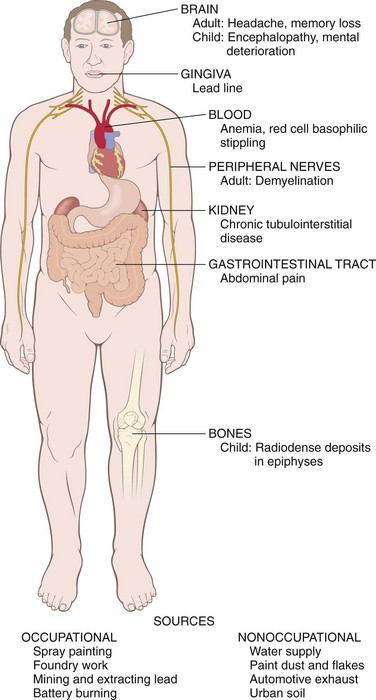
Most of the absorbed lead (80% to 85%) is taken up into bone and developing teeth; lead competes with calcium, binds phosphates, and has a half-life in bone of 20 to 30 years. About 5% to 10% of the absorbed lead remains in the blood, and the remainder is distributed throughout soft tissues. Excess lead causes neurologic effects in adults and children; peripheral neuropathies predominate in adults, while central effects are more common in children. The effects of chronic lead exposure in children include a lower intellectual capacity manifested by low intelligence quotient (IQ), behavioral problems such as hyperactivity, and poor organizational skills. Lead-induced peripheral neuropathies in adults generally remit with elimination of exposure, but both peripheral and CNS abnormalities in children usually are irreversible. Excess lead interferes with the normal remodeling of calcified cartilage and primary bone trabeculae in the epiphyses in children, causing increased bone density detected as radiodense “lead lines” (Fig. 7–5). Lead lines of a different sort also may occur in the gums, where excess lead stimulates hyperpigmentation. Lead inhibits the healing of fractures by increasing chondrogenesis and delaying cartilage mineralization. Excretion of lead occurs by way of the kidneys, and acute exposures may cause damage to proximal tubules.
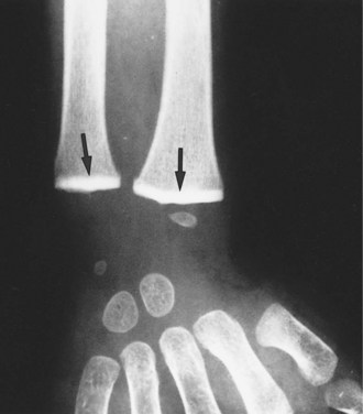
Impaired remodeling of calcified cartilage in the epiphyses (arrows) of the wrist has caused a marked increase in their radiodensity, so that they are as radiopaque as the cortical bone.
(Courtesy of Dr. G.W. Dietz, Department of Radiology, University of Texas Southwestern Medical School, Dallas, Texas.)
Lead has a high affinity for sulfhydryl groups and interferes with two enzymes involved in heme synthesis, aminolevulinic acid dehydratase and delta ferrochelatase. Iron incorporation into heme is impaired, leading to anemia. Lead also inhibits sodium- and potassium-dependent ATPases in cell membranes, an effect that may increase the fragility of red cells, causing hemolysis. The diagnosis of lead poisoning requires constant vigilance. It may be suspected on the basis of neurologic changes in children or unexplained anemia with basophilic stippling in red cells. Elevated blood lead and red cell free protoporphyrin levels (greater than 50 µg/dL) or, alternatively, zinc-protoporphyrin levels, are required for definitive diagnosis. In milder cases of lead exposure, anemia may be the only obvious abnormality.
Morphology
The major anatomic targets of lead toxicity are the blood, bone marrow, nervous system, GI tract, and kidneys (Fig. 7–4).
Blood changes are one of the earliest signs of lead accumulation and are characteristic, consisting of a microcytic, hypochromic anemia associated with a distinctive punctate basophilic stippling of red cells. These changes in the blood stem from the inhibition of heme synthesis in marrow erythroid progenitors. Another consequence of this blockade is that zinc-protoporphyrin is formed instead of heme. Thus, elevated blood levels of zinc-protoporphyrin or its product, free red cell protoporphyrin, are important indicators of lead poisoning.
Brain damage is prone to occur in children. It may be subtle, producing mild dysfunction, or it may be massive and lethal. In young children, sensory, motor, intellectual, and psychologic impairments have been described, including reduced IQ, learning disabilities, retarded psychomotor development, and, in more severe cases, blindness, psychoses, seizures, and coma. Lead toxicity in the mother may be the cause of impairment of prenatal brain development. The anatomic changes underlying the more subtle functional deficits are ill defined, but some of the defects may be permanent. At the more severe end of the spectrum are brain edema, demyelination of the cerebral and cerebellar white matter, and necrosis of cortical neurons accompanied by diffuse astrocytic proliferation. In adults, the CNS is less often affected, but frequently a peripheral demyelinating neuropathy appears, typically involving motor neurons innervating the most commonly used muscles. Thus, the extensor muscles of the wrist and fingers are often the first to be affected, followed by paralysis of the peroneal muscles (wristdrop and footdrop).
The GI tract also is a locus for major clinical manifestations. Lead “colic” is characterized by extremely severe, poorly localized abdominal pain.
The kidneys may develop proximal tubular damage with intranuclear lead inclusions. Chronic renal damage leads eventually to interstitial fibrosis and possibly renal failure and findings suggestive of gout (“saturnine gout”). Other features of lead poisoning are shown in Figure 7–4.
Mercury
Humans have used mercury in many ways throughout history, including as a pigment in cave paintings, a cosmetic, a remedy for syphilis, and a component of diuretics. Poisoning from inhalation of mercury vapors has long been recognized and is associated with tremor, gingivitis, and bizarre behavior, such as that of the “Mad Hatter” in Lewis Carroll’s Alice in Wonderland (mercury formerly was used in hat-making).
Today, the main sources of exposure to mercury are contaminated fish and dental amalgams, which release mercury vapors. In some areas of the world, mercury used in gold mining has contaminated rivers and streams. Inorganic mercury from the natural degassing of the earth’s crust or from industrial contamination is converted to organic compounds such as methyl mercury by bacteria. Methyl mercury enters the food chain, and in carnivorous fish such as swordfish, shark, and bluefish, mercury levels may be a million times higher than in the surrounding water. The consumption of contaminated fish from the release of methyl mercury in Minamata Bay and the Agano River in Japan, and the consumption of bread containing grain treated with a methyl mercury–based fungicide in Iraq, caused widespread morbidity and many deaths.
The medical disorders associated with the Minamata episode became known as “Minamata disease” and include cerebral palsy, deafness, blindness, and major CNS defects in children exposed in utero. The developing brain is extremely sensitive to methyl mercury; for this reason, the Centers for Disease Control and Prevention (CDC) in the United States has recommended that pregnant women avoid the consumption of fish known to contain mercury. There has been much publicity about a possible relationship between thimerosal (a compound that contains ethyl mercury, used until recently as a preservative in some vaccines) and the development of autism, but several large studies have failed to detect any association.
Arsenic
Arsenic was the favorite poison in Renaissance Italy, and this application had some skilled practitioners among the Borgias and Medicis. Deliberate poisoning by arsenic is exceedingly rare today, but exposure to arsenic is an important health problem in many areas of the world. Arsenic is found naturally in soil and water and is used in wood preservatives, herbicides, and other agricultural products. It may be released into the environment by the mining and smelting industries. Large concentrations of inorganic arsenic are present in ground water in countries such as Bangladesh, Chile, and China. As many as 20 million people in Bangladesh drink water contaminated by arsenic, constituting one of the largest environmental cancer risks yet identified.
The most toxic forms of arsenic are the trivalent compounds arsenic trioxide, sodium arsenite, and arsenic trichloride. If ingested in large quantities, arsenic causes acute toxicity manifesting as severe gastrointestinal, cardiovascular, and central nervous system disturbances, often progressing to death. These effects may be attributed to the interference with mitochondrial oxidative phosphorylation. Chronic exposure to arsenic causes hyperpigmentation and hyperkeratosis of the skin, which may be followed by the development of basal and squamous cell carcinomas (but not melanomas). Arsenic-induced skin tumors differ from those induced by sunlight by appearing on palms and soles, and by occurring as multiple lesions. Arsenic exposure also is associated with an increased risk of lung carcinoma. The mechanisms of arsenic carcinogenesis in skin and lung are uncertain.
Cadmium
In contrast with the metals already discussed, cadmium is a relatively modern toxic agent. It is used mainly in nickel-cadmium batteries, which generally are disposed of as household waste. It can contaminate soil and plants directly or through fertilizers and irrigation water. Food is the most important source of exposure for the general population. Excessive cadmium intake can lead to obstructive lung disease and renal toxicity, initially as tubular damage that may progress to end-stage renal disease. Cadmium exposure can also cause skeletal abnormalities associated with calcium loss. Cadmium-contaminated water used to irrigate rice fields in Japan caused a disease in postmenopausal women known as “itai-itai” (ouch-ouch), a combination of osteoporosis and osteomalacia associated with renal disease. A recent survey showed that 5% of persons aged 20 years and older in the U.S. population have urinary cadmium levels that, according to research data, may produce subtle kidney injury and increased calcium loss.
Summary
Toxic Effects of Heavy Metals
• Lead, mercury, arsenic, and cadmium are the heavy metals most commonly associated with toxic effects in humans.
• Children absorb more ingested lead than adults; the main source of exposure for children is lead-containing paint.
• Excess lead causes CNS defects in children and peripheral neuropathy in adults. Excess lead competes with calcium in bones and interferes with the remodeling of cartilage; it also causes anemia.
• The major source of exposure to mercury is contaminated fish. The developing brain is highly sensitive to methyl mercury, which accumulates in the brain and blocks ion channels.
• Exposure of the fetus to high levels of mercury in utero may lead to Minamata disease, characterized by cerebral palsy, deafness, and blindness.
• Arsenic is naturally found in soil and water and is a component of some wood preservatives and herbicides. Excess arsenic interferes with mitochondrial oxidative phosphorylation and causes toxic effects in the GI tract, CNS, and cardiovascular system; long-term exposure causes skin lesions and carcinomas.
• Cadmium from nickel-cadmium batteries and chemical fertilizers can contaminate soil. Excess cadmium causes obstructive lung disease and kidney damage.
Industrial and Agricultural Exposures
More than 10 million occupational injuries occur annually in the United States, and approximately 65,000 people die as a consequence of occupational injuries and illnesses. Industrial exposures to toxic agents are as varied as the industries themselves. They range from merely annoying irritations of respiratory airways by formaldehyde or ammonia fumes to fatal lung cancers arising from exposure to asbestos, arsenic, or uranium mining. Human diseases associated with occupational exposures are listed in Table 7–2. In addition to the toxic metals (which have already been discussed), other important agents that contribute to environmental diseases include the following:
• Organic solvents are widely used in huge quantities worldwide. Some, such as chloroform and carbon tetrachloride, are found in degreasing and dry cleaning agents and paint removers. Acute exposure to high levels of vapors from these agents can cause dizziness and confusion, leading to CNS depression and even coma. Lower levels have toxicity for the liver and kidneys. Occupational exposure of rubber workers to benzene and 1,3-butadiene increases the risk of leukemia. Benzene is oxidized to an epoxide through hepatic CYP2E1, a component of the P-450 enzyme system already mentioned. The epoxide and other metabolites disrupt progenitor cell differentiation in the bone marrow, causing marrow aplasia and acute myeloid leukemia.
• Polycyclic hydrocarbons may be released during the combustion of coal and gas, particularly at the high temperatures used in steel foundries, and also are present in tar and soot. (Pott identified soot as the cause of scrotal cancers in chimney sweeps in 1775, as mentioned in Chapter 5.) Polycyclic hydrocarbons are among the most potent carcinogens, and industrial exposures have been implicated in the causation of lung and bladder cancer.
• Organochlorines (and halogenated organic compounds in general) are synthetic products that resist degradation and are lipophilic. Important organochlorines used as pesticides are DDT (dichlorodiphenyltrichloroethane) and its metabolites and agents such as lindane, aldrin, and dieldrin. Nonpesticide organochlorines include polychlorinated biphenyls (PCBs) and dioxin (TCDD [2,3,7,8-tetrachlorodibenzo-p-dioxin]). DDT was banned in the United States in 1973, but more than half of the population have detectable serum levels of p,p′-DDE, a long-lasting DDT metabolite, including those born after the ban on DDT went into effect. PCB and TCDD also are present in the blood of most of the U.S. population. Acute DDT poisoning in humans causes neurologic toxicity. Most organochlorines are endocrine disruptors and have antiestrogenic or antiandrogenic activity in laboratory animals, but long-term health effects in humans have not been firmly established.
• Dioxins and PCBs can cause skin disorders such as folliculitis and acneiform dermatosis known as chloracne, which consists of acne, cyst formation, hyperpigmentation, and hyperkeratosis, generally around the face and behind the ears. It can be accompanied by abnormalities in the liver and CNS. Because PCBs induce the P-450 enzyme system, workers exposed to these substances may show altered drug metabolism. Environmental disasters in Japan and China in the late 1960s caused by the consumption of rice oil contaminated by PCBs during its production poisoned about 2000 people in each episode. The primary manifestations of the disease (yusho in Japan, yu-cheng in China) were chloracne and hyperpigmentation of the skin and nails.
• Bisphenol A (BPA) is used in the synthesis of polycarbonate food and water containers and of epoxy resins that line almost all food bottles and cans; as a result, exposure to BPA is virtually ubiquitous in humans. BPA has long been known as a potential endocrine disruptor. Several large retrospective studies have linked elevated urinary BPA levels to heart disease in adult populations. In addition, infants who drink from BPA-containing containers may be particularly susceptible to its endocrine effects. In 2010, Canada was the first country to list BPA as a toxic substance, and the largest makers of baby bottles and “sippy” cups have stopped using BPA in the manufacturing process. The extent of the human health risks associated with BPA remains uncertain, however, and requires further study.
• Exposure to vinyl chloride, used in the synthesis of polyvinyl resins, was found to cause angiosarcoma of the liver, a rare type of liver tumor.
• Inhalation of mineral dusts causes chronic, non-neoplastic lung diseases called pneumoconioses. This group of disorders includes diseases induced by organic and inorganic particulates as well as chemical fume- and vapor-induced non-neoplastic lung diseases. The most common pneumoconioses are caused by exposures to mineral dust: coal dust (in mining of hard coal), silica (in sandblasting and stone cutting), asbestos (in mining, fabrication, and insulation work), and beryllium (in mining and fabrication). Exposure to these agents nearly always occurs in the workplace. The increased risk of cancer as a result of asbestos exposure, however, extends to family members of asbestos workers and to other persons exposed outside the workplace. Pneumoconioses and their pathogenesis are discussed in Chapter 12.
Table 7–2 Human Diseases Associated With Occupational Exposures
| Organ/System | Effect(s) | Toxicant(s) |
|---|---|---|
| Cardiovascular system | Heart disease | Carbon monoxide, lead, solvents, cobalt, cadmium |
| Respiratory system | Nasal cancer | Isopropyl alcohol, wood dust |
| Lung cancer | Radon, asbestos, silica, bis(chloromethyl)ether, nickel, arsenic, chromium, mustard gas | |
| Chronic obstructive lung disease | Grain dust, coal dust, cadmium | |
| Hypersensitivity | Beryllium, isocyanates | |
| Irritation | Ammonia, sulfur oxides, formaldehyde | |
| Fibrosis | Silica, asbestos, cobalt | |
| Nervous system | Peripheral neuropathies | Solvents, acrylamide, methyl chloride, mercury, lead, arsenic, DDT |
| Ataxic gait | Chlordane, toluene, acrylamide, mercury | |
| Central nervous system depression | Alcohols, ketones, aldehydes, solvents | |
| Cataracts | Ultraviolet radiation | |
| Urinary system | Toxicity | Mercury, lead, glycol ethers, solvents |
| Bladder cancer | Naphthylamines, 4-aminobiphenyl, benzidine, rubber products | |
| Reproductive system | Male infertility | Lead, phthalate plasticizers |
| Female infertility | Cadmium, lead | |
| Teratogenesis | Mercury, polychlorinated biphenyls | |
| Hematopoietic system | Leukemia | Benzene, radon, uranium |
| Skin | Folliculitis and acneiform dermatosis | Polychlorinated biphenyls, dioxins, herbicides |
| Cancer | Ultraviolet radiation | |
| Gastrointestinal tract | Liver angiosarcoma | Vinyl chloride |
DDT, dichlorodiphenyltrichloroethane.
Data from Leigh JP, Markowitz SB, Fahs M, et al: Occupational injury and illness in the United States. Estimates of costs, morbidity, and mortality. Arch Intern Med 157:1557, 1997; Mitchell FL: Hazardous waste. In Rom WN (ed): Environmental and Occupational Medicine, 2nd ed. Boston, Little, Brown, 1992, p 1275; and Levi PE: Classes of toxic chemicals. In Hodgson E, Levi PE (eds): A Textbook of Modern Toxicology. Stamford, CT, Appleton & Lange, 1997, p 229.
Effects of Tobacco
Tobacco is the most common exogenous cause of human cancers, being responsible for 90% of lung cancers. The main culprit is cigarette smoking, but smokeless tobacco in its various forms (snuff, chewing tobacco) also is harmful to health and is an important cause of oral cancer. Not only does the use of tobacco products create personal risk, but passive tobacco inhalation from the environment (“second-hand smoke”) can cause lung cancer in nonsmokers. Cigarette smoking causes, worldwide, more than 4 million deaths annually, mostly from cardiovascular disease, various types of cancers, and chronic respiratory problems. It is expected that there will be 8 million tobacco-related deaths yearly by 2020, the major increase occurring in developing countries. Of people alive today, an estimated 500 million will die from tobacco-related illnesses. In the United States alone, tobacco is responsible for more than 400,000 deaths per year, one third of these attributable to lung cancer.
Smoking is the most preventable cause of human death. It reduces overall survival in a dose-dependent fashion. While 80% of nonsmokers are alive at age 70, only about 50% of smokers survive to this age (Fig. 7–6). Cessation of smoking greatly reduces the risk of death from lung cancer, and it even has an effect, albeit reduced, on people who stop smoking at age 60. During the period 1998 to 2007 in the United States, the incidence of smoking declined modestly, but approximately 20% of adults remained smokers. More disturbing, smoking in the world’s most populous country, China, is becoming the rule rather than the exception. It is estimated that more than 1 million people in China die each year of smoking-related diseases.
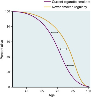
Figure 7–6 The effects of smoking on survival. The study compared age-specific death rates for current cigarette smokers with that of individuals who never smoke regularly (British Doctors Study). The difference in survival, measured at age 75, between smokers and nonsmokers is 7.5 years.
(Modified from Stewart BW, Kleihues P [eds]: World Cancer Report. Lyon, IARC Press, 2003.)
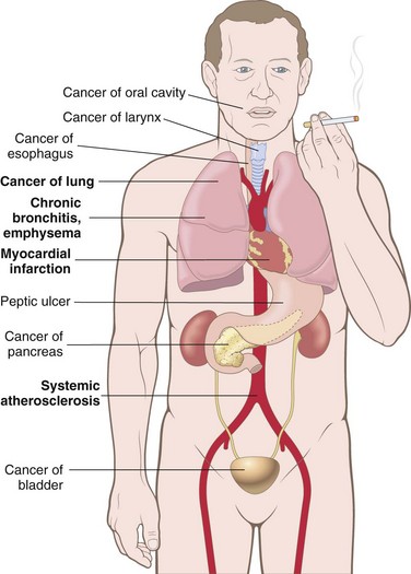
Discussed next are some of the agents contained in tobacco and diseases associated with tobacco consumption. Adverse effects of smoking in various organ systems are shown in Figure 7–7.
The number of potentially noxious chemicals in tobacco smoke is vast; Table 7–3 presents only a partial list and includes the type of injury produced by these agents. Nicotine, an alkaloid present in tobacco leaves, is not a direct cause of tobacco-related diseases, but it is highly addictive. Nicotine binds to receptors in the brain and, through the release of catecholamines, is responsible for the acute effects of smoking, such as increased heart rate and blood pressure, and increased cardiac contractility and output.
Table 7–3 Effects of Selected Tobacco Smoke Constituents
| Substance | Effect(s) |
|---|---|
| Tar | Carcinogenesis |
| Polycyclic aromatic hydrocarbons | Carcinogenesis |
| Nicotine | Ganglionic stimulation and depression, tumor promotion |
| Phenol | Tumor promotion; mucosal irritation |
| Benzopyrene | Carcinogenesis |
| Carbon monoxide | Impaired oxygen transport and utilization |
| Formaldehyde | Toxicity to cilia; mucosal irritation |
| Oxides of nitrogen | Toxicity to cilia; mucosal irritation |
| Nitrosamine | Carcinogenesis |
The most common diseases caused by cigarette smoking involve the lung and include emphysema, chronic bronchitis, and lung cancer, all discussed in Chapter 12. The mechanisms responsible for some tobacco-induced diseases are outlined next.
• Agents in smoke have a direct irritant effect on the tracheobronchial mucosa, producing inflammation and increased mucus production (bronchitis). Cigarette smoke also causes the recruitment of leukocytes to the lung, increasing local elastase production and subsequent injury to lung tissue that leads to emphysema.
• Components of cigarette smoke, particularly polycyclic hydrocarbons and nitrosamines (Table 7–4), are potent carcinogens in animals and probably are involved in the causation of lung carcinomas in humans (see Chapter 12). The risk of developing lung cancer is related to the intensity of exposure, frequently expressed in terms of “pack years” (e.g., one pack daily for 20 years equals 20 pack years) or in cigarettes smoked per day (Fig. 7–8). Moreover, smoking multiplies the risk of disease associated with other carcinogens; well-recognized examples are the 10-fold higher incidence of lung carcinomas in asbestos workers and uranium miners who smoke than that in those who do not, and the interaction between tobacco consumption and alcohol in the risk for oral cancers as described later on.
• Atherosclerosis and its major complication, myocardial infarction, are strongly linked to cigarette smoking. The causal mechanisms probably relate to several factors, including increased platelet aggregation, decreased myocardial oxygen supply (because of lung disease coupled with hypoxia related to CO in cigarette smoke) accompanied by increased oxygen demand, and a decreased threshold for ventricular fibrillation. Almost one third of all heart attacks are associated with cigarette smoking. Smoking has a multiplicative effect on risk when combined with hypertension and hypercholesterolemia.
• In addition to lung cancers, tobacco smoke contributes to the development of cancers of the oral cavity, esophagus, pancreas, and bladder. Table 7–4 lists organ-specific carcinogens contained in tobacco smoke.
• The combination of tobacco (chewed or smoked) and alcohol consumption has multiplicative effects on the risks of oral, laryngeal, and esophageal cancers. An example of the carcinogenic interaction of these all too common vices is shown below for laryngeal cancer (Fig. 7–9).
• Maternal smoking increases the risk of spontaneous abortions and preterm births and results in intrauterine growth retardation (Chapter 6); however, birth weights of infants born to mothers who stopped smoking before pregnancy are normal.
• Exposure to environmental tobacco smoke (passive smoke inhalation) is also associated with detrimental effects. It is estimated that the relative risk of lung cancer in nonsmokers exposed to environmental smoke is about 1.3 times that in nonsmokers who are not exposed to smoke. In the United States, approximately 3000 lung cancer deaths in nonsmokers over the age of 35 years can be attributed each year to environmental tobacco smoke. Even more striking is the increased risk of coronary atherosclerosis and fatal myocardial infarction. Studies report that every year, 30,000 to 60,000 cardiac deaths in the United States are associated with passive exposure to smoke. Children living in a household with an adult who smokes have an increased frequency of respiratory illnesses and asthma. Passive smoke inhalation in nonsmokers can be estimated by measuring the blood levels of cotinine, a metabolite of nicotine. In the United States, median cotinine levels in nonsmokers have decreased by more than 60% during the last 15 years due to adoption of non-smoking policies in public places. However, passive exposure to tobacco smoke in the home remains a major public health concern, particularly for children. It is clear that the transient pleasure a puff may give comes with a heavy long-term price.
Table 7–4 Organ-Specific Carcinogens in Tobacco Smoke
| Organ | Carcinogen(s) |
|---|---|
| Lung, larynx | Polycyclic aromatic hydrocarbons |
| 4-(Methylnitrosoamino)-1-(3-pyridyl)-1-butanone (NNK) | |
| 210Polonium | |
| Esophagus | N′-Nitrosonornicotine (NNN) |
| Pancreas | NNK (?) |
| Bladder | 4-Aminobiphenyl, 2-naphthylamine |
| Oral cavity: smoking | Polycyclic aromatic hydrocarbons, NNK, NNN |
| Oral cavity: snuff | NNK, NNN, 210polonium |
Data from Szczesny LB, Holbrook JH: Cigarette smoking. In Rom WH (ed): Environmental and Occupational Medicine, 2nd ed. Boston, Little, Brown, 1992, p 1211.
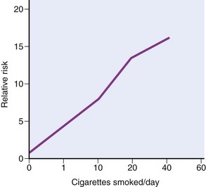
Figure 7–8 The risk of lung cancer is determined by the number of cigarettes smoked.
(Data from Stewart BW, Kleihues P [eds]: World Cancer Report. Lyon, IARC Press, 2003.)
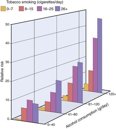
Figure 7–9 Multiplicative increase in the risk of laryngeal cancer from the interaction between cigarette smoking and alcohol consumption.
(Data from Stewart BW, Kleihues P [eds]: World Cancer Report. Lyon, IARC Press, 2003.)
Summary
Health Effects of Tobacco
Effects of Alcohol
Ethanol is consumed, at least partly, for its mood-altering properties, but when used in moderation its effects are socially acceptable and not injurious. When excessive amounts are used, alcohol can cause marked physical and psychologic damage. Here we describe the lesions that are directly associated with the abuse of alcohol.
Despite all the attention given to illegal drugs, alcohol abuse is a more widespread hazard and claims many more lives. Fifty percent of adults in the Western world drink alcohol, and approximately 5% to 10% have chronic alcoholism. It is estimated that there are more than 10 million chronic alcoholics in the United States and that alcohol consumption is responsible for more than 100,000 deaths annually. Almost 50% of these deaths result from accidents caused by drunken driving and alcohol-related homicides and suicides, and about 25% are a consequence of cirrhosis of the liver.
After consumption, ethanol is absorbed unaltered in the stomach and small intestine and then distributes to all of the tissues and fluids of the body in direct proportion to the blood level. Less than 10% is excreted unchanged in the urine, sweat, and breath. The amount exhaled is proportional to the blood level and forms the basis for the breath test used by law enforcement agencies. A concentration of 80 mg/dL in the blood constitutes the legal definition of drunk driving in most states. For an average individual, this alcohol concentration may be reached after consumption of about eight bottles of beer (6 to 16 g of alcohol per bottle), 12 ounces of wine (9 to 18 g of alcohol per glass), or 6 ounces of whiskey (about 11 g of alcohol per ounce). Drowsiness occurs at 200 mg/dL, stupor at 300 mg/dL, and coma, with possible respiratory arrest, at higher levels. The rate of metabolism affects the blood alcohol level. Persons with chronic alcoholism can tolerate levels as high as 700 mg/dL, due in part to accelerated ethanol metabolism caused by a 5- to 10-fold increase in induction of the hepatic cytochrome P-450 system, discussed next.
Most of the alcohol in the blood is metabolized to acetaldehyde in the liver by three enzyme systems: alcohol dehydrogenase, cytochrome P-450 isoenzymes, and catalase (Fig. 7–10). Of these, the main enzyme involved in alcohol metabolism is alcohol dehydrogenase, located in the cytosol of hepatocytes. At high blood alcohol levels, however, the microsomal ethanol-oxidizing system also has an important role. This system involves cytochrome P-450 enzymes, particularly the CYP2E1 isoform, located in the smooth ER. Induction of P-450 enzymes by alcohol explains the increased susceptibility of alcoholics to other compounds metabolized by the same enzyme system, which include drugs (acetaminophen, cocaine), anesthetics, carcinogens, and industrial solvents. Of note, however, when alcohol is present in the blood at high concentrations, it competes with other CYP2E1 substrates and may delay the catabolism of other drugs, thereby potentiating their effects. Catalase is of minor importance, being responsible for only about 5% of alcohol metabolism. Acetaldehyde produced by these systems is in turn converted by acetaldehyde dehydrogenase to acetate, which is utilized in the mitochondrial respiratory chain.

Figure 7–10 Metabolism of ethanol: oxidation of ethanol to acetaldehyde by three different routes, and the generation of acetic acid. Note that oxidation by alcohol dehydrogenase (ADH) takes place in the cytosol; the cytochrome P-450 system and its CYP2E1 isoform are located in the ER (microsomes), and catalase is located in peroxisomes. Oxidation of acetaldehyde by aldehyde dehydrogenase (ALDH) occurs in mitochondria.
(Data from Parkinson A: Biotransformation of xenobiotics. In Klassen CD [ed]: Casarett and Doull’s Toxicology: The Basic Science of Poisons, 6th ed. New York, McGraw-Hill, 2001, p 133.)
Several toxic effects result from ethanol metabolism. Listed here are only the most important of these:
• Alcohol oxidation by alcohol dehydrogenase causes a decrease in nicotinamide adenine dinucleotide (NAD+) and an increase in NADH (the reduced form of NAD+). NAD+ is required for fatty acid oxidation in the liver. Its deficiency is a main cause of fat accumulation in the liver of alcoholics. The increase in the NADH/NAD+ ratio in alcoholics also causes lactic acidosis.
• Acetaldehyde has many toxic effects and may be responsible for some of the acute effects of alcohol. Acetaldehyde metabolism differs between populations because of genetic variation. Most notably, about 50% of Asians express a defective form of acetaldehyde dehydrogenase. After ingesting alcohol, such persons experience flushing, tachycardia, and hyperventilation owing to the accumulation of acetaldehyde.
• Metabolism of ethanol in the liver by CYP2E1 produces reactive oxygen species and causes lipid peroxidation of cell membranes. Nevertheless, the precise mechanisms that account for alcohol-induced cellular injury have not been well defined.
• Alcohol may cause the release of endotoxin (lipopolysaccharide), a product of gram-negative bacteria, from the intestinal flora. Endotoxin stimulates the release of tumor necrosis factor (TNF) and other cytokines from circulating macrophages and from Kupffer cells in the liver, causing cell injury.
The adverse effects of ethanol abuse can be categorized as acute or chronic. Acute alcoholism exerts its effects mainly on the CNS but also may induce reversible hepatic and gastric injuries. Even with moderate intake of alcohol, multiple fat droplets accumulate in the cytoplasm of hepatocytes (fatty change or hepatic steatosis). Gastric damage occurs in the form of acute gastritis and ulceration. In the CNS, alcohol is a depressant, first affecting subcortical structures that modulate cerebral cortical activity. Consequently there is stimulation and disordered cortical, motor, and intellectual behavior. At progressively higher blood levels, cortical neurons and then lower medullary centers are depressed, including those that regulate respiration. Respiratory arrest may follow.
Chronic alcoholism affects not only the liver and stomach but virtually all other organs and tissues as well. Chronic alcoholics suffer significant morbidity and have a shortened life span, related principally to damage to the liver, GI tract, CNS, cardiovascular system, and pancreas.
• The liver is the main site of chronic injury. In addition to fatty change, mentioned earlier, chronic alcoholism causes alcoholic hepatitis and cirrhosis (described in Chapter 15). Cirrhosis is associated with portal hypertension and an increased risk of hepatocellular carcinoma.
• In the GI tract, chronic alcoholism can cause massive bleeding from gastritis, gastric ulcer, or esophageal varices (associated with cirrhosis), which may prove fatal.
• Thiamine deficiency is common in chronic alcoholic patients; the principal lesions resulting from this deficiency are peripheral neuropathies and the Wernicke-Korsakoff syndrome (see Table 7–9 and Chapter 22). Cerebral atrophy, cerebellar degeneration, and optic neuropathy may also occur.
• Alcohol has diverse effects on the cardiovascular system. Injury to the myocardium may produce dilated congestive cardiomyopathy (alcoholic cardiomyopathy), discussed in Chapter 10. Moderate amounts of alcohol (one drink per day) have been reported to increase serum levels of high-density lipoproteins (HDLs) and inhibit platelet aggregation, thus protecting against coronary heart disease. However, heavy consumption, with attendant liver injury, results in decreased levels of HDL, increasing the likelihood of coronary heart disease. Chronic alcoholism also is associated with an increased incidence of hypertension.
• Excess alcohol intake increases the risk of acute and chronic pancreatitis (Chapter 16).
• The use of ethanol during pregnancy—reportedly even in low amounts—can cause fetal alcohol syndrome. It consists of microcephaly, growth retardation and facial abnormalities in the newborn and reduction in mental functions in older children. It is difficult to establish the amount of alcohol consumption that can cause fetal alcohol syndrome, but consumption during the first trimester of pregnancy is particularly harmful.
• Chronic alcohol consumption is associated with an increased incidence of cancers of the oral cavity, esophagus, liver, and, possibly, breast in females. The mechanisms of the carcinogenic effect are uncertain.
• Ethanol is a substantial source of energy, but is often consumed at the expense of food (empty calories). Chronic alcoholism is thus associated with malnutrition and deficiencies, particularly of the B vitamins.
Summary
Alcohol—Metabolism and Health Effects
• Acute alcohol abuse causes drowsiness at blood levels of approximately 200 mg/dL. Stupor and coma develop at higher levels.
• Alcohol is oxidized to acetaldehyde in the liver by alcohol dehydrogenase, by the cytochrome P-450 system, and by catalase, which is of minor importance. Acetaldehyde is converted to acetate in mitochondria and utilized in the respiratory chain.
• Alcohol oxidation by alcohol dehydrogenase depletes NAD, leading to accumulation of fat in the liver and metabolic acidosis.
• The main effects of chronic alcoholism are fatty liver, alcoholic hepatitis, and cirrhosis, which leads to portal hypertension and increases the risk for development of hepatocellular carcinoma.
• Chronic alcoholism can cause bleeding from gastritis and gastric ulcers, peripheral neuropathy associated with thiamine deficiency, and alcoholic cardiomyopathy and increases the risk for development of acute and chronic pancreatitis.
• Chronic alcoholism is a major risk factor for cancers of the oral cavity, larynx, and esophagus. The risk is greatly increased by concurrent smoking or use of smokeless tobacco.
Injury by Therapeutic Drugs and Drugs of Abuse
Injury by Therapeutic Drugs: Adverse Drug Reactions
Adverse drug reactions (ADRs) are untoward effects of drugs that are given in conventional therapeutic settings. These reactions are extremely common in the practice of medicine and are believed to affect 7% to 8% of patients admitted to a hospital. About 10% of such reactions prove fatal. Table 7–5 lists common pathologic findings in ADRs and the drugs most frequently involved. As can be seen, many of the drugs involved in ADRs, such as the antineoplastic agents, are highly potent, and the ADR is a calculated risk for the dosage assumed to achieve the maximum therapeutic effect. Commonly used drugs such as long-acting tetracyclines, which are used to treat diverse conditions, including acne, may produce localized or systemic reactions (Fig. 7–11). Because they are widely used, estrogens and oral contraceptives (OCs) are discussed next in more detail. In addition, acetaminophen and aspirin, which are nonprescription drugs but are important causes of accidental or intentional overdose, merit special comment.
Table 7–5 Some Common Adverse Drug Reactions and Their Agents
| Reaction | Major Offenders |
|---|---|
| Blood Dyscrasias* | |
| Granulocytopenia, aplastic anemia, pancytopenia | Antineoplastic agents, immunosuppressives, and chloramphenicol |
| Hemolytic anemia, thrombocytopenia | Penicillin, methyldopa, quinidine |
| Cutaneous | |
| Urticaria, macules, papules, vesicles, petechiae, exfoliative dermatitis, fixed drug eruptions, abnormal pigmentation | Antineoplastic agents, sulfonamides, hydantoins, some antibiotics, and many other agents |
| Cardiac | |
| Arrhythmias | Theophylline, hydantoins |
| Cardiomyopathy | Doxorubicin, daunorubicin |
| Renal | |
| Glomerulonephritis | Penicillamine |
| Acute tubular necrosis | Aminoglycoside antibiotics, cyclosporine, amphotericin B |
| Tubulointerstitial disease with papillary necrosis | Phenacetin, salicylates |
| Pulmonary | |
| Asthma | Salicylates |
| Acute pneumonitis | Nitrofurantoin |
| Interstitial fibrosis | Busulfan, nitrofurantoin, bleomycin |
| Hepatic | |
| Fatty change | Tetracycline |
| Diffuse hepatocellular damage | Halothane, isoniazid, acetaminophen |
| Cholestasis | Chlorpromazine, estrogens, contraceptive agents |
| Systemic | |
| Anaphylaxis | Penicillin |
| Lupus erythematosus syndrome (drug-induced lupus) | Hydralazine, procainamide |
| Central Nervous System | |
| Tinnitus and dizziness | Salicylates |
| Acute dystonic reactions and parkinsonian syndrome | Phenothiazine antipsychotics |
| Respiratory depression | Sedatives |
* Feature in almost half of all drug-related deaths.
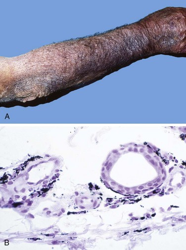
Figure 7–11 Adverse reaction to minocycline, a long-acting tetracycline derivative. A, Diffuse blue-gray pigmentation of the forearm, secondary to minocycline administration. B, Deposition of drug metabolite/iron/melanin pigment particles in the dermis.
(A and B, Courtesy of Dr. Zsolt Argenyi, Department of Pathology, University of Washington, Seattle, Washington.)
Exogenous Estrogens and Oral Contraceptives
Exogenous Estrogens
Estrogen therapy, once used primarily for distressing menopausal symptoms (e.g., hot flashes), has been widely used in postmenopausal women, with or without added progestins, to prevent or slow the progression of osteoporosis (Chapter 20) and to reduce the likelihood of myocardial infarction. Such therapy is referred to as hormone replacement therapy (HRT). In view of the fact that endogenous hyperestrinism increases the risk of endometrial carcinoma and, probably, breast carcinoma, from the outset there has been understandable concern about the use of HRT. The main focus of controversy is the potential benefit of HRT as protection against ischemic myocardial disease. Recent data have confirmed the adverse effects of HRT on endometrial and breast cancers but do not support the view that HRT offers protection against ischemic heart disease. Here is a summary of the main adverse effects of HRT.
• Results from randomized control trials show that HRT with estrogen alone increases the risk of endometrial cancer. Unopposed estrogen therapy increases the risk of endometrial carcinoma 3- to 6-fold after 5 years of use and more than 10-fold after 10 years, but the risk is drastically reduced or eliminated when progestins are added to the therapeutic regimen. On the other hand, long-term HRT with estrogens and progestins is associated with an increased risk of breast cancer. Of note, these findings led to a decrease in HRT prescriptions from 16 million in 2001 to 6 million in 2006, a drop that was accompanied by an apparent decrease in the number of newly diagnosed breast cancers. It is sobering to note that at 3 years of follow-up after cessation of estrogen-progestin HRT, women receiving these hormones continued to develop breast cancer at an increased rate.
• HRT with estrogen, with or without progestins, increases the risk of thromboembolism, including deep vein thrombosis, pulmonary embolism, and stroke, by several-fold. The increase is more pronounced during the first 2 years of treatment and in association with other risk factors such as immobilization or factor V or prothrombin mutations.
• Estrogens and progestins increase blood levels of high-density lipoprotein and decrease levels of low-density lipoprotein. On the basis of retrospective epidemiologic data, it was thought that HRT would be beneficial in protecting against atherosclerosis and ischemic heart disease. However, large well-controlled prospective studies did not demonstrate a protective effect of HRT against myocardial infarction.
Oral Contraceptives
Although OCs have been used for over 35 years, disagreement continues about their safety and adverse effects. They nearly always contain a synthetic estradiol and a variable amount of a progestin (“combination OCs”), but a few preparations contain only progestins. Currently prescribed OCs contain a smaller amount of estrogens (less than 50 µg/day) and clearly have fewer side effects than those reported for earlier formulations. Hence, the results of epidemiologic studies must be interpreted in the context of the dosage. Nevertheless, there is reasonable evidence to support the following conclusions:
• Breast carcinoma: The prevailing opinion is that OCs do not cause an increase in breast cancer risk.
• Endometrial cancer and ovarian cancers: OCs have a protective effect against these tumors.
• Cervical cancer: OCs may increase risk of cervical carcinomas in women infected with human papillomavirus, although it is unclear whether the increased risk results from sexual activity.
• Thromboembolism: Most studies indicate that OCs, including the newer low-dose (less than 50 µg of estrogen) preparations, are associated with a three- to six-fold increased risk of venous thrombosis and pulmonary thromboembolism resulting from increased hepatic synthesis of coagulation factors. This risk may be even higher with newer “third-generation” OCs that contain synthetic progestins, particularly in women who are carriers of the factor V Leiden mutation. To put this complication into context, however, the risk of thromboembolism associated with OC use is two to six times lower than the risk of thromboembolism associated with pregnancy.
• Cardiovascular disease: There is considerable uncertainty about the risk of atherosclerosis and myocardial infarction in users of OCs. It seems that OCs do not increase the risk of coronary artery disease in women younger than 30 years or in older women who are nonsmokers, but the risk does approximately double in women older than 35 years who smoke.
• Hepatic adenoma: There is a well-defined association between the use of OCs and this rare benign hepatic tumor, especially in older women who have used OCs for prolonged periods. The tumor appears as a large, solitary, and well-encapsulated mass.
Obviously, the pros and cons of OCs must be viewed in the context of their wide applicability and acceptance as a form of contraception that protects against unwanted pregnancies.
Acetaminophen
At therapeutic doses, acetaminophen, a widely used nonprescription analgesic and antipyretic, is mostly conjugated in the liver with glucuronide or sulfate. About 5% or less is metabolized to NAPQI (N-acetyl-p-benzoquinoneimine) through the hepatic P-450 system. With very large doses, however, NAPQI accumulates, leading to centrilobular hepatic necrosis. The mechanisms of injury produced by NAPQI include (1) covalent binding to hepatic proteins and (2) depletion of reduced glutathione (GSH). The depletion of GSH makes the hepatocytes more susceptible to cell death caused by reactive oxygen species. The window between the usual therapeutic dose (0.5 g) and the toxic dose (15 to 25 g) is large, and the drug ordinarily is very safe. Nevertheless, accidental overdoses occur in children, and suicide attempts using acetaminophen are not uncommon, particularly in the United Kingdom. Toxicity begins with nausea, vomiting, diarrhea, and sometimes shock, followed in a few days by appearance of jaundice. Overdoses of acetaminophen can be treated in early stages by administration of N-acetylcysteine, which restores GSH. With serious overdoses, liver failure ensues, and centrilobular necrosis may extend to involve entire lobules; patients often require liver transplantation for survival. Some patients also show evidence of concurrent renal damage.
Aspirin (Acetylsalicylic Acid)
Aspirin overdose may result from accidental ingestion in young children or suicide attempts in adults. The major untoward consequences are metabolic, with few morphologic changes. At first, respiratory alkalosis develops, followed by a metabolic acidosis that often proves fatal. Fatal doses may be as little as 2 to 4 gm in children and 10 to 30 gm in adults, but survival has been reported after doses five times larger.
Chronic aspirin toxicity (salicylism) may develop in persons who take 3 gm or more daily (the dose used to treat chronic inflammatory conditions). Chronic salicylism is manifested by headache, dizziness, ringing in the ears (tinnitus), difficulty in hearing, mental confusion, drowsiness, nausea, vomiting, and diarrhea. The CNS changes may progress to convulsions and coma. The morphologic consequences of chronic salicylism are varied. Most often, there is an acute erosive gastritis (Chapter 14), which may produce overt or covert GI bleeding and lead to gastric ulceration. A bleeding tendency may appear concurrently with chronic toxicity, because aspirin irreversibly inhibits platelet cyclooxygenase and blocks the ability to make thromboxane A2, an activator of platelet aggregation. Petechial hemorrhages may appear in the skin and internal viscera, and bleeding from gastric ulcerations may be exaggerated.
Proprietary analgesic mixtures of aspirin and phenacetin or its active metabolite, acetaminophen, when taken over several years, can cause tubulointerstitial nephritis with renal papillary necrosis. This clinical entity is referred to as analgesic nephropathy (Chapter 13).
Injury by Nontherapeutic Toxic Agents (Drug Abuse)
Drug abuse generally involves the use of mind-altering substances beyond therapeutic or social norms. Drug addiction and overdose are serious public health problems. Common drugs of abuse are listed in Table 7–6. Considered here are cocaine, heroin, and marijuana, with a brief mention of a few other drugs.
Table 7–6 Common Drugs of Abuse
| Class | Molecular Target | Examples |
|---|---|---|
| Opioid narcotics | Mu opioid receptor (agonist) | |
| Sedative-hypnotics | GABAA receptor (agonist) | |
| Psychomotor stimulants | Dopamine transporter (antagonist) Serotonin receptors (toxicity) |
|
| Phencyclidine-like drugs | NMDA glutamate receptor channel (antagonist) | |
| Cannabinoids | CB1 cannabinoid receptors (agonist) | |
| Nicotine | Nicotine acetylcholine receptor (agonist) | |
| Hallucinogens | Serotonin 5-HT2 receptors (agonist) |
CB1, cannabinoid receptor type 1; GABA, γ-aminobutyric acid; 5-HT2, 5-hydroxytryptamine; NMDA, N-methyl-d-aspartate; PCP, 1-(1-phenylcyclohexyl)piperidine.
Data from Hyman SE: A 28-year-old man addicted to cocaine. JAMA 286:2586, 2001.
Cocaine
In 2008, the National Survey on Drug Use and Health estimated that there were 1.9 million users of cocaine in the United States, of which approximately 15% to 20% were users of “crack” cocaine. Use is highest among adults 18 to 25 years of age, of whom 1.5% reported taking cocaine within the past month. Extracted from the leaves of the coca plant, cocaine usually is prepared as a water-soluble powder, cocaine hydrochloride, but when sold on the street it is liberally diluted with talcum powder, lactose, or other look-alikes. Crystallization of the pure alkaloid from cocaine hydrochloride yields nuggets of crack (so called because of the popping sound it makes when heated). The pharmacologic actions of cocaine and crack are identical, but crack is far more potent. Both forms can be snorted, smoked after mixing with tobacco, ingested, or injected subcutaneously or intravenously.
Cocaine produces a sense of intense euphoria and mental alertness, making it one of the most addictive of all drugs. Experimental animals will press a lever more than 1000 times and forgo food and drink to obtain the drug. In cocaine users, although physical dependence seems not to occur, the psychologic dependence is profound. Intense cravings are particularly severe in the first several months after abstinence and can recur for years. Acute overdose produces seizures, cardiac arrhythmias, and respiratory arrest. Following are the important manifestations of cocaine toxicity:
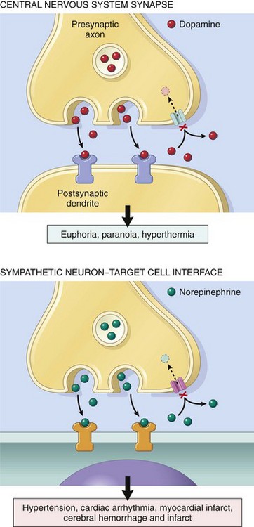
• Cardiovascular effects. The most serious physical effects of cocaine relate to its acute action on the cardiovascular system. Cocaine is a sympathomimetic agent (Fig. 7–12), both in the CNS, where it blocks the reuptake of dopamine, and at adrenergic nerve endings, where it blocks the reuptake of both epinephrine and norepinephrine while stimulating the presynaptic release of norepinephrine. The net effect is the accumulation of these neurotransmitters in synapses and excessive stimulation, manifested by tachycardia, hypertension, and peripheral vasoconstriction. Cocaine also induces myocardial ischemia, the basis for which is multifactorial. It causes coronary artery vasoconstriction and promotes thrombus formation by facilitating platelet aggregation. Cigarette smoking potentiates cocaine-induced coronary vasospasm. Thus, by increasing myocardial oxygen demand by its sympathomimetic action and, at the same time, reducing coronary blood flow, cocaine often triggers myocardial ischemia, which may lead to myocardial infarction. Cocaine also can precipitate lethal arrhythmias by enhanced sympathetic activity as well as by disrupting normal ion (K+, Ca2+, Na+) transport in the myocardium. These toxic effects are not necessarily dose-related, and a fatal event may occur in a first-time user with what is a typical mood-altering dose.
• CNS effects. The most common CNS findings are hyperpyrexia (thought to be caused by aberrations of the dopaminergic pathways that control body temperature) and seizures.
• Effects on the fetus. In pregnant women, cocaine may cause decreased blood flow to the placenta, resulting in fetal hypoxia and spontaneous abortion. Neurologic development may be impaired in the fetuses of pregnant women who are chronic drug users.
• Chronic cocaine use. Chronic use may cause (1) perforation of the nasal septum in snorters, (2) decrease in lung diffusing capacity in users who inhale the smoke, and (3) the development of dilated cardiomyopathy.
Heroin
Heroin is an addictive opioid derived from the poppy plant and is closely related to morphine. Its effects are even more harmful than those of cocaine. Nevertheless, it is estimated that almost 4 million people in the United States have used heroin at least once, and that in 2008 more than 400,000 people used the drug at some time during the year. As sold on the street, it is cut (diluted) with an agent (often talc or quinine); thus, the size of the dose not only is variable but also usually is unknown to the buyer. Heroin along with any contaminating substances usually is self-administered intravenously or subcutaneously. Effects are varied and include euphoria, hallucinations, somnolence, and sedation. Heroin has a wide range of adverse physical effects that can be categorized etiologically according to (1) the pharmacologic action of the agent, (2) reactions to the cutting agents or contaminants, (3) hypersensitivity reactions to the drug or its adulterants, and (4) diseases contracted through sharing of needles. Some of the most important adverse effects of heroin are the following:
• Sudden death. Sudden death, usually related to overdose, is an ever-present risk, because drug purity generally is unknown and may range from 2% to 90%. The yearly incidence of sudden death among chronic users in the United States is estimated to be between 1% and 3%. Sudden death sometimes is due to loss of tolerance for the drug, such as after a period of incarceration. The mechanisms of death include profound respiratory depression, arrhythmia and cardiac arrest, and pulmonary edema.
• Pulmonary disease. Pulmonary complications include edema, septic embolism, lung abscess, opportunistic infections, and foreign body granulomas from talc and other adulterants. Although granulomas occur principally in the lung, they also are sometimes found in the spleen, liver, and lymph nodes that drain the upper extremities. Examination under polarized light often highlights trapped talc crystals, sometimes enclosed within foreign body giant cells.
• Infections. Infectious complications are common. The sites most commonly affected are the skin and subcutaneous tissue, heart valves, liver, and lungs. In a series of addicted patients admitted to the hospital, more than 10% had endocarditis, which often takes a distinctive form involving right-sided heart valves, particularly the tricuspid. Most cases are caused by Staphylococcus aureus, but fungi and a multitude of other organisms have also been implicated. Viral hepatitis is the most common infection among addicts and is acquired by the sharing of dirty needles. In the United States, this practice has also led to a very high incidence of human immunodeficiency virus (HIV) infection in intravenous drug abusers.
• Skin lesions. Cutaneous lesions probably are the most frequent telltale sign of heroin addiction. Acute changes include abscesses, cellulitis, and ulcerations due to subcutaneous injections. Scarring at injection sites, hyperpigmentation over commonly used veins, and thrombosed veins are the usual sequelae of repeated intravenous inoculations.
• Renal problems. Kidney disease is a relatively common hazard. The two forms most frequently encountered are amyloidosis (generally secondary to skin infections) and focal glomerulosclerosis; both induce heavy proteinuria and the nephrotic syndrome.
Marijuana
Marijuana, or “pot,” is the most widely used illegal drug. As of 2008, it was estimated that over 100 million people in the United States had used marijuana during their lifetimes, with more than 15 million people (6.1% of the population) admitting use during the previous month. It is made from the leaves of the Cannabis sativa plant, which contain the psychoactive substance Δ9-tetrahydrocannabinol (THC). When marijuana is smoked, about 5% to 10% of the THC content is absorbed. Despite numerous studies, whether the drug has persistent adverse physical and functional effects remains unresolved. Some of the untoward anecdotal effects may be allergic or idiosyncratic reactions or are possibly related to contaminants in the preparations, rather than to marijuana’s pharmacologic effects. On the other hand, beneficial effects of THC include its capacity to decrease intraocular pressure in glaucoma and to combat intractable nausea secondary to cancer chemotherapy.
The functional and organic CNS consequences of marijuana have received great scrutiny. Marijuana use is well recognized to distort sensory perception and impair motor coordination, but these acute effects generally clear in 4 to 5 hours. With continued use, these changes may progress to cognitive and psychomotor impairments, such as inability to judge time, speed, and distance. Among adolescents, such impairment often leads to automobile accidents. Marijuana increases the heart rate and sometimes blood pressure, and it may cause angina in a person with coronary artery disease.
The lungs are affected by chronic marijuana smoking; laryngitis, pharyngitis, bronchitis, cough, hoarseness, and asthma-like symptoms all have been described, along with mild but significant airway obstruction. Smoking a marijuana cigarette, compared with a tobacco cigarette, is associated with a three-fold increase in the amount of tar inhaled and retained in the lungs, as a consequence of deeper inhalation and longer breath holding.
Other Illicit Drugs
The variety of drugs that have been tried by those seeking “new experiences” (highs, lows, “out-of-body experiences”) defies belief. These drugs include various stimulants, depressants, analgesics, and hallucinogens. Among these are PCP (1-(1-phenylcyclohexyl) piperidine), or phenylcyclidine, and ketamine (related anesthetic agents); lysergic acid diethylamide (LSD), the most potent hallucinogen known; “ecstasy” (3,4-methylenedioxymethamphetamine [MDMA]); and oxycodone (an opiate). Not much is known about the long-time deleterious effects of any of these agents. Acutely, LSD has unpredictable effects on mood, affect, and thought, sometimes leading to bizarre and dangerous behaviors. Chronic use of ecstasy may deplete the CNS of serotonin, potentially leading to sleep disorders, depression, anxiety, and aggressive behavior.
Summary
Drug Injury
• Drug injury may be caused by therapeutic drugs (adverse drug reactions) or non-therapeutic agents (drug abuse).
• Antineoplastic agents, long-acting tetracyclines and other antibiotics, HRT preparations and OCs, acetaminophen, and aspirin are the drugs most frequently involved.
• HRT increases the risk of endometrial and breast cancers and thromboembolism but does not appear to protect against ischemic heart disease. OCs have a protective effect against endometrial and ovarian cancers but increase the risk of thromboembolism and hepatic adenomas.
• Overdose of acetaminophen may cause centrilobular liver necrosis, leading to liver failure. Early treatment with agents that restore GSH levels may limit toxicity. Aspirin blocks the production of thromboxane A2, which may produce gastric ulceration and bleeding.
• The common drugs of abuse include sedative-hypnotics (barbiturates, ethanol), psychomotor stimulants (cocaine, amphetamine, ecstasy), opioid narcotics (heroin, methadone, oxycodone), hallucinogens (LSD, mescaline), and cannabinoids (marijuana, hashish).
Injury by Physical Agents
Injury induced by physical agents is divided into the following categories: mechanical trauma, thermal injury, electrical injury, and injury produced by ionizing radiation. Each type is considered separately.
Mechanical Trauma
Mechanical forces may inflict a variety of forms of damage. The type of injury depends on the shape of the colliding object, the amount of energy discharged at impact, and the tissues or organs that bear the impact. Bone and head injuries result in unique damage and are discussed elsewhere (Chapter 22). All soft tissues react similarly to mechanical forces, and the patterns of injury can be divided into abrasions, contusions, lacerations, incised wounds, and puncture wounds (Fig. 7–13).
Figure 7–13 A, Laceration of the scalp: The bridging strands of fibrous tissues are evident. B, Contusion resulting from blunt trauma. The skin is intact, but hemorrhage of subcutaneous vessels has produced extensive discoloration.
(A, B, From the teaching collection of the Department of Pathology, University of Texas Southwestern Medical School, Dallas, Texas.)
Morphology
An abrasion is a wound produced by scraping or rubbing the skin surface, damaging the superficial layer. Typical skin abrasions remove only the epidermal layer. A contusion, or bruise, is a wound usually produced by a blunt trauma and is characterized by damage to vessel and extravasation of blood into tissues. A laceration is a tear or disruptive stretching of tissue caused by the application of force by a blunt object. In contrast with an incision, most lacerations have intact bridging blood vessels and jagged, irregular edges. An incised wound is one inflicted by a sharp instrument. The bridging blood vessels are severed. A puncture wound is typically caused by a long, narrow instrument and is termed penetrating when the instrument pierces the tissue and perforating when it traverses a tissue to also create an exit wound. Gunshot wounds are special forms of puncture wounds that demonstrate distinctive features important to the forensic pathologist. For example, a wound from a bullet fired at close range leaves powder burns, whereas one fired from more than 4 or 5 feet away does not.
One of the most common causes of mechanical injury is vehicular accident. Injuries typically sustained result from (1) hitting a part of the interior of the vehicle or being hit by objects that enter the passenger compartment during the crash, such as engine parts; (2) being thrown from the vehicle; or (3) being trapped in a burning vehicle. The pattern of injury relates to whether one or all three of these mechanisms are operative. For example, in a head-on collision, a common pattern of injury sustained by a driver who is not wearing a seat belt includes trauma to the head (windshield impact), chest (steering column impact), and knees (dashboard impact). Common chest injuries stemming from such accidents include sternal and rib fractures, heart contusions, aortic lacerations, and (less commonly) lacerations of the spleen and liver. Thus, in caring for an automobile injury victim, it is essential to recognize that internal wounds often accompany superficial abrasions, contusions, and lacerations. Indeed, in many cases, external evidence of serious internal damage is completely absent.
Thermal Injury
Both excess heat and excess cold are important causes of injury. Burns are all too common and are discussed first; a brief discussion of hyperthermia and hypothermia follows.
Thermal Burns
In the United States, burns cause 5000 deaths per year and result in the hospitalization of more than 10 times that many persons. Many victims are children, in whom the cause of injury often is scalding by hot liquids. Fortunately, since the 1970s marked decreases have been seen in both mortality rates and the length of hospitalizations. These improvements have been achieved through better understanding of the systemic effects of massive burns and discovery of better ways to prevent wound infection and facilitate the healing of skin surfaces.
The clinical severity of burns depends on the following important variables:
• Percentage of body surface involved
• Whether internal injuries from inhalation of hot and toxic fumes are present
• Promptness and efficacy of therapy, especially fluid and electrolyte management and prevention or control of wound infections
A full-thickness burn produces total destruction of the epidermis and dermis, including the dermal appendages that harbor cells needed for epithelial regeneration. Both third- and fourth-degree burns are in this category. In partial-thickness burns, at least the deeper portions of the dermal appendages are spared. Partial-thickness burns include first-degree burns (epithelial involvement only) and second-degree burns (involving both epidermis and superficial dermis).
Morphology
On gross inspection, full-thickness burns are white or charred, dry, and anesthetic (as a result of destruction of nerve endings), whereas partial-thickness burns, depending on the depth, are pink or mottled, blistered and painful. Histologic examination of devitalized tissue reveals coagulative necrosis adjacent to vital tissue, which quickly accumulates inflammatory cells and marked exudation.
Despite continuous improvement in therapy, any burn exceeding 50% of the total body surface, whether superficial or deep, is grave and potentially fatal. With burns of more than 20% of the body surface, there is a rapid shift of body fluids into the interstitial compartments, both at the burn site and systemically, which can result in hypovolemic shock (Chapter 3). Because protein from the blood is lost into interstitial tissue, generalized edema, including pulmonary edema, may become severe.
Another important consideration is the degree of injury to the airways and lungs. Inhalation injury is frequent in persons trapped in burning buildings and may result from the direct effect of heat on the mouth, nose, and upper airways or from the inhalation of heated air and gases in the smoke. Water-soluble gases, such as chlorine, sulfur oxides, and ammonia, may react with water to form acids or alkalis, particularly in the upper airways, resulting in inflammation and swelling, which may lead to partial or complete airway obstruction. Lipid-soluble gases, such as nitrous oxide and products of burning plastics, are more likely to reach deeper airways, producing pneumonitis. Unlike in shock, which develops within hours, pulmonary manifestations may not develop for 24 to 48 hours.
Organ system failure resulting from sepsis continues to be the leading cause of death in burned patients. The burn site is ideal for growth of microorganisms; the serum and debris provide nutrients, and the burn injury compromises blood flow, blocking effective inflammatory responses. The most common offender is the opportunist Pseudomonas aeruginosa, but antibiotic-resistant strains of other common hospital-acquired bacteria, such as S. aureus, and fungi, particularly Candida spp., also may be involved. Furthermore, cellular and humoral defenses against infections are compromised, and both lymphocyte and phagocyte functions are impaired. Direct bacteremic spread and release of toxic substances such as endotoxin from the local site have dire consequences. Pneumonia or septic shock accompanied by renal failure and/or the acute respiratory distress syndrome (ARDS) (Chapter 12) are the most common serious sequelae.
Another very important pathophysiologic effect of burns is the development of a hypermetabolic state, with excess heat loss and an increased need for nutritional support. It is estimated that when more than 40% of the body surface is burned, the resting metabolic rate may approach twice normal.
Hyperthermia
Prolonged exposure to elevated ambient temperatures can result in heat cramps, heat exhaustion, or heat stroke.
• Heat cramps result from loss of electrolytes through sweating. Cramping of voluntary muscles, usually in association with vigorous exercise, is the hallmark sign. Heat-dissipating mechanisms are able to maintain normal core body temperature.
• Heat exhaustion is probably the most common hyperthermic syndrome. Its onset is sudden, with prostration and collapse, and it results from a failure of the cardiovascular system to compensate for hypovolemia, secondary to water depletion. After a period of collapse, which is usually brief, equilibrium is spontaneously reestablished.
• Heat stroke is associated with high ambient temperatures and high humidity. Thermoregulatory mechanisms fail, sweating ceases, and core body temperature rises. In the clinical setting, a rectal temperature of 106°F or higher is considered a grave prognostic sign, and the mortality rate for such patients exceeds 50%. The underlying mechanism is marked generalized peripheral vasodilation with peripheral pooling of blood and a decreased effective circulating blood volume. Necrosis of the muscles and myocardium may occur. Arrhythmias, disseminated intravascular coagulation, and other systemic effects are common. Elderly people, persons with cardiovascular disease, and otherwise healthy people undergoing physical stress (such as young athletes and military recruits) are prime candidates for heat stroke.
• Malignant hyperthermia, although similar sounding, is not caused by exposure to high temperature. It is a genetic condition resulting from mutations in genes such as RYR1 that control calcium levels in skeletal muscle cells. In affected individuals, exposure to certain anesthetics during surgery may trigger a rapid rise in calcium levels in skeletal muscle, which in turn leads to muscle rigidity and increased heat production. The resulting hyperthermia has a mortality rate of approximately 80% if untreated, but this falls to less than 5% if the condition is recognized and muscle relaxants are given promptly.
Hypothermia
Prolonged exposure to low ambient temperature leads to hypothermia. The condition is seen all too frequently in homeless alcoholics, in whom wet or inadequate clothing and dilation of superficial blood vessels occurring as a result of the ingestion of alcohol hasten the lowering of body temperature. At about 90°F, loss of consciousness occurs, followed by bradycardia and atrial fibrillation at lower core temperatures.
Chilling or freezing of cells and tissues causes injury by two mechanisms:
• Direct effects probably are mediated by physical disruptions within cells and high salt concentrations incident to the crystallization of the intra- and extracellular water.
• Indirect effects are the result of circulatory changes, which vary depending on the rate and the duration of the temperature drop. Slowly developing, prolonged chilling may induce vasoconstriction and increased permeability, leading to edema. Such changes are typical of “trench foot.” Atrophy and fibrosis may follow. Alternatively, with sudden sharp drops in temperature, the vasoconstriction and increased viscosity of the blood in the local area may cause ischemic injury and degenerative changes in peripheral nerves. In this situation, the vascular injury and increased permeability with exudation only become evident with rewarming. If the period of ischemia is prolonged, hypoxic changes and infarction of the affected tissues (e.g., gangrene of toes or feet) may result.
Electrical Injury
Electrical injuries, which may be fatal, can arise from low-voltage currents (i.e., in the home and workplace) or from high-voltage currents carried in power lines or by lightning. Injuries are of two types: (1) burns and (2) ventricular fibrillation or cardiac and respiratory center failure resulting from disruption of normal electrical impulses. The type of injury and the severity and extent of burning depend on the amperage of the electric current and its path within the body.
Voltage in the household and the workplace (120 or 220 V) is high enough that with low resistance at the site of contact (as when the skin is wet), sufficient current can pass through the body to cause serious injury, including ventricular fibrillation. If current flow continues long enough, it generates enough heat to produce burns at the site of entry and exit as well as in internal organs. An important characteristic of alternating current, the type available in most homes, is that it induces tetanic muscle spasm, so that when a live wire or switch is grasped, irreversible clutching is likely to occur, prolonging the period of current flow. This results in a greater likelihood of extensive electrical burns and, in some cases, spasm of the chest wall muscles, producing death from asphyxia. Currents generated from high-voltage sources cause similar damage; however, because of the large current flows generated, these injuries are more likely to produce paralysis of medullary centers and extensive burns. Lightning is a classic cause of high-voltage electrical injury.
Injury Produced by Ionizing Radiation
Radiation is energy that travels in the form of waves or high-speed particles. Radiation has a wide range of energies that span the electromagnetic spectrum; it can be divided into nonionizing and ionizing radiation. The energy of nonionizing radiation, such as ultraviolet (UV) and infrared light, microwaves, and sound waves, can move atoms in a molecule or cause them to vibrate but is not sufficient to displace electrons from atoms. By contrast, ionizing radiation has sufficient energy to remove tightly bound electrons. Collision of these free electrons with other atoms releases additional electrons, in a reaction cascade referred to as ionization. The main sources of ionizing radiation are (1) x-rays and gamma rays, which are electromagnetic waves of very high frequencies, and (2) high-energy neutrons, alpha particles (composed of two protons and two neutrons), and beta particles, which are essentially electrons. About 18% of the total dose of ionizing radiation received by the U.S. population is related to health care, originating for the most part in use of medical devices and radioisotopes.
Ionizing radiation is indispensable in medical practice, but this application constitutes a two-edged sword. Radiation in this form is used in the treatment of cancer, in diagnostic imaging, and as therapeutic or diagnostic radioisotopes. However, it also is mutagenic, carcinogenic, and teratogenic. The following terms are used to express exposure, absorption, and dose of ionizing radiation:
• Curie (Ci) represents the disintegrations per second of a spontaneously disintegrating radionuclide (radioisotope). One Ci is equal to 3.7 × 10¹⁰ disintegrations per second.
• Gray (Gy) is a unit that expresses the energy absorbed by a target tissue. It corresponds to the absorption of 104 ergs per gram of tissue. A centigray (cGy), which is the absorption of 100 ergs per gram of tissue, is equivalent to the exposure of tissue to 100 rads (R) (“radiation absorbed dose”). The cGy nomenclature has now replaced the rad in medical parlance.
• Sievert (Sv) is a unit of equivalent dose that depends on the biologic rather than the physical effects of radiation (it replaced a unit called the rem). For the same absorbed dose, various types of radiation differ in the extent of damage they produce. The equivalent dose controls for this variation and provides a uniform measuring unit. The equivalent dose (expressed in sieverts) corresponds to the absorbed dose (expressed in grays) multiplied by the relative biologic effectiveness of the radiation. The relative biologic effectiveness depends on the type of radiation, the type and volume of the exposed tissue, and the duration of the exposure, as well as other biologic factors (discussed next). The effective dose of x-rays, computed tomography (CT), and other imaging and nuclear medicine procedures are commonly expressed in millisieverts (mSv).
In addition to the physical properties of the radiation, its biologic effects depend heavily on the following variables:
• Rate of delivery. The rate of delivery significantly modifies the biologic effect. Although the effect of radiant energy is cumulative, delivery in divided doses may allow cells to repair some of the damage in the intervals. Thus, fractional doses of radiant energy have a cumulative effect only to the extent that repair during the intervals is incomplete. Radiotherapy of tumors exploits the capability of normal cells to repair themselves and recover more rapidly than tumor cells.
• Field size. The size of the field exposed to radiation has a great influence on its consequences. The body can sustain relatively high doses of radiation when they are delivered to small, carefully shielded fields, whereas smaller doses delivered to larger fields may be lethal.
• Cell proliferation. Because ionizing radiation damages DNA, rapidly dividing cells are more vulnerable to injury than are quiescent cells. Except at extremely high doses that impair DNA transcription, DNA damage is compatible with survival in nondividing cells, such as neurons and muscle cells. However, in dividing cells, chromosome abnormalities and other types of mutations are recognized by cell cycle checkpoint mechanisms, which lead to growth arrest and apoptosis. Understandably, therefore, tissues with a high rate of cell turnover, such as gonads, bone marrow, lymphoid tissue, and the mucosa of the GI tract, are extremely vulnerable to radiation, and the injury is manifested early after exposure.
• Hypoxia. The production of reactive oxygen species by the radiolysis of water is the most important mechanism of DNA damage by ionizing radiation. Tissue hypoxia, such as may exist in the center of rapidly growing poorly vascularized tumors, may thus reduce the extent of damage and the effectiveness of radiotherapy directed against tumors.
• Vascular damage. Damage to endothelial cells, which are moderately sensitive to radiation, may cause narrowing or occlusion of blood vessels, leading to impaired healing, fibrosis, and chronic ischemic atrophy. These changes may appear months or years after exposure. Despite the low sensitivity of brain cells to radiation, vascular damage after irradiation can lead to late manifestations of radiation injury in this tissue.
DNA Damage and Carcinogenesis
The most important cellular target of ionizing radiation is DNA. Damage to DNA caused by ionizing radiation that is not precisely repaired leads to mutations, which can manifest years or decades later as cancer. Ionizing radiation can cause many types of damage in DNA, including base damage, single- and double-strand breaks, and cross-links between DNA and protein (Fig. 7–14). In surviving cells, simple defects may be reparable by various enzyme repair systems contained in mammalian cells (see Chapter 5). These repair systems are linked to cell cycle regulation through proteins such as ATM (ataxia-telangiectasia mutated) that initiate signal transduction after the damage, and p53, which can transiently slow down the cell cycle to allow for DNA repair or trigger apoptosis of cells that are irreparable. However, double-strand breaks may persist without repair, or the repair of lesions may be imprecise (error-prone), creating mutations. If cell cycle checkpoints are not functioning (for instance, because of a mutation in P53), cells with abnormal and unstable genomes survive and may expand as abnormal clones to eventually form tumors.
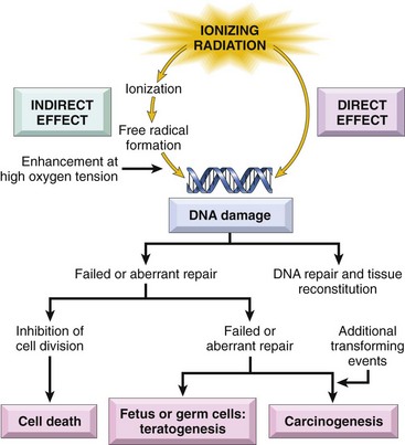
Fibrosis
A common consequence of cancer radiotherapy is the development of fibrosis in the irradiated field (Fig. 7–15). Fibrosis may occur weeks or months after irradiation, leading to the replacement of dead parenchymal cells by connective tissue and the formation of scars and adhesions (see Chapter 2). As already mentioned, ionizing radiation causes vascular damage and consequent tissue ischemia. Vascular damage, the killing of tissue stem cells by ionizing radiation, and the release of cytokines and chemokines that promote an inflammatory reaction and fibroblast activation are the main contributors to the development of radiation-induced fibrosis.
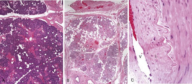
Figure 7–15 Vascular changes and fibrosis of salivary glands produced by radiation therapy of the neck region. A, Normal salivary gland; B, fibrosis caused by radiation; C, fibrosis and vascular changes consisting of fibrointimal thickening and arteriolar sclerosis. V, vessel lumen; I, thickened intima.
(A–C, Courtesy of Dr. Melissa Upton, Department of Pathology, University of Washington, Seattle, Washington.)
Morphology
Cells surviving radiant energy damage show a wide range of structural changes in chromosomes, including deletions, breaks, translocations, and fragmentation. The mitotic spindle often becomes disorderly, and polyploidy and aneuploidy may be encountered. Nuclear swelling and condensation and clumping of chromatin may appear; breaks in the nuclear membrane also may be noted. Apoptosis may occur. Cells with abnormal nuclear morphology may be produced and persist for years, including giant cells with pleomorphic nuclei or more than one nucleus. At extremely high dose levels of radiant energy, features that foretell impending cell death, such as nuclear pyknosis, appear quickly.
In addition to affecting DNA and nuclei, radiant energy may induce a variety of cytoplasmic changes, including cytoplasmic swelling, mitochondrial distortion, and degeneration of the ER. Plasma membrane breaks and focal defects may appear. The histologic constellation of cellular pleomorphism, giant cell formation, changes in nuclei, and mitotic figures creates a more than passing similarity between radiation-injured cells and cancer cells, a problem that plagues the pathologist when evaluating post-irradiation tissues for the possible persistence of tumor cells.
At the light microscopic level, vascular changes and interstitial fibrosis are prominent in irradiated tissues (Fig. 7–15). During the immediate post-irradiation period, vessels may show only dilation. Later, or with higher doses, a variety of degenerative changes appear, including endothelial cell swelling and vacuolation, or even dissolution with total necrosis of the walls of small vessels such as capillaries and venules. Affected vessels may rupture or undergo thrombosis. Still later, endothelial cell proliferation and collagenous hyalinization with thickening of the media layer are seen in irradiated vessels, resulting in marked narrowing or even obliteration of the vascular lumina. At this time, an increase in interstitial collagen in the irradiated field, leading to scarring and contractions, usually becomes evident.
Effects on Organ Systems
Figure 7–16 depicts the main consequences of radiation injury. As already mentioned, the most sensitive organs and tissues are the gonads, the hematopoietic and lymphoid systems, and the lining of the GI tract. Estimated threshold doses for the effects of acute exposure to radiation in various organs are shown in Table 7–7. The changes in the hematopoietic and lymphoid systems, along with cancers induced by environmental or occupational exposure to ionizing radiation, are summarized as follows:
• Hematopoietic and lymphoid systems. The hematopoietic and lymphoid systems are extremely susceptible to radiation injury and deserve special mention. With high dose levels and large exposure fields, severe lymphopenia may appear within hours of irradiation, along with shrinkage of the lymph nodes and spleen. Radiation directly destroys lymphocytes, both in the circulating blood and in tissues (nodes, spleen, thymus, gut). With sublethal doses of radiation, regeneration from viable progenitors is prompt, leading to restoration of a normal lymphocyte count in the blood within weeks to months. The circulating granulocyte count may first rise but begins to fall toward the end of the first week. Levels near zero may be reached during the second week. If the patient survives, recovery of the normal granulocyte count may require 2 to 3 months. Platelets are affected as well, with the nadir of the count occurring somewhat later than that for granulocytes; recovery is similarly delayed. Hematopoietic cells in the bone marrow, including red cell precursors, also are quite sensitive to radiant energy. Red cells are radioresistant, but red cell progenitors are not; as a result, anemia appears after 2 to 3 weeks and may persist for months.
• Environmental exposure and cancer development. Any cell capable of division that has sustained mutations has the potential to become cancerous. Thus, an increased incidence of neoplasms may occur in any organ after exposure to ionizing radiation. The level of radiation required to increase the risk of cancer development is difficult to determine, but there is little doubt that acute or prolonged exposures that result in doses of 100 mSv cause serious consequences, including cancer. This is documented by the increased incidence of leukemias and tumors at various sites (such as thyroid, breast, and lung) in survivors of the atomic bombings of Hiroshima and Nagasaki, the increase in thyroid cancers in survivors of the Chernobyl accident, and the frequent occurrence of “second cancers” in patients, particularly children, treated with radiotherapy for neoplastic disease. It is feared that radiation leaks from the Fukushima nuclear power plant in Japan in the aftermath of the tsunami in 2010 will increase the incidence of cancer in the affected area.
• Occupational exposure and cancer development. Radon is a ubiquitous product of the spontaneous decay of uranium. The carcinogenic agents are two radon decay byproducts (polonium-214 and -218, or “radon daughters”), which emit alpha particles and have a short half-life. These particulates are deposited in the lung, and chronic exposure in uranium miners may give rise to lung carcinomas. Risks also are present in those homes in which the levels of radon are very high, comparable to those found in mines. However, there is little or no evidence to suggest that radon may be a contributor to the risk of lung cancer in the average household.
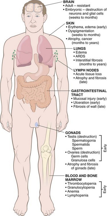
Figure 7–16 Overview of the major morphologic consequences of radiation injury. Early changes occur in hours to weeks; late changes occur in months to years. ARDS, acute respiratory distress syndrome.
Table 7–7 Estimated Threshold Doses for Acute Radiation Effects on Specific Organs
| Health Effect | Organ/Structure | Dose (Sv) |
|---|---|---|
| Temporary sterility | Testes | 0.15 |
| Depression of hematopoiesis | Bone marrow | 0.50 |
| Reversible skin effects (e.g., erythema) | Skin | 1.0–2.0 |
| Permanent sterility | Ovaries | 2.5–6.0 |
| Temporary hair loss | Skin | 3.0–5.0 |
| Permanent sterility | Testis | 3.5 |
| Cataract | Lens of eye | 5.0 |
Total-Body Irradiation
Exposure of large areas of the body to even very small doses of radiation may have devastating effects. Dosages below 1 Sv produce minimal or no symptoms. Higher levels of exposure, however, cause health effects known as acute radiation syndromes, which at progressively higher doses involve the hematopoietic system, GI system, and CNS. The syndromes associated with total-body exposure to ionizing radiation are summarized in Table 7–8.
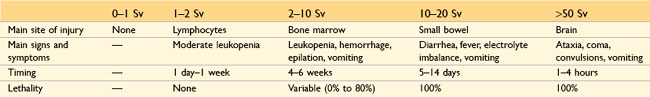
Summary
Radiation Injury
• Ionizing radiation may injure cells directly or indirectly by generating free radicals from water or molecular oxygen.
• Ionizing radiation damages DNA; therefore, rapidly dividing cells such as germ cells, and those in the bone marrow and GI tract are very sensitive to radiation injury.
• DNA damage that is not adequately repaired may result in mutations that predispose affected cells to neoplastic transformation.
• Ionizing radiation may cause vascular damage and sclerosis, resulting in ischemic necrosis of parenchymal cells and their replacement by fibrous tissue.
Nutritional Diseases
Millions of people in developing nations starve or live on the cruel edge of starvation, while those in the developed world struggle to avoid calories and the attendant obesity or fear that what they eat may contribute to atherosclerosis and hypertension. So both lack of nutrition and overnutrition are major health concerns.
Malnutrition
A healthy diet provides (1) sufficient energy, in the form of carbohydrates, fats, and proteins, for the body’s daily metabolic needs; (2) essential (as well as nonessential) amino acids and fatty acids, used as building blocks for synthesis of structural and functional proteins and lipids; and (3) vitamins and minerals, which function as coenzymes or hormones in vital metabolic pathways or, as in the case of calcium and phosphate, as important structural components. In primary malnutrition, one or all of these components are missing from the diet. By contrast, in secondary, or conditional, malnutrition, the dietary intake of nutrients is adequate, and malnutrition results from nutrient malabsorption, impaired utilization or storage, excess losses, or increased requirements. The causes of secondary malnutrition can be grouped into three general but overlapping categories: GI diseases, chronic wasting diseases, and acute critical illness.
Malnutrition is widespread and may be gross or subtle. Some common causes of dietary insufficiencies are listed here.
• Poverty. Homeless people, elderly persons, and children of the poor often suffer from protein-energy malnutrition (PEM) as well as trace nutrient deficiencies. In poor countries, poverty, together with droughts, crop failure, and livestock deaths, creates the setting for malnourishment of children and adults.
• Ignorance. Even the affluent may fail to recognize that infants, adolescents, and pregnant women have increased nutritional needs. Ignorance about the nutritional content of various foods also contributes to malnutrition, as follows: (1) iron deficiency often develops in infants fed exclusively artificial milk diets; (2) polished rice used as the mainstay of a diet may lack adequate amounts of thiamine; and (3) iodine often is lacking from food and water in regions removed from the oceans, unless supplementation is provided.
• Chronic alcoholism. Alcoholic persons may sometimes suffer from PEM but are more frequently lacking in several vitamins, especially thiamine, pyridoxine, folate, and vitamin A, as a result of dietary deficiency, defective GI absorption, abnormal nutrient utilization and storage, increased metabolic needs, and an increased rate of loss. A failure to recognize thiamine deficiency in patients with chronic alcoholism may result in irreversible brain damage (e.g., Korsakoff psychosis, discussed in Chapter 22).
• Acute and chronic illnesses. The basal metabolic rate becomes accelerated in many illnesses (in patients with extensive burns, it may double), resulting in increased daily requirements for all nutrients. Failure to recognize these nutritional needs may delay recovery. PEM is often present in patients with metastatic cancers (discussed later).
• Self-imposed dietary restriction. Anorexia nervosa, bulimia, and less overt eating disorders affect a large population of persons who are concerned about body image or suffer from an unreasonable fear of cardiovascular disease (anorexia and bulimia are discussed in a separate section in this chapter).
• Other causes. Additional causes of malnutrition include GI diseases, acquired and inherited malabsorption syndromes, specific drug therapies (which block uptake or utilization of particular nutrients), and total parenteral nutrition.
The remainder of this section presents a general overview of nutritional disorders. Particular attention is devoted to PEM, anorexia nervosa and bulimia, deficiencies of vitamins and trace minerals, and obesity, with a brief consideration of the relationships of diet to atherosclerosis and cancer. Other nutrients and nutritional issues are discussed in the context of specific diseases throughout the text.
Protein-Energy Malnutrition
Severe PEM is a serious, often lethal disease. It is common in poor countries, where as many as 25% of children may be affected and where it is a major contributor to the high death rates among the very young. For example, in the drought-prone western African country of Niger, the United Nations estimated in 2009 that 800,000 children younger than 5 years were malnourished, and that malnutrition was the major cause of death in infancy and childhood in this population.
PEM manifests as a range of clinical syndromes, all resulting from a dietary intake of protein and calories that is inadequate to meet the body’s needs. The two ends of the spectrum of syndromes are known as marasmus and kwashiorkor. In considering these conditions, an important point is that from a functional standpoint, there are two protein compartments in the body: the somatic compartment, represented by proteins in skeletal muscles, and the visceral compartment, represented by protein stores in the visceral organs, primarily the liver. These two compartments are regulated differently, as detailed subsequently. The somatic compartment is affected more severely in marasmus and the visceral compartment is depleted more severely in kwashiorkor. Clinical assessment of undernutrition is discussed next, followed by descriptions of the clinical presentations of marasmus and kwashiorkor.
The most common victims of PEM worldwide are children. A child whose weight falls to less than 80% of normal is considered malnourished. The diagnosis of PEM is obvious in its most severe forms. In mild to moderate forms, the usual approach is to compare the body weight for a given height against standard tables; other helpful parameters are fat stores, muscle mass, and serum proteins. With a loss of fat, measured skinfold thickness (which includes skin and subcutaneous tissue) is reduced. If the somatic protein compartment is catabolized, the resultant reduction in muscle mass is reflected by reduced circumference of the midarm. Measurement of levels of serum proteins (albumin, transferrin, and others) provides a measure of the adequacy of the visceral protein compartment.
Marasmus
A child is considered to have marasmus when weight level falls to 60% of normal for sex, height, and age (Fig. 7–17, A). A marasmic child suffers growth retardation and loss of muscle mass as a result of catabolism and depletion of the somatic protein compartment. This seems to be an adaptive response that provides the body with amino acids as a source of energy. Of interest, the visceral protein compartment, which presumably is more precious and critical for survival, is depleted only marginally, so serum albumin levels are either normal or only slightly reduced. In addition to muscle proteins, subcutaneous fat is also mobilized and used as fuel. Leptin (discussed later under “Obesity”) production is low, which may stimulate the hypothalamic-pituitary-adrenal axis to produce high levels of cortisol that contribute to lipolysis. With such losses of muscle and subcutaneous fat, the extremities are emaciated; by comparison, the head appears too large for the body. Anemia and manifestations of multivitamin deficiencies are present, and there is evidence of immune deficiency, particularly of T cell–mediated immunity. Hence, concurrent infections are usually present that impose an additional stress on an already weakened body.
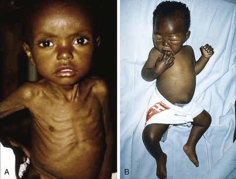
Figure 7–17 Childhood malnutrition. A, Marasmus. Note the loss of muscle mass and subcutaneous fat; the head appears to be too large for the emaciated body. B, Kwashiorkor. The infant shows generalized edema, seen as ascites and puffiness of the face, hands, and legs.
(A, From Clinic Barak, Reisebericht Kenya.)
Kwashiorkor
Kwashiorkor occurs when protein deprivation is relatively greater than the reduction in total calories (Fig. 7–17, B). This is the most common form of PEM seen in African children who have been weaned too early and subsequently fed, almost exclusively, a carbohydrate diet (the name kwashiorkor, from the Ga language in Ghana, describes the illness in a baby that appears after the arrival of another child). The prevalence of kwashiorkor also is high in impoverished countries of Southeast Asia. Less severe forms may occur worldwide in persons with chronic diarrheal states, in which protein is not absorbed, or in those with chronic protein loss (e.g., protein-losing enteropathies, the nephrotic syndrome, or the aftermath of extensive burns). Rare cases of kwashiorkor resulting from fad diets or replacement of milk by rice-based beverages have been reported in the United States.
In kwashiorkor, unlike in marasmus, marked protein deprivation is associated with severe loss of the visceral protein compartment, and the resultant hypoalbuminemia gives rise to generalized or dependent edema (Fig. 7–17). The weight of children with severe kwashiorkor typically is 60% to 80% of normal. However, the true loss of weight is masked by the increased fluid retention (edema). In further contrast with marasmus, there is relative sparing of subcutaneous fat and muscle mass. The modest loss of these compartments may also be masked by edema.
Children with kwashiorkor have characteristic skin lesions with alternating zones of hyperpigmentation, desquamation, and hypopigmentation, giving a “flaky paint” appearance. Hair changes include loss of color or alternating bands of pale and darker color, straightening, fine texture, and loss of firm attachment to the scalp. Other features that distinguish kwashiorkor from marasmus include an enlarged, fatty liver (resulting from reduced synthesis of the carrier protein component of lipoproteins) and the development of apathy, listlessness, and loss of appetite. As in marasmus, vitamin deficiencies are likely to be present, as are defects in immunity and secondary infections. In kwashiorkor, the inflammation caused by infection produces a catabolic state that aggravates the malnutrition. Of note, marasmus and kwashiorkor represent two ends of a spectrum, and considerable overlap exists.
Secondary Protein-Energy Malnutrition
Secondary PEM is common in chronically ill or hospitalized patients. A particularly severe form of secondary PEM, called cachexia, often develops in patients with advanced cancer (Chapter 5). The wasting is all too apparent and often presages death. Although loss of appetite may partly explain it, cachexia may appear before appetite decreases. The underlying mechanisms are complex, but appear to involve “cachectins” such as proteolysis-inducing factor, which are secreted by tumor cells, and cytokines, particularly TNF, which are released as part of the host response to advanced tumors. Both types of factors directly stimulate the degradation of skeletal muscle proteins, and cytokines such as TNF also stimulate fat mobilization from lipid stores.
Morphology
The hallmark anatomic changes in PEM are (1) growth failure, (2) peripheral edema in kwashiorkor, and (3) loss of body fat and atrophy of muscle, more marked in marasmus.
The liver in kwashiorkor, but not in marasmus, is enlarged and fatty; superimposed cirrhosis is rare.
In kwashiorkor (rarely in marasmus) the small bowel shows a decrease in the mitotic index in the crypts of the glands, associated with mucosal atrophy and loss of villi and microvilli. In such cases concurrent loss of small intestinal enzymes occurs, most often manifested as disaccharidase deficiency. Hence, infants with kwashiorkor are lactate intolerant initially and may not respond well to full-strength, milk-based diets. With treatment, the mucosal changes are reversible.
The bone marrow in both kwashiorkor and marasmus may be hypoplastic, mainly as a result of decreased numbers of red cell precursors. How much of this derangement is due to a deficiency of protein and folates and how much to reduced synthesis of transferrin and ceruloplasmin is uncertain. Thus, anemia is usually present, most often hypochromic, microcytic anemia, but a concurrent deficiency of folates may lead to a mixed microcytic-macrocytic anemia.
The brain in infants who are born to malnourished mothers and who suffer from PEM during the first 1 or 2 years of life has been reported by some investigators to show cerebral atrophy, a reduced number of neurons, and impaired myelination of white matter.
Many other changes may be present, including (1) thymic and lymphoid atrophy (more marked in kwashiorkor than in marasmus), (2) anatomic alterations induced by intercurrent infections, particularly with endemic helminths and other parasites, and (3) deficiencies of other required nutrients such as iodine and vitamins.
Anorexia Nervosa and Bulimia
Anorexia nervosa is a state of self-induced starvation resulting in marked weight loss; bulimia is a condition in which the patient binges on food and then induces vomiting. Bulimia is more common than anorexia nervosa and carries a better prognosis. It is estimated to occur in 1% to 2% of women and 0.1% of men, with an average age at onset of 20 years. Anorexia nervosa also occurs primarily in previously healthy young women who have acquired an obsession with attaining or maintaining thinness.
The clinical findings in anorexia nervosa generally are similar to those in severe PEM. In addition, effects on the endocrine system are prominent. Amenorrhea, resulting from decreased secretion of gonadotropin-releasing hormone (and consequent decreased secretion of luteinizing and follicle-stimulating hormones), is so common that its presence is almost a diagnostic feature. Other common findings, related to decreased thyroid hormone release, include cold intolerance, bradycardia, constipation, and changes in the skin and hair. In addition, dehydration and electrolyte abnormalities are frequent findings. The skin becomes dry and scaly and may be yellow-tinged as a result of excess carotene in the blood. Body hair may be increased but usually is fine and pale (lanugo). Bone density is decreased, most likely because of low estrogen levels, which mimics the postmenopausal acceleration of osteoporosis. As expected with severe PEM, anemia, lymphopenia, and hypoalbuminemia may be present. A major complication of anorexia nervosa is an increased susceptibility to cardiac arrhythmia and sudden death, both due to hypokalemia.
In bulimia, binge eating is the norm. Huge amounts of food, principally carbohydrates, are ingested, only to be followed by induced vomiting. Although menstrual irregularities are common, amenorrhea occurs in less than 50% of bulimic patients, probably because weight and gonadotropin levels are maintained near normal. The major medical complications are related to continual induced vomiting and chronic use of laxatives and diuretics. These include (1) electrolyte imbalances (hypokalemia), which predispose the patient to cardiac arrhythmias; (2) pulmonary aspiration of gastric contents; and (3) esophageal and stomach rupture. Nevertheless, there are no specific signs and symptoms for this syndrome, and the diagnosis must rely on a comprehensive psychologic assessment of the patient.
Vitamin Deficiencies
Before we summarize the functions of individual vitamins and the consequence of their deficiency, some general comments are in order.
• Thirteen vitamins are necessary for health; four—A, D, E, and K—are fat-soluble and the remainder water-soluble. The distinction between fat- and water-soluble vitamins is important; although the former are more readily stored in the body, they may be poorly absorbed in fat malabsorption disorders, caused by disturbances of digestive functions (discussed in Chapter 14).
• Certain vitamins can be synthesized endogenously—vitamin D from precursor steroids, vitamin K and biotin by the intestinal microflora, and niacin from tryptophan, an essential amino acid. Notwithstanding this endogenous synthesis, a dietary supply of all vitamins is essential for health.
• Deficiency of a single vitamin is uncommon, and single- or multiple-vitamin deficiencies may be submerged in concurrent PEM.
In the following sections, vitamins A, D, and C are presented in some detail because of their wide-ranging functions and the morphologic changes of deficient states. This is followed by a summary in tabular form of the main consequences of deficiencies of the remaining vitamins—E, K, and the B complex—and some essential minerals.
Vitamin A
Vitamin A is a generic name for a group of related fat-soluble compounds that include retinol, retinal, and retinoic acid, which have similar biologic activities. Retinol is the chemical name for vitamin A. It is the transport form and, as retinol ester, also the storage form. A widely used term, retinoids, refers to both natural and synthetic chemicals that are structurally related to vitamin A but may not necessarily have vitamin A activity. Animal-derived foods such as liver, fish, eggs, milk, and butter are important dietary sources of pre-formed vitamin A. Yellow and leafy green vegetables such as carrots, squash, and spinach supply large amounts of carotenoids, many of which are provitamins that are metabolized to active vitamin A in the body. Carotenoids contribute approximately 30% of the vitamin A in human diets; the most important of these is β-carotene, which is efficiently converted to vitamin A. The recommended dietary allowance for vitamin A is expressed in retinol equivalents, to take into account both pre-formed vitamin A and β-carotene.
As with all fats, the digestion and absorption of carotenes and retinoids require bile and pancreatic enzymes. Retinol (generally ingested as retinol ester) and β-carotene are absorbed through the intestinal wall, where β-carotene is converted to retinol (Fig. 7–18). Retinol is then transported in chylomicrons, where it is taken up into liver cells through the apolipoprotein E receptor. More than 90% of the body’s vitamin A reserves are stored in the liver, predominantly in the perisinusoidal stellate (Ito) cells. In healthy persons who consume an adequate diet, these reserves are sufficient to support the body’s needs for at least 6 months. Retinol esters stored in the liver can be mobilized; before release, retinol binds to a specific retinol-binding protein (RBP), synthesized in the liver. The uptake of retinol and RBP in peripheral tissues is dependent on cell surface RBP receptors. After uptake by cells, retinol is released, and the RBP is recycled back into the blood. Retinol may be stored in peripheral tissues as retinyl ester or be oxidized to form retinoic acid.
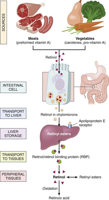
Function
In humans, the best-defined functions of vitamin A are the following:
• Maintaining normal vision in reduced light
• Potentiating the differentiation of specialized epithelial cells, mainly mucus-secreting cells
• Enhancing immunity to infections, particularly in children with measles
In addition, the retinoids, β-carotene, and some related carotenoids can function as photoprotective and antioxidant agents. Retinoids have broad biologic effects, including effects on embryonic development, cellular differentiation and proliferation, and lipid metabolism.
• The visual process involves four forms of vitamin A–containing pigments: rhodopsin, the most light-sensitive pigment and therefore important in reduced light, which is located in rod cells; and three iodopsins, each responsive to a specific color in bright light, which are located in cone cells. The synthesis of rhodopsin from retinol involves (1) oxidation to all-trans-retinal, (2) isomerization to 11-cis-retinal, and (3) interaction with opsin to form rhodopsin. A photon of light causes the isomerization of 11-cis-retinal to all-trans-retinal, and a sequence of configuration changes in rhodopsin, which produce a visual signal. In the process, a nerve impulse is generated (by changes in membrane potential) and transmitted by means of neurons from the retina to the brain. During dark adaptation, some of the all-trans-retinal is reconverted to 11-cis-retinal, but most is reduced to retinol and lost to the retina, explaining the need for continuous supply of retinol.
• Vitamin A and retinoids play an important role in the orderly differentiation of mucus-secreting epithelium. When a deficiency state exists, the epithelium undergoes squamous metaplasia and differentiation to a keratinizing epithelium. All-trans–retinoic acid (ATRA), a potent acid derivative of vitamin A, exerts its effects by binding to retinoic acid receptors (RARs), which regulate the differentiation of myeloid cells. This coupling is the basis for the remarkable ability of ATRA to induce remission of acute promyelocytic leukemia (APML). In this leukemia, a t(15 : 17) translocation (Chapter 11) results in the fusion of a truncated RARA gene on chromosome 17 with the PML gene on chromosome 15. The fusion gene encodes an abnormal RAR that blocks the expression of genes that are required for myeloid cell differentiation. Pharmacologic doses of ATRA overcome the block, causing the malignant promyelocytes to differentiate into neutrophils and die. When combined with other conventional chemotherapeutic agents or arsenic salts, ATRA therapy is often curative in APML. Retinoic acid, it should be noted, has no effect on vision.
• Vitamin A plays a role in host resistance to infections. Vitamin A supplementation can reduce morbidity and mortality rates for some forms of diarrhea. Similarly, supplementation in preschool children with measles, particularly those who are malnourished, can reduce mortality and complications of the disease, including eye damage and blindness.
The effects of vitamin A on infections probably derive in part from its ability to stimulate the immune system through unclear mechanisms. Infections may reduce the bioavailability of vitamin A, possibly by inducing the acute phase response, which appears to inhibit RBP synthesis in the liver. The drop in hepatic RBP causes a decrease in circulating retinol, which reduces the tissue availability of vitamin A. The beneficial effect of vitamin A in diarrheal diseases may be related to the maintenance and restoration of the integrity of the epithelium of the gut.
Deficiency States
Vitamin A deficiency occurs worldwide as a consequence of either poor nutrition or fat malabsorption. In children, stores of vitamin A are depleted by infections, and the absorption of the vitamin is poor in newborn infants. In adults, vitamin A deficiency, in conjunction with depletion of other fat-soluble vitamins, may develop in conjunction with malabsorption syndromes, such as celiac disease, Crohn disease, and colitis. Bariatric surgery and continuous use of mineral oil laxatives also may lead to deficiency. The multiple effects of vitamin A deficiency are discussed next.
• As was already discussed, vitamin A is a component of rhodopsin and other visual pigments. Not surprisingly, one of the earliest manifestations of vitamin A deficiency is impaired vision, particularly in reduced light (night blindness).
• Other effects of vitamin A deficiency are related to the role of vitamin A in maintaining the differentiation of epithelial cells (Fig. 7–19). Persistent deficiency gives rise to a series of changes involving epithelial metaplasia and keratization. The most devastating changes occur in the eyes and result in the clinical entity referred to as xerophthalmia (dry eye). First, there is dryness of the conjunctiva (xerosis conjunctivae) as the normal lachrymal and mucus-secreting epithelium is replaced by keratinized epithelium. This is followed by a buildup of keratin debris in small opaque plaques (Bitot spots) and, eventually, the erosion of the roughened corneal surface, leading to softening and destruction of the cornea (keratomalacia) and total blindness.
• Vitamin A deficiency also leads to replacement of the epithelium lining the upper respiratory passage and urinary tract by keratinizing squamous cells (squamous metaplasia). Loss of the mucociliary epithelium of the airways predisposes affected patients to pulmonary infections, and desquamation of keratin debris in the urinary tract predisposes to renal and bladder stones. Hyperplasia and hyperkeratinization of the epidermis with plugging of the ducts of the adnexal glands may produce follicular or papular dermatosis.
• Another serious consequence of lack of vitamin A is immune deficiency. This impairment of immunity leads to higher mortality rates from common infections such as measles, pneumonia, and infectious diarrhea. In parts of the world with high prevalence of vitamin A deficiency, dietary supplements reduce mortality rates for infectious disorders by 20% to 30%.
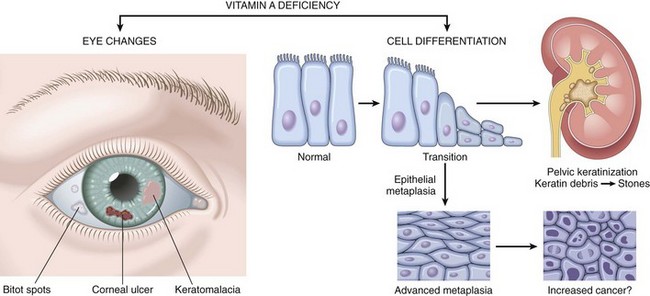
Vitamin A Toxicity
Both short- and long-term excesses of vitamin A may produce toxic manifestations—a point of concern because of the megadoses being touted by certain sellers of supplements. The consequences of acute hypervitaminosis A were first described in 1597 by Gerrit de Veer, a ship’s carpenter stranded in the Arctic, who recounted in his diary the serious symptoms that he and other crew members developed after eating polar bear liver. With this cautionary tale in mind, the adventurous eater should note that acute vitamin A toxicity also has been described in persons who ingested the livers of whales, sharks, and even tuna!
The signs and symptoms of acute toxicity include headache, dizziness, vomiting, stupor, and blurred vision—all of which may be confused with those of a brain tumor. Chronic toxicity is associated with weight loss, anorexia, nausea, vomiting, and bone and joint pain. Retinoic acid stimulates osteoclast production and activity, which lead to increased bone resorption and consequent high risk of fractures. Although synthetic retinoids used for the treatment of acne are not associated with these complications, their use in pregnancy must be avoided because of the well-established teratogenic effect of retinoids.
Vitamin D
The major function of the fat-soluble vitamin D is the maintenance of normal plasma levels of calcium and phosphorus. In this capacity, it is required for the prevention of bone diseases known as rickets (in children whose epiphyses have not already closed), osteomalacia (in adults), and hypocalcemic tetany. With respect to tetany, vitamin D maintains the correct concentration of ionized calcium in the extracellular fluid compartment. When deficiency develops, the drop in ionized calcium in the extracellular fluid results in continuous excitation of muscle (tetany). Our attention here is focused on the function of vitamin D in the regulation of serum calcium levels.
Metabolism
The major source of vitamin D for humans is its endogenous synthesis in the skin by photochemical conversion of a precursor, 7-dehydrocholesterol, powered by the energy of solar or artificial UV light. Irradiation of this compound forms cholecalciferol, known as vitamin D3; in the following discussion, for the sake of simplicity, the term vitamin D is used to refer to this compound.
Under usual conditions of sun exposure, approximately 90% of the vitamin D needed is endogenously derived from 7-dehydrocholesterol present in the skin. However, blacks may have a lower level of vitamin D production in the skin because of melanin pigmentation (perhaps a small price to pay for protection against UV-induced cancers). The small remainder comes from dietary sources, such as deep sea fish, plants, and grains. In plant sources, vitamin D is present in a precursor form, ergosterol, that is converted to vitamin D in the body.
The metabolism of vitamin D can be outlined as follows (Fig. 7–20):
1 Absorption of vitamin D along with other fats in the gut or synthesis from precursors in the skin
2 Binding to plasma α1-globulin (vitamin D–binding protein) and transport to liver
3 Conversion to 25-hydroxyvitamin D (25-OH-D) by 25-hydroxylase in the liver
4 Conversion of 25-OH-D to 1,25-dihydroxyvitamin D [1,25-(OH)2-D] (biologically the most active form of vitamin D) by α1-hydroxylase in the kidney
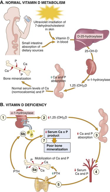
Figure 7–20 A, Normal vitamin D metabolism. B, Vitamin D deficiency. There is inadequate substrate for the renal hydroxylase (1), yielding a deficiency of 1,25-(OH)2D (2), and deficient absorption of calcium and phosphorus from the gut (3), with consequent depressed serum levels of both (4). The hypocalcemia activates the parathyroid glands (5), causing mobilization of calcium and phosphorus from bone (6a). Simultaneously, parathyroid hormone (PTH) induces wasting of phosphate in the urine (6b) and calcium retention. Consequently, the serum levels of calcium are normal or nearly normal, but the phosphate is low; hence, mineralization is impaired (7).
Renal production of 1,25-(OH)2-D is regulated by three mechanisms:
• Hypocalcemia stimulates secretion of parathyroid hormone (PTH), which in turn augments the conversion of 25-OH-D to 1,25-(OH)2-D by activating α1-hydroxylase.
• Hypophosphatemia directly activates α1-hydroxylase, thereby increasing the formation of 1,25(OH)2-D.
• In a feedback loop, increased levels of 1,25-(OH)2-D downregulate the synthesis of this metabolite by inhibiting the action of α1-hydroxylase (decreases in 1,25-(OH)2-D have the opposite effect).
Functions
Like retinoids and steroid hormones, 1,25-(OH)2-D acts by binding to a high-affinity nuclear receptor that in turn binds to regulatory DNA sequences, thereby inducing transcription of specific target genes. The receptors for 1,25-(OH)2-D are present in most nucleated cells of the body, and they transduce signals that result in various biologic activities, beyond those involved in calcium and phosphorus homeostasis. Nevertheless, the best-understood functions of vitamin D relate to the maintenance of normal plasma levels of calcium and phosphorus, through action on the intestines, bones, and kidneys (Fig. 7–20).
• Stimulates intestinal absorption of calcium through upregulation of calcium transport, in enterocytes
• Stimulates calcium resorption in renal distal tubules.
• Collaborates with PTH to regulate blood calcium. This occurs in part through upregulation of RANK ligand on osteoblasts, which in turn activates RANK receptors on osteoclast precursors. RANK activation produces signals that increase osteoclast differentiation and bone resorptive activities (Chapter 20).
• Promotes the mineralization of bone. Vitamin D is needed for the mineralization of osteoid matrix and epiphyseal cartilage during the formation of flat and long bones. It stimulates osteoblasts to synthesize the calcium-binding protein osteocalcin, which promotes calcium deposition.
Of note, effects of vitamin D on bone depend on the plasma levels of calcium: On the one hand, in hypocalcemic states 1,25-(OH)2-D together with PTH increases the resorption of calcium and phosphorus from bone to support blood levels. On the other hand, in normocalcemic states vitamin D also is required for calcium deposition in epiphyseal cartilage and osteoid matrix.
Deficiency States
Rickets in growing children and osteomalacia in adults are skeletal diseases with worldwide distribution. They may result from diets deficient in calcium and vitamin D, but probably more important is limited exposure to sunlight (for instance, in heavily veiled women; children born to mothers who have frequent pregnancies followed by lactation, which leads to vitamin D deficiency; and inhabitants of northern climates with scant sunlight). Other, less common causes of rickets and osteomalacia include renal disorders causing decreased synthesis of 1,25-(OH)2-D or phosphate depletion, and malabsorption disorders. Although rickets and osteomalacia rarely occur outside high-risk groups, milder forms of vitamin D deficiency (also called vitamin D insufficiency) leading to bone loss and hip fractures are common among elderly persons. Studies also suggest that vitamin D may be important for preventing demineralization of bones. It appears that certain genetically determined variants of the vitamin D receptor are associated with an accelerated loss of bone minerals with aging and certain familial forms of osteoporosis (Chapter 20).
Whatever the basis, a deficiency of vitamin D tends to cause hypocalcemia. This in turn stimulates PTH production, which (1) activates renal α1-hydroxylase, increasing the amount of active vitamin D and calcium absorption; (2) mobilizes calcium from bone; (3) decreases renal calcium excretion; and (4) increases renal excretion of phosphate. Thus, the serum level of calcium is restored to near normal, but hypophosphatemia persists, so mineralization of bone is impaired or there is high bone turnover.
An understanding of the morphologic changes in rickets and osteomalacia is facilitated by a brief summary of normal bone development and maintenance. The development of flat bones in the skeleton involves intramembranous ossification, while the formation of long tubular bones proceeds by endochondral ossification. With intramembranous bone formation, mesenchymal cells differentiate directly into osteoblasts, which synthesize the collagenous osteoid matrix on which calcium is deposited. By contrast, with endochondral ossification, growing cartilage at the epiphyseal plates is provisionally mineralized and then progressively resorbed and replaced by osteoid matrix, which undergoes mineralization to create bone (Fig. 7–21, A).
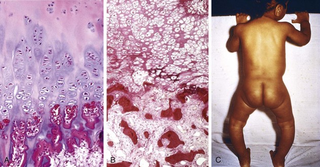
A, Normal costochondral junction of a young child. Note cartilage palisade formation and orderly transition from cartilage to new bone. B, Rachitic costochondral junction in which the palisade of cartilage is absent. Darker trabeculae are well-formed bone; paler trabeculae consist of uncalcified osteoid. C, Note bowing of legs as a consequence of the formation of poorly mineralized bone in a child with rickets.
(B, Courtesy of Dr. Andrew E. Rosenberg, Massachusetts General Hospital, Boston, Massachusetts.)
Morphology
The basic derangement in both rickets and osteomalacia is an excess of unmineralized bone matrix. The changes that occur in the growing bones of children with rickets, however, are complicated by inadequate provisional calcification of epiphyseal cartilage, deranging endochondral bone growth. The following sequence ensues in rickets:
• Overgrowth of epiphyseal cartilage due to inadequate provisional calcification and failure of the cartilage cells to mature and disintegrate
• Persistence of distorted, irregular masses of cartilage, many of which project into the marrow cavity
• Deposition of osteoid matrix on inadequately mineralized cartilaginous remnants
• Disruption of the orderly replacement of cartilage by osteoid matrix, with enlargement and lateral expansion of the osteochondral junction (Fig. 7–21, B)
• Abnormal overgrowth of capillaries and fibroblasts in the disorganized zone resulting from microfractures and stresses on the inadequately mineralized, weak, poorly formed bone
• Deformation of the skeleton due to the loss of structural rigidity of the developing bones
The gross skeletal changes depend on the severity of the rachitic process; its duration; and, in particular, the stresses to which individual bones are subjected. During the nonambulatory stage of infancy, the head and chest sustain the greatest stresses. The softened occipital bones may become flattened, and the parietal bones can be buckled inward by pressure; with the release of the pressure, elastic recoil snaps the bones back into their original positions (craniotabes). An excess of osteoid produces frontal bossing and a squared appearance to the head. Deformation of the chest results from overgrowth of cartilage or osteoid tissue at the costochondral junction, producing the “rachitic rosary.” The weakened metaphyseal areas of the ribs are subject to the pull of the respiratory muscles, causing them to bend inward and creating anterior protrusion of the sternum (pigeon breast deformity). The inward pull at the margin of the diaphragm creates the Harrison groove, girdling the thoracic cavity at the lower margin of the rib cage. The pelvis may become deformed. When an ambulating child develops rickets, deformities are likely to affect the spine, pelvis, and long bones (e.g., tibia), causing, most notably, lumbar lordosis and bowing of the legs (Fig. 7–21, C).
In adults, the lack of vitamin D deranges the normal bone remodeling that occurs throughout life. The newly formed osteoid matrix laid down by osteoblasts is inadequately mineralized, producing the excess of persistent osteoid that is characteristic of osteomalacia. Although the contours of the bone are not affected, the bone is weak and vulnerable to gross fractures or microfractures, which are most likely to affect vertebral bodies and femoral necks. On histologic examination, the unmineralized osteoid can be visualized as a thickened layer of matrix (which stains pink in hematoxylin and eosin preparations) arranged about the more basophilic, normally mineralized trabeculae.
Toxicity
Prolonged exposure to normal sunlight does not produce an excess of vitamin D, but megadoses of orally administered vitamin can lead to hypervitaminosis. In children, hypervitaminosis D may take the form of metastatic calcifications of soft tissues such as the kidney; in adults, it causes bone pain and hypercalcemia. As a point of some interest, the toxic potential of this vitamin is so great that in sufficiently large doses it is a potent rodenticide!
Vitamin C (Ascorbic Acid)
A deficiency of water-soluble vitamin C leads to the development of scurvy, characterized principally by bone disease in growing children and by hemorrhages and healing defects in both children and adults. Sailors of the British Royal Navy were nicknamed “limeys” because at the end of the 18th century the Navy began to provide lime and lemon juice to them to prevent scurvy during their long sojourn at sea. It was not until 1932 that ascorbic acid was identified and synthesized. Unlike vitamin D, ascorbic acid is not synthesized endogenously in humans, who therefore are entirely dependent on the diet for this nutrient. Vitamin C is present in milk and some animal products (liver, fish) and is abundant in a variety of fruits and vegetables. All but the most restricted diets provide adequate amounts of vitamin C.
Function
Ascorbic acid acts in a variety of biosynthetic pathways by accelerating hydroxylation and amidation reactions. The most clearly established function of vitamin C is the activation of prolyl and lysyl hydroxylases from inactive precursors, allowing for hydroxylation of procollagen. Inadequately hydroxylated procollagen cannot acquire a stable helical configuration or be adequately cross-linked, so it is poorly secreted from the fibroblasts. Those molecules that are secreted lack tensile strength, are more soluble, and are more vulnerable to enzymatic degradation. Collagen, which normally has the highest content of hydroxyproline, is most affected, particularly in blood vessels, accounting for the predisposition to hemorrhages in scurvy. In addition, a deficiency of vitamin C suppresses the synthesis of collagen polypeptides, independent of effects on proline hydroxylation.
Vitamin C also has antioxidant properties. These include an ability to scavenge free radicals directly and participation in metabolic reactions that regenerate the antioxidant form of vitamin E.
Deficiency States
Consequences of vitamin C deficiency are illustrated in Figure 7–22. Fortunately, because of the abundance of ascorbic acid in foods, scurvy has ceased to be a global problem. It is sometimes encountered even in affluent populations as a secondary deficiency, particularly among elderly persons, people who live alone, and chronic alcoholics—groups often characterized by erratic and inadequate eating patterns. Occasionally, scurvy appears in patients undergoing peritoneal dialysis and hemodialysis and among food faddists.
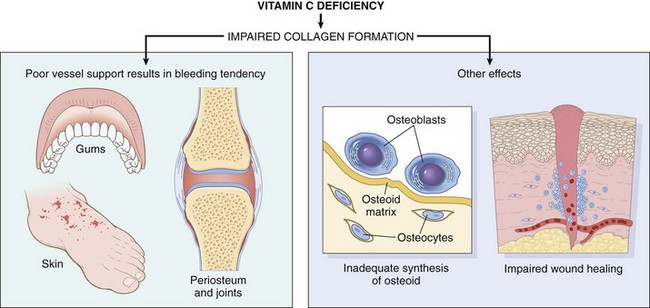
Toxicity
The popular notion that megadoses of vitamin C protect against the common cold or at least allay the symptoms has not been borne out by controlled clinical studies. Such slight relief as may be experienced probably is a result of the mild antihistamine action of ascorbic acid. The large excess of vitamin C is promptly excreted in the urine but may cause uricosuria and increased absorption of iron, with the potential for iron overload.
Other vitamins and some essential minerals are listed and briefly described in Tables 7-9 and 7-10. Folic acid and vitamin B12 are discussed in Chapter 11.
Table 7–9 Vitamins: Major Functions and Deficiency Syndromes
| Vitamin | Functions | Deficiency Syndromes |
|---|---|---|
| Fat-Soluble | ||
| Vitamin A | A component of visual pigment | Night blindness, xerophthalmia, blindness |
| Maintenance of specialized epithelia | Squamous metaplasia | |
| Maintenance of resistance to infection | Vulnerability to infection, particularly measles | |
| Vitamin D | Facilitates intestinal absorption of calcium and phosphorus and mineralization of bone | Rickets in children Osteomalacia in adults |
| Vitamin E | Major antioxidant; scavenges free radicals | Spinocerebellar degeneration |
| Vitamin K | Cofactor in hepatic carboxylation of procoagulants—factors II (prothrombin), VII, IX, and X; and protein C and protein S | Bleeding diathesis |
| Water-Soluble | ||
| Vitamin B1 (thiamine) | As pyrophosphate, is coenzyme in decarboxylation reactions | Dry and wet beriberi, Wernicke syndrome, Korsakoff syndrome |
| Vitamin B2 (riboflavin) | Converted to coenzymes flavin mononucleotide and flavin adenine dinucleotide, cofactors for many enzymes in intermediary metabolism | Cheilosis, stomatitis, glossitis, dermatitis, corneal vascularization |
| Niacin | Incorporated into nicotinamide adenine dinucleotide (NAD) and NAD phosphate; involved in a variety of oxidation–reduction (redox) reactions | Pellagra—“three Ds”: dementia, dermatitis, diarrhea |
| Vitamin B6 (pyridoxine) | Derivatives serve as coenzymes in many intermediary reactions | Cheilosis, glossitis, dermatitis, peripheral neuropathy |
| Vitamin B12 | Required for normal folate metabolism and DNA synthesis Maintenance of myelinization of spinal cord tracts |
Combined system disease (megaloblastic anemia and degeneration of posterolateral spinal cord tracts) |
| Vitamin C | Serves in many redox reactions and hydroxylation of collagen | Scurvy |
| Folate | Essential for transfer and use of one-carbon units in DNA synthesis | Megaloblastic anemia, neural tube defects |
| Pantothenic acid | Incorporated in coenzyme A | No nonexperimental syndrome recognized |
| Biotin | Cofactor in carboxylation reactions | No clearly defined clinical syndrome |
Summary
Nutritional Diseases
• Primary PEM is a common cause of childhood deaths in poor countries. The two main primary PEM syndromes are marasmus and kwashiorkor. Secondary PEM occurs in the chronically ill and in patients with advanced cancer (as a result of cachexia).
• Kwashiorkor is characterized by hypoalbuminemia, generalized edema, fatty liver, skin changes, and defects in immunity. It is caused by diets low in protein but normal in calories.
• Marasmus is characterized by emaciation resulting from loss of muscle mass and fat with relative preservation of serum albumin. It is caused by diets severely lacking in calories—both protein and nonprotein.
• Anorexia nervosa is self-induced starvation; it is characterized by amenorrhea and multiple manifestations of low thyroid hormone levels. Bulimia is a condition in which food binges alternate with induced vomiting.
• Vitamins A and D are fat-soluble vitamins with a wide range of activities. Vitamin C and members of the vitamin B family are water-soluble (Table 7–9 lists vitamin functions and deficiency syndromes).
Obesity
In the United States, obesity has reached epidemic proportions. The prevalence of obesity increased from 13% to 34% between 1960 and 2008, and as of 2009, 68% of Americans between 20 and 75 years of age were overweight. Equally alarming, childhood obesity, a strong predictor of obesity in adults, also increased two- to three-fold over the same period. Recent studies suggest that the epidemic of obesity also is rapidly spreading in developing countries such as India. Globally, the World Health Organization (WHO) estimates that by 2015, 700 million adults will be obese. The causes of this epidemic are complex but undoubtedly are related to societal changes in diet and levels of physical activity. Obesity is associated with an increased risk of several important diseases (e.g., diabetes, hypertension), making it a major public health concern. Indeed, in 2009 it was estimated that the health care cost of obesity had risen to $147 billion annually in the United States, a price tag that appears bound to rise as the nation’s collective waistline expands.
Obesity is defined as a state of increased body weight, due to adipose tissue accumulation, that is of sufficient magnitude to produce adverse health effects. How does one measure fat accumulation? Several high-tech methods have been devised, but for practical purposes the following measures are commonly used:
• Some expression of weight in relation to height, such as the measurement referred to as the body mass index (BMI) = (weight in kilograms)/(height in meters)2, or kg/m2
• Various body circumferences, particularly the waist-to-hip circumference ratio
The BMI is closely correlated with body fat. BMIs in the range 18.5 to 25 kg/m2 are considered normal, while BMIs between 25 and 30 kg/m2 identify the overweight, and BMIs greater than 30 kg/m2, the obese. It is generally agreed that a BMI higher than 30 kg/m2 imparts a health risk. In the following discussion, for the sake of simplicity, the term obesity is applied to both the overweight and the truly obese.
The untoward effects of obesity are related not only to the total body weight but also to the distribution of the stored fat. Central, or visceral, obesity, in which fat accumulates in the trunk and in the abdominal cavity (in the mesentery and around viscera), is associated with a much higher risk for several diseases than is excess accumulation of fat in a diffuse distribution in subcutaneous tissue.
The etiology of obesity is complex and incompletely understood. Involved are genetic, environmental, and psychologic factors. However, simply put, obesity is a disorder of energy balance. The two sides of the energy equation, intake and expenditure, are finely regulated by neural and hormonal mechanisms, so that body weight is maintained within a narrow range for many years. Apparently, this fine balance is controlled by an internal set point, or “lipostat,” that senses the quantity of energy stores (adipose tissue) and appropriately regulates food intake as well as energy expenditure. In recent years, several “obesity genes” have been identified. As might be expected, they encode the molecular components of the physiologic system that regulates energy balance. A key player in energy homeostasis is the LEP gene and its product, leptin. This unique member of the cytokine family, secreted by adipocytes, regulates both sides of the energy equation—intake of food and expenditure of energy. As discussed later, the net effect of leptin is to reduce food intake and enhance the expenditure of energy.
In a simplified way the neurohumoral mechanisms that regulate energy balance and body weight may be divided into three components (Fig. 7–23):
• The peripheral or afferent system generates signals from various sites. Its main components are leptin and adiponectin produced by fat cells, insulin from the pancreas, ghrelin from the stomach, and peptide YY from the ileum and colon. Leptin reduces food intake and is discussed in detail further on. Ghrelin secretion stimulates appetite, and it may function as a “meal-initiating” signal. Peptide YY, which is released postprandially by endocrine cells in the ileum and colon, is a satiety signal.
• The arcuate nucleus in the hypothalamus, which processes and integrates the peripheral signals and generates new signals that are transmitted by (1) POMC (pro-opiomelanocortin) and CART (cocaine- and amphetamine-regulated transcript) neurons; and (2) NPY (neuropeptide Y) and AgRP (agouti-related peptide) neurons.
• The efferent system, which consists of hypothalamic neurons regulated by the arcuate nucleus. POMC/CART neurons activate efferent neurons that enhance energy expenditure and weight loss, while NPY/AgRP neurons activate efferent neurons that promote food intake and weight gain. Signals transmitted by efferent neurons also communicate with forebrain and midbrain centers that control the autonomic nervous system.
Figure 7–23 Energy balance regulatory circuitry.
When sufficient energy is stored in adipose tissue and the individual is well fed, afferent adiposity signals (insulin, leptin, ghrelin, peptide YY) are delivered to the central neuronal processing units, in the hypothalamus. Here the adiposity signals inhibit anabolic circuits and activate catabolic circuits. The effector arms of these central circuits then influence energy balance by inhibiting food intake and promoting energy expenditure. This in turn reduces the energy stores, and pro-adiposity signals are blunted. Conversely, when energy stores are low, the available anabolic circuits take over, at the expense of catabolic circuits, to generate energy stores in the form of adipose tissue.
Discussed next are three important components of the afferent system that regulate appetite and satiety: leptin, adipose tissue, and gut hormones.
Leptin
Through complex, incompletely understood mechanisms, the output of leptin is regulated by the adequacy of fat stores. With abundant adipose tissue, leptin secretion is stimulated, and the hormone travels to the hypothalamus, where it reduces food intake by stimulating POMC/CART neurons and inhibiting NPY/AgRP neurons. The opposite sequence of events occurs when there are inadequate stores of body fat: Leptin secretion is diminished and food intake is increased. In persons of stable weight, the activities of these pathways are balanced. Leptin also increases energy expenditure by stimulating physical activity, energy expenditure, and thermogenesis, which may be the most important catabolic effects mediated by leptin through the hypothalamus. Thermogenesis seems to be controlled in part by efferent hypothalamic signals that increase the release of norepinephrine from sympathetic nerve endings in adipose tissue. Fat cells express β3-adrenergic receptors that, when stimulated by norepinephrine, cause fatty acid hydrolysis and also uncouple energy production from storage.
In rodents and humans, loss-of-function mutations affecting components of the leptin pathway give rise to massive obesity. Mice with mutations that disable the leptin gene or its receptor fail to sense the adequacy of fat stores, so they behave as if they are undernourished, eating ravenously. As in mice, mutations of the leptin gene or receptor in humans, although rare, may cause massive obesity. More common are mutations in the melanocortin receptor-4 gene (MC4R) gene, found in 4% to 5% of patients with massive obesity. These monogenic traits underscore the importance of the leptin pathway in the control of body weight, and it is possible that more common types of defects in this pathway will be discovered in the obese. For example, many obese persons have high blood leptin levels, suggesting that leptin resistance is prevalent among humans.
Adipose Tissue
In addition to leptin, adipose tissue produces other mediators, such as adiponectin, cytokines, chemokines, and steroid hormones, which allow adipose tissue to function as a link between lipid metabolism, nutrition, and inflammatory responses. The total number of adipocytes is established by adolescence and is higher in people who were obese as children, providing another reason for concern about childhood obesity. Although in adults about 10% of adipocytes turn over annually, the number of adipocytes remains constant, regardless of individual body mass. Diets fail in part because loss of fat from adipocytes causes leptin levels to fall, stimulating the appetite and diminishing energy expenditure.
Gut Hormones
Gut hormones are rapidly acting initiators and terminators of volitional eating. Prototypical examples are ghrelin and peptide YY (PYY). Ghrelin is produced in the stomach and is the only known gut peptide that increases food intake. It probably acts by stimulating the NPY/AgRP neurons in the hypothalamus. Ghrelin levels normally rise before meals and fall 1 to 2 hours afterward, but this drop is attenuated in obese persons. PYY is secreted from endocrine cells in the ileum and colon in response to consumption of food. It presumably acts by stimulating POMC/CART neurons in the hypothalamus, thereby decreasing food intake.
Clinical Consequences of Obesity
Obesity, particularly central obesity, is a known risk factor for a number of conditions, including type 2 diabetes, cardiovascular disease, and cancer. Central obesity also stands at the center of a cluster of alterations known as the metabolic syndrome, characterized by abnormalities of glucose and lipid metabolism coupled with hypertension and evidence of a systemic pro-inflammatory state. The mechanisms underlying these associations are complex and probably interrelated. The following associations are worthy of note:
• Obesity is associated with insulin resistance and hyperinsulinemia, important features of type 2 diabetes (formerly known as non–insulin-dependent diabetes). It has been speculated that excess insulin, in turn, may play a role in the retention of sodium, expansion of blood volume, production of excess norepinephrine, and smooth muscle proliferation that are the hallmarks of hypertension. Whatever the mechanism, the risk of developing hypertension among previously normotensive persons increases proportionately with weight.
• Obese persons generally have hypertriglyceridemia and low HDL cholesterol levels, factors that increase the risk of coronary artery disease. The association between obesity and heart disease is not straightforward, however, and such linkage as there is relates more to the associated diabetes and hypertension than to weight per se.
• There is an increased incidence of certain cancers in the overweight, including cancers of the esophagus, thyroid, colon, and kidney in men and cancers of the esophagus, endometrium, gallbladder, and kidney in women. Overall, obesity is associated with approximately 20% of cancer deaths in women and 14% of deaths in men. The underlying mechanisms are unknown and are likely to be multiple. One suspect is hyperinsulinemia. Insulin increases levels of insulin-like growth factor-1 (IGF-1), which can stimulate the growth and survival of many types of cancer cells by activating its cognate receptor, IGF1R. The association of obesity and endometrial cancer may be indirect: High estrogen levels are associated with an increased risk of endometrial cancer (Chapter 18), and obesity is known to raise estrogen levels. With breast cancer, the data are controversial.
• Nonalcoholic steatohepatitis is commonly associated with obesity and type 2 diabetes. This condition, also referred to as nonalcoholic fatty liver disease, can progress to fibrosis and cirrhosis (Chapter 15).
• Cholelithiasis (gallstones) is six times more common in obese than in lean subjects. The mechanism is mainly an increase in total body cholesterol, increased cholesterol turnover, and augmented biliary excretion of cholesterol in the bile, which in turn predisposes affected persons to the formation of cholesterol-rich gallstones (Chapter 15).
• Hypoventilation syndrome is a constellation of respiratory abnormalities in very obese persons. It has been called the pickwickian syndrome, after the fat lad who was constantly falling asleep in Charles Dickens’ The Pickwick Papers. Hypersomnolence, both at night and during the day, is characteristic and is often associated with apneic pauses during sleep, polycythemia, and eventual right-sided heart failure.
• Marked adiposity is a predisposing factor for the development of degenerative joint disease (osteoarthritis). This form of arthritis, which typically appears in older persons, is attributed in large part to the cumulative effects of wear and tear on joints. The greater the body burden of fat, the greater the trauma to joints with passage of time.
• Markers of inflammation, such as C-reactive protein (CRP) and pro-inflammatory cytokines like TNF, are often elevated in obese persons. The basis for the inflammation is uncertain; both a direct pro-inflammatory effect of excess circulating lipids and increased release of cytokines from fat-laden adipocytes have been proposed. Whatever the cause, it is thought that chronic inflammation may contribute to many of the complications of obesity, including insulin resistance, metabolic abnormalities, thrombosis, cardiovascular disease, and cancer.
Summary
Obesity
• Obesity is a disorder of energy regulation. It increases the risk for a number of important conditions such as insulin resistance, type 2 diabetes, hypertension, and hypertriglyceridemia, which are associated with the development of coronary artery disease.
• The regulation of energy balance is very complex. It has three main components: (1) afferent signals, provided mostly by insulin, leptin, ghrelin, and peptide YY; (2) the central hypothalamic system, which integrates afferent signals and triggers the efferent signals; and (3) efferent signals, which control energy balance.
• Leptin plays a key role in energy balance. Its output from adipose tissues is regulated by the abundance of fat stores. Leptin binding to its receptors in the hypothalamus reduces food intake by stimulating POMC/CART neurons and inhibiting NPY/AgRP neurons.
• In addition to diabetes and cardiovascular disease, obesity also is associated with increased risk for certain cancers, nonalcoholic fatty liver disease, and gallstones.
Diet and Systemic Diseases
The problems of under- and overnutrition, as well as specific nutrient deficiencies, have been discussed; however, the composition of the diet, even in the absence of any of these problems, may make a significant contribution to the causation and progression of a number of diseases. A few examples suffice here.
Currently, one of the most important and controversial issues is the contribution of diet to atherogenesis. The central question is whether dietary modification—specifically, reduction in the consumption of foods high in cholesterol and saturated animal fats (e.g., eggs, butter, beef)—can reduce serum cholesterol levels and prevent or retard the development of atherosclerosis (of most importance, coronary heart disease). The average adult in the United States consumes a large amount of fat and cholesterol daily, with a ratio of saturated fatty acids to polyunsaturated fatty acids of about 3 : 1. Lowering the level of saturates to the level of the polyunsaturates causes a 10% to 15% reduction in serum cholesterol level within a few weeks. Vegetable oils (e.g., corn and safflower oils) and fish oils contain polyunsaturated fatty acids and are good sources of such cholesterol-lowering lipids. Fish oil fatty acids belonging to the omega-3, or n-3, family have more double bonds than do the omega-6, or n-6, fatty acids found in vegetable oils. One study of Dutch men whose usual daily diet contained 30 gm of fish revealed a substantially lower frequency of death from coronary heart disease than that among comparable control subjects, providing some hope (but no definitive proof) that long-term supplementation of food with omega-3 fatty acids may reduce coronary artery disease.
Other specific effects of diet on disease have been recognized:
• Hypertension is reduced by restricting sodium intake.
• Dietary fiber, or roughage, resulting in increased fecal bulk, is thought by some investigators to have a preventive effect against diverticulosis of the colon.
• Caloric restriction has been convincingly demonstrated to increase life span in experimental animals, including monkeys. The basis for this striking observation is not clear (Chapter 1).
• Even lowly garlic has been touted to protect against heart disease (and also, alas, against kisses—and the devil), although research has yet to prove this effect unequivocally.
Diet and Cancer
With respect to carcinogenesis, three aspects of the diet are of concern: (1) the content of exogenous carcinogens, (2) the endogenous synthesis of carcinogens from dietary components, and (3) the lack of protective factors.
• An example of an exogenous carcinogen is aflatoxin, which is an important factor in the development of hepatocellular carcinomas in parts of Asia and Africa. Exposure to aflatoxin causes a specific mutation (codon 249) in the P53 gene in tumor cells. The mutation can be used as a molecular signature for aflatoxin exposure in epidemiologic studies. Debate continues about the carcinogenicity of food additives, artificial sweeteners, and contaminating pesticides. Some artificial sweeteners (cyclamates and saccharin) have been implicated in the pathogenesis of bladder cancers, but convincing evidence is lacking.
• The concern about endogenous synthesis of carcinogens or promoters from components of the diet relates principally to gastric carcinomas. Nitrosamines and nitrosamides are suspected to generate these tumors in humans, as they induce gastric cancer in animals. These compounds are formed in the body from nitrites and amines or amides derived from digested proteins. Sources of nitrites include sodium nitrite, added to foods as a preservative, and nitrates, present in common vegetables, which are reduced in the gut by bacterial flora. There is, then, the potential for endogenous production of carcinogenic agents from dietary components, which might well have an effect on the stomach.
• High animal fat intake combined with low fiber intake has been implicated in the causation of colon cancer. The most convincing explanation for this association is as follows: High fat intake increases the level of bile acids in the gut, which in turn modifies intestinal flora, favoring the growth of microaerophilic bacteria. The bile acids or bile acid metabolites produced by these bacteria might serve as carcinogens or promoters. The protective effect of a high-fiber diet might relate to (1) increased stool bulk and decreased transit time, which decreases the exposure of mucosa to putative offenders, and (2) the capacity of certain fibers to bind carcinogens and thereby protect the mucosa. Attempts to document these theories in clinical and experimental studies have, on the whole, led to contradictory results.
• Vitamins C and E, β-carotenes, and selenium have been assumed to have anticarcinogenic effects because of their antioxidant properties. To date, however, no convincing evidence has emerged to show that these antioxidants act as chemopreventive agents. As already mentioned, retinoic acid promotes epithelial differentiation and is believed to reverse squamous metaplasia.
Thus, despite many tantalizing trends and proclamations by “diet gurus,” to date there is no definite proof that diet in general can cause or protect against cancer. Nonetheless, concern persists that carcinogens lurk in things as pleasurable as a juicy steak and rich ice cream.
Bellinger DC. Lead. Pediatrics. 2004;113:1016. [An excellent overview of the subject.]
Boffetta P, Hecht S, Gray N, et al. Smokeless tobacco and cancer. Lancet Oncol. 2009;9:667. [A review of cancer risks associated with smokeless tobacco worldwide.]
Centers for Disease Control and Prevention: Third National Report on Human Exposure to Environmental Chemicals, 2005. [A very important survey of environmental chemicals, with comments on exposure and health risk trends.]
Casals-Casas C, Desvergne B. Endocrine disruptors: from endocrine to metabolic disruption. Annu Rev Phys. 2011;73:135. [An update discussing the scope and possible consequences of human exposure to this class of chemical.]
Clarkson TW, Magos L, Myers GJ. The toxicology of mercury—current exposures and clinical manifestations. N Engl J Med. 2003;349:1731. [An excellent overview of the subject.]
Gregor MF, Hotamisligil GS. Inflammatory mechanisms in obesity. Annu Rev Immunol. 2011;29:445. [A concise discussion of current views of the pro-inflammatory state associated with obesity.]
Heiss G, Wallace R, Anderson GL, et al. Health risks and benefits 3 years after stopping randomized treatment with estrogen and progestin. JAMA. 2008;299:1036. [A paper describing a persistently elevated risk of breast cancer in women 3 years after stopping HRT.]
Hollick MF. Vitamin D deficiency. N Engl J Med. 2007;357:266. [A comprehensive review of vitamin D deficiency.]
Jornayvaz FR, Samuel VT, Shulman GI. The role of muscle insulin resistance in the pathogenesis of atherogenic dyslipidemia and nonalcoholic fatty liver disease associated with the metabolic syndrome. Annu Rev Nutr. 2010;30:273. [An interesting perspective on the metabolic syndrome focused on the role of insulin resistance in skeletal muscle.]
Manson JE, Hsia J, Johnson KC, et al. Estrogen plus progestin and the risk of coronary heart disease. N Engl J Med. 2003;349:523. [A landmark study from the Women’s Health Initiative.]
Pope CA, Ezzati M, Dockery DW. Fine-particulate air pollution and life expectancy in the United States. N Engl J Med. 2009;360:376. [An interesting paper correlating increases in life expectancy in major U.S. cities with decreases in fine-particulate air pollution.]
Ravdin PM, Cronin KA, Howlader N, et al. The decrease in breast-cancer incidence in 2003 in the United States. N Engl J Med. 2007;356:1670. [An important paper documenting the decrease in breast cancer that followed its linkage to HRT.]
Roberts DL, Dive C, Renehan AG. Biological mechanisms linking obesity and cancer risk: new perspectives. Annu Rev Med. 2010;61:301. [A discussion of the possible interactions between obesity and cancer risk.]
Suzuki K, Simpson KA, Minnion JS, et al. The role of gut hormones and the hypothalamus in appetite regulation. Endocr J. 2010;57:359. [An excellent review of the interplay between the gut and the hypothalamus in regulating food consumption.]
Tang X-H, Gudas LJ. Retinoids, retinoic acid receptors, and cancer. Annu Rev Pathol. 2011;6:345. [A review of the role of retinoids in cancer, with a focus on solid tumors.]