The small intestine extends from the duodenum to the ileocaecal valve. It is approximately 3–6 m in length, and 300 m2 in surface area. The upper 40% is the duodenum and jejunum, the remainder is the ileum. Its surface area is enormously increased by circumferential mucosal folds that have on them multiple finger-like projections called villi. On the villi the surface area is further increased by microvilli on the luminal side of the epithelial cells (enterocytes) (Fig. 6.23).
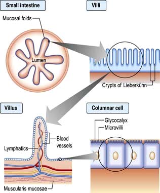
Each villus consists of a core containing blood vessels, lacteals (lymphatics) and cells (e.g. plasma cells and lymphocytes). The lamina propria contains plasma cells, lymphocytes, macrophages, eosinophils and mast cells. The crypts of Lieberkühn are the spaces between the bases of the villi.
Enterocytes are formed at the bottom of the crypts and migrate toward the tops of the villi, where they are shed. This process takes 3–4 days. On its luminal side, the enterocyte is covered by microvilli and a gelatinous layer called the glycocalyx. Scattered between the epithelial cells are mucin-secreting goblet cells and occasional intraepithelial lymphocytes and Paneth cells. Most of the blood supply to the small intestine is via branches of the superior mesenteric artery. The terminal branches are end arteries – there are no local anastomotic connections.
This controls the functioning of the small bowel; it is an independent system that coordinates absorption, secretion, blood flow and motility. It is estimated to contain 108 neurones (as many as the spinal cord) contained in two major ganglionated plexuses: the myenteric plexus between the muscular layers of the intestinal wall, and the submucosal plexuses associated with the mucosa. The ENS communicates with the central nervous system via autonomic afferent and efferent pathways but can operate autonomously.
Coordination of small intestinal function involves a complex and poorly understood interplay between many neuroactive mediators and their receptors, ion channels, GI hormones, nitric oxide and other transmitters. Acetylcholine, adrenaline, ATP, vasoactive intestinal peptide (VIP) and other hormones and opioids have been shown to have actions in the small bowel, but the exact role for each is far from being understood.
The contractile patterns of the small intestinal muscular layers are primarily determined by the enteric nervous system. The CNS and gut hormones also have a modulatory role on motility. The interstitial cells of Cajal which lie within the smooth muscle appear to govern rhythmic contractions.
During fasting, a distally migrating sequence of motor events termed the migrating motor complex (MMC) occurs in a cyclical fashion. The MMC consists of
A period of motor quiescence (phase I),
followed by a period of irregular contractile activity (phase II),
culminating in a short (5–10 min) burst of regular phasic contractions (phase III).
Each MMC cycle lasts for approximately 90 min. In the duodenum, phase III is associated with increased gastric, pancreatic and biliary secretions. The role of the MMC is unclear, but the strong phase III contractions propel secretions, residual food and desquamated cells towards the colon. It is named the ‘intestinal housekeeper’.
After a meal, the MMC pattern is disrupted and replaced by irregular contractions. This seemingly chaotic pattern lasts typically for 2–5 hours after feeding, depending on the size and nutrient content of the meal. The irregular contractions of the fed pattern have a mixing function, moving intraluminal contents to and fro, aiding the digestive process.
The hormone-producing cells of the gut are scattered diffusely throughout its length and also occur in the pancreas. The cells that synthesize hormones are derived from neural ectoderm and are known as APUD (amine precursor uptake and decarboxylation) cells. Many of these hormones have very similar structures and their action is probably local.
Gut hormones play a part in the regulation and integration of the functions of the small bowel and other metabolic activities. Their actions are complex and interacting, both with each other and with the ENS (Table 6.8).
Table 6.8 Gut regulatory peptides
Peptide | Localization | Main actions |
---|---|---|
Gastrin/cholecystokinin family |
|
|
Cholecystokinin (CCK) |
|
|
Multiple forms from CCK8 (8 amino acids) to CCK83; 8, 33 and 58 are predominant. Terminal 5 amino acids same as gastrin |
Duodenum and jejunum (I cells) |
Causes gall bladder contraction and sphincter of Oddi relaxation. Trophic effects on duodenum and pancreas. Pancreatic secretion (minor role). Role in satiety – acting in CNS |
Gastrin |
G cells in gastric antrum and duodenum |
Stimulates acid secretion. |
Secretin-glucagon family |
|
|
Secretin |
Duodenum and jejunum (S cells) |
Stimulates pancreatic bicarbonate secretion |
Glucagon |
Alpha cells of pancreas |
Opposes insulin in blood glucose control |
Vasoactive polypeptide (VIP) |
Enteric nerves |
Intestinal secretion of water and electrolytes. |
Glucose-dependent insulinotropic peptide (GIP) |
Duodenum (K cells) |
Release by intraduodenal glucose causes greater insulin release by islets than i.v. glucose (incretin effect) |
Glucagon-like peptide-1 (GLP-1) |
Ileum and colon (L cells) |
Incretin. Stimulates insulin synthesis. Trophic to islet cells. Inhibits glucagon secretion and gastric emptying |
Glicentin |
L cells |
Stimulates insulin secretion and gut growth, inhibits gastric secretion |
Growth hormone-releasing factor (GHRF) |
Small intestine |
Unclear |
Pancreatic polypeptide family |
|
|
Pancreatic polypeptide (PP) |
Pancreas (PP cells) |
Inhibits pancreatic and biliary secretion |
Peptide YY (PYY) |
Ileum and colon (L cells) |
Inhibits pancreatic exocrine secretion. Slows gastric and small bowel transit (‘ileal brake’). Reduces food intake and appetite |
Neuropeptide Y (NPY) |
Enteric nerves |
Stimulates feeding. Regulates intestinal blood flow |
Other |
|
|
Motilin |
Whole gut |
Increases gastric emptying and small bowel contraction |
Ghrelin |
Stomach |
Stimulates appetite, increases gastric emptying |
Obestatin |
Stomach and small intestine |
Opposes ghrelin |
Oxyntomodulin |
Colon |
Inhibits appetite |
Gastrin releasing-polypeptide (bombesin) |
Whole gut and pancreas |
Stimulates pancreatic exocrine secretion and gastric acid secretion |
Somatostatin |
Stomach and pancreas (D cells) |
Inhibits secretion and action of most hormones |
Substance P |
Enteric nerves |
Enhances gastric acid secretion, smooth muscle contraction |
Neurotensin |
Ileum |
Affects gut motility. Increases jejunal and ileal fluid secretion |
Insulin |
Pancreatic β cells |
Increases glucose utilization |
Chromogranins |
Neuroendocrine cells |
Precursor for other regulatory peptides that inhibit neuroendocrine secretion |
In the small bowel digestion and absorption of nutrients and ions takes place, as does the regulation of fluid absorption and secretion. The epithelial cells of the small bowel form a physical barrier that is selectively permeable to ions, small molecules and macromolecules. Digestive enzymes such as proteases and disaccharidases are produced by intestinal cells and expressed on the surface of microvilli; others such as lipases produced by the pancreas are associated with the glycocalyx. Some nutrients are absorbed most actively in specific parts of the small intestine; iron and folate in the duodenum and jejunum, vitamin B12 and bile salts in the terminal ileum where they have specific receptors.
This process is nonspecific, requires no carrier molecule or energy and takes place if there is a concentration gradient from the intestinal lumen (high concentration) to the bloodstream (low concentration). Vitamin B12 can be absorbed from the jejunum by this means.
Absorption takes place down a concentration gradient, but a membrane carrier protein is involved, conferring specificity on the process. Fructose is an example.
Absorption occurs via a specific carrier protein, powered by cellular energy, and thus a substance can be transported against a concentration gradient. Many carrier proteins are powered by ion gradients across the enterocyte wall. For example, glucose crosses the enterocyte microvillous membrane from the lumen into the cell against a concentration gradient by using a co-transporter carrier molecule. This is the sodium/glucose co-transporter, SGLT1 (Fig. 6.24). The process is powered by the energy derived from the flow of Na+ ions from a high concentration outside the cell to a low concentration inside. The sodium gradient across the cell wall is maintained by a separate ATP-consuming Na+/K+ exchanger in the basolateral membrane. Glucose leaves the cell on the serosal side by facilitated diffusion via a sodium-independent carrier (GLUT-2) in the basolateral membrane.
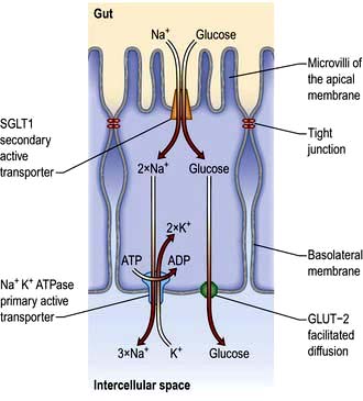
Figure 6.24 Transcellular uptake of glucose across the intestinal epithelia. Glucose is co-transported across the apical membrane with sodium ions by the sodium-dependent glucose transporter (SGLT). This is secondary active transport as the sodium is travelling down its electrochemical gradient. The sodium gradient is maintained by the primary active transport of sodium across the basolateral membrane by the Na+K+ ATPase (thus intracellular Na+ is kept low). Transcellular transport of glucose is achieved by facilitated diffusion across the basolateral membrane as glucose is moved down its concentration gradient by GLUT-2.
Another active transport mechanism operates for Na+ absorption in the ileum using an Na+/H+ exchange mechanism powered by the outwardly directed gradient of H+ across the cell membrane.
Dietary carbohydrate consists mainly of starch, with some sucrose and a small amount of lactose. Starch is a polysaccharide made up of numerous glucose units. In order to have a nutrient value starch must be digested into smaller oligo-, di- and finally monosaccharides which may then be absorbed. Polysaccharide hydrolysis begins in the mouth catalysed by salivary amylase, though the majority takes place under the action of pancreatic amylase in the upper intestine. The breakdown products of starch digestion are maltose and maltotriose, together with sucrose and lactose. These are further hydrolysed on the microvillous membrane by specific oligo- and disaccharidases to form glucose, galactose and fructose. These monosaccharides are then able to be transported across the enterocytes into the blood (Fig. 6.24).
Dietary protein is digested by pancreatic proteolytic enzymes to amino acids and peptides prior to absorption. These enzymes are secreted by the pancreas as pro-enzymes and transformed to active forms in the lumen. Protein in the duodenal lumen stimulates the enzymatic conversion of trypsinogen to trypsin, and this in turn activates the other pro-enzymes, chymotrypsin and elastase.
These enzymes break down protein into oligopeptides. Some di- and tri-peptides are absorbed intact by carrier-mediated processes, while the remainder are broken down into free amino acids by peptidases on the microvillous membranes of the enterocytes, prior to absorption into the cell by a variety of amino acid and peptide carrier systems.
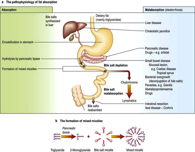
Dietary fat consists mainly of triglycerides with some cholesterol and fat-soluble vitamins. Fat is emulsified by mechanical action in the stomach. Bile containing the amphipathic detergents, bile acids and phospholipids enters the duodenum following gall bladder contraction. They act to solubilize fat and promote hydrolysis of triglycerides in the duodenum by pancreatic lipase to yield fatty acids and monoglycerides. Bile acids, phospholipids and the products of fat digestion cluster together with their hydrophilic ends on the outside to form aggregations called mixed micelles. Trapped in the centre of the micelles are the hydrophobic monoglycerides, fatty acids and cholesterol. At the cell membrane the lipid contents of the micelles are absorbed, while the bile salts remain in the lumen. Inside the cell the monoglycerides and fatty acids are re-esterified to triglycerides. The triglycerides and other fat-soluble molecules (e.g. cholesterol, phospholipids) are then incorporated into chylomicrons to be transported into the lymph.
By contrast, another mechanism exists for medium-chain triglycerides (MCT; fatty acids of chain length 6–12) which are transported via the portal vein with a small amount of long-chain fatty acid. Patients with pancreatic exocrine or bile salt insufficiency can therefore supplement their fat absorption with MCT.
Bile salts are not absorbed in the jejunum, so the intraluminal concentration in the upper gut is high. They pass down the intestine to be absorbed in the terminal ileum and are transported back to the liver. This enterohepatic circulation prevents excess loss of bile salts (see p. 306).
The pathophysiology of fat absorption is shown in Figure 6.25. Interference with absorption can occur at all stages, as indicated, giving rise to steatorrhoea (>17 mmol or 6 g of faecal fat per day).
Large amounts of water and electrolytes, partly dietary, but mainly from intestinal secretions, are absorbed coupled with absorption of monosaccharides, amino acids and bicarbonate in the upper jejunum. Water and electrolytes are also absorbed paracellularly (between the enterocytes) down electrochemical and osmotic gradients. Additional water and electrolytes are absorbed in the ileum and colon, where active sodium transport is not coupled to solute absorption. Secretion of fluid and electrolytes occur together to maintain the normal functioning of the gut. Secretory diarrhoea (see p. 292) can occur because of defects in intestinal secretory mechanisms.
These are all absorbed in the small intestine. Vitamin B12 (see p. 382) and bile salts are absorbed by specific transport mechanisms in the terminal ileum; malabsorption of both these substances often occurs following ileal resection.
The small bowel has a number of mechanisms to prevent colonization and invasion by pathogens while simultaneously preventing inappropriate responses to foreign antigens or the indigenous bacterial population. At the same time commensal bacteria maintain the integrity of the small bowel and play a major role in host physiology.
Continuous shedding of surface epithelial cells
The physical movement of the luminal contents
Colonization resistance – the ability of the indigenous microbiota to outcompete pathogens for a survival niche in the gut.
Enzymes such as lysozyme and phospholipase A2 secreted by Paneth cells at the base of the crypts help ensure an infection-free environment in the gut, even in the presence of commensal bacteria.
Antimicrobial peptides are secreted from enterocytes and Paneth cells in response to pathogenic bacteria. These include defensins, which are 15–20 amino acid peptides with potent activity against a broad range of pathogens including Gram-positive and Gram-negative bacteria, fungi and viruses.
Trefoil peptides are a family of small proteins secreted by goblet cells. They consist of a three-loop structure with intra-chain disulphide bonds which makes the molecules highly resistant to digestion. Their actions include stabilization of mucus, promotion of cell migration over injured areas, and promotion of repair. Three trefoil factors (TFF) are found in humans (TFF1, TFF2 and TFF3) all of which have been implicated in the response to gastrointestinal injury in experimental models. Their molecular mode of action is not yet known.
Humoral. IgA is the principal mucosal antibody. IgA mediates mucosal immunity by agglutinating and neutralizing pathogens in the lumen and preventing colonization of the epithelial surface (Fig. 6.26). IgA is secreted from immunocytes in the lamina propria as dimers joined by a protein called the ‘Joining chain’ (J-chain); in this form it is known as polymeric IgA (pIgA). This pIgA is internalized by endocytosis at the basolateral membrane of enterocytes. It crosses the cell as a complex of pIgA/pIgAR and is secreted onto the mucosal surface.
B-cell sensitization. Antigens from the lumen of the bowel are transported by M cells and dendritic cells in the follicle-associated epithelium (FAE). This covers Peyer’s patches in the ‘dome’ region which contain abundant virgin B cells, helper T cells and antigen-presenting cells. Activated B cells then produce IgA locally and are programmed to home back to the lamina propria. They travel through mesenteric lymph nodes and then via the thoracic duct to the blood and back to the small bowel and other mucosal surfaces (such as the airways) where they undergo terminal differentiation into plasma cells. Homing back to the gut is facilitated by the α4β7-integrin on gut-derived lymphocytes binding to MAdCAM-1, uniquely expressed on blood vessels in the gut.
Cellular defence. T lymphocytes also provide host defence and initiate, activate and regulate adaptive immune responses. Intestinal T lymphocytes occur principally in three major compartments:
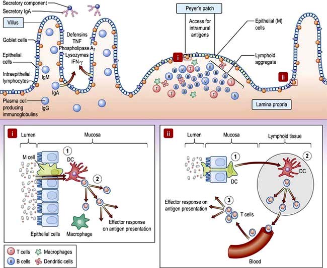
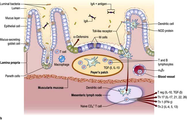
Figure 6.26 Small intestinal mucosa with a Peyer’s patch, showing the gut-associated lymphoid tissue (GALT). Inset i: (1) Specialized ‘M’ cells within a lymphoid follicle pass antigen to underlying APC such as DC. (2) DC present antigen to T cells resulting in activation and effector responses. Inset ii: (1) DC pass dendrites through epithelial tight junctions to sample luminal antigen. (2) DC traffic to the mesenteric lymph node and present antigen to naive T cells which differentiate into effector or regulatory subtypes and are imprinted with a gut homing integrin. (3) On subsequent antigen presentation, activated T cells home to lamina propria and exert regulatory or inflammatory effect depending on their phenotype. DC, dendritic cell.
The relationship between the hundred thousand billion microbes in the human gut and the host are only beginning to be appreciated. Germ-free mice have essentially no mucosal immune system showing that the abundant and activated immune system seen in healthy individuals is driven by the flora, without adverse effects. Bacteria also release chemical signals such as LPS and lipoteichoic acid that are recognized by Toll-like receptors (TLRs) (see p. 55) present on a variety of intestinal cells, priming repair processes and enhancing the ability of the epithelium to respond to injury.
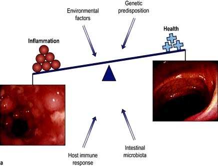
The immune system must guard against pathogens and toxins while avoiding an excessive response to the multiplicity of food antigens and commensal bacteria. The mechanisms by which tolerance occurs are undoubtedly multiple, including maintaining barrier function to prevent excess antigen uptake, active inhibition via regulatory T cells, and dendritic cells which promote tolerogenic rather than immunogenic T cell responses. All of these are likely to play a role in diseases such as coeliac disease, caused by an excessive T cell response to gluten, or Crohn’s disease, where tolerance to the indigenous bacterial population is defective.
Regardless of the cause, the common presenting features of small bowel disease are listed below. However, 10–20% of patients will have no diarrhoea or any other gastrointestinal symptoms.
Diarrhoea is common and may be watery.
Steatorrhoea occurs when the stool fat is >17 mmol/day (or 6 g/day). The stools are pale, bulky, offensive, float (because of their increased air content), leave a fatty film on the water in the pan and are difficult to flush away.
Abdominal pain and discomfort. Abdominal distension can cause discomfort and flatulence. The pain has no specific character or periodicity and is not usually severe.
Weight loss. Weight loss is largely due to the anorexia that invariably accompanies small bowel disease. The calorie deficit due to malabsorption is small relative to the reduction in intake.
Nutritional deficiencies. Deficiencies of iron, B12, folate or all of these, leading to anaemia, are the only common deficiencies. Occasionally malabsorption of other vitamins or minerals occurs, causing bruising (vitamin K deficiency), tetany (calcium deficiency), osteomalacia (vitamin D deficiency), or stomatitis, sore tongue and aphthous ulceration (multiple vitamin deficiencies). Oedema due to hypoproteinaemia is due to low intake and intestinal loss of albumin (protein-losing enteropathy).
Physical signs are few and nonspecific. If present, they are usually associated with anaemia and the nutritional deficiencies described above.
Abdominal examination is often normal, but sometimes distension and, rarely, hepatomegaly or an abdominal mass are found. Visible peristalsis and high pitched bowel sounds can indicate chronic subacute obstruction of the small intestine, e.g. due to stricturing Crohn’s disease. Gross weight loss, oedema and muscle wasting is seen only in severe cases. A neuropathy, not always due to B12 deficiency, can be present.
The emphasis in the investigation of malabsorption is on the structural features of the underlying disorder, rather than on the documentation of malabsorption itself.
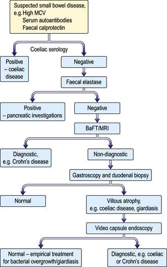
Figure 6.27 Flow diagram for investigation of patients with suspected small bowel disease. BaFT, barium follow-through; MRI, magnetic resonance imaging.
Full blood count and film. Anaemia can be microcytic, macrocytic or normocytic and the blood film may be dimorphic. Other abnormal cells (e.g. Howell–Jolly bodies, p. 406) may be seen in splenic atrophy associated with coeliac disease.
Low serum calcium and raised alkaline phosphatase may indicate the presence of osteomalacia due to vitamin D deficiency.
Liver biochemistry and serum albumin and prothrombin time.
Immunological tests. Measurement of serum antibodies to endomysium and tissue transglutaminase is useful for the diagnosis of coeliac disease. These should always be accompanied by an assessment of total immunoglobulin levels.
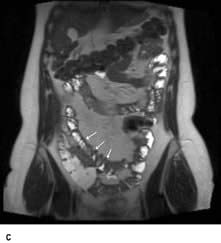
MRI enteroclysis. This is cross-sectional imaging which does not involve radiation. Using oral loading with water or a hyperonic solution to distend the small bowel lumen.
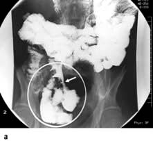
Small bowel barium follow-through (see p. 234). This detects gross anatomical defects such as diverticula, strictures and Crohn’s disease. Dilatation of the bowel and a changed fold pattern may suggest malabsorption but these are nonspecific findings. Gross dilatation is seen in myopathic pseudo-obstruction.
Small bowel biopsy. This is used to assess the microanatomy of the small bowel mucosa. Biopsies are usually obtained via an endoscope passed into the duodenum and should be well-orientated for correct evaluation. The histological appearances are described in the sections on individual diseases. A smear of the jejunal juice or a mucosal impression should also be made when Giardia intestinalis is suspected.
Ultrasound is a useful preliminary investigation which can show thickened small bowel or distended loops.
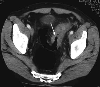
CT scanning is used to look for small bowel wall thickening, diverticula and for extraintestinal features such as abscesses (e.g. in Crohn’s disease).
Video-capsule enteroscopy is increasingly widely used to directly visualize the small bowel lumen and mucosa along its entire length. It is particularly useful in the diagnosis of occult GI bleeding.
These are required only in complicated cases.
Fat malabsorption. The confirmation of the presence of steatorrhoea is only occasionally necessary. Three-day faecal fat analysis, triglyceride breath tests and serum β carotene are now rarely performed. In rare cases when it is essential to confirm steatorrhoea, Sudan III staining of a faecal sample can be used.
Lactose tolerance test. Testing is of little use in adults because lactose intolerance is rarely a clinical problem; patients who are upset by milk usually avoid it. Formal testing involves giving an oral dose of 50 g of lactose and serial measurement of blood glucose over 2 hours. (Note: 500 mL of milk contain 20 g of lactose). There is a high incidence of lactase deficiency in many parts of the world (e.g. the Mediterranean countries, and parts of Africa and Asia).
Hydrogen breath test. This is frequently used as a screening test to measure transit time and to detect small bowel bacterial overgrowth. Bacteria are present in the oral cavity so the mouth should be rinsed out with an antiseptic mouthwash beforehand. The appearance of a breath hydrogen peak after oral lactulose is used to estimate mouth to caecum transit time. An earlier rise in the breath hydrogen after lactulose indicates bacterial breakdown in the small intestine. This test is simple to perform and it does not involve radioisotopes. However, interpretation is often difficult with a low sensitivity and specificity.
Tests for pancreatic insufficiency are used in the differential diagnosis of steatorrhoea. Human pancreatic elastase 1 (E1) remains undegraded during intestinal transit so its concentration in faeces reflects exocrine pancreatic function. The faecal elastase test quantifies E1 in stool, allowing the diagnosis or exclusion of severe pancreatic exocrine insufficiency (see p. 360).
Other blood tests. Serum immunoglobulins are measured to exclude immune deficiencies in particular IgA deficiency which may lead to false-negative coeliac antibody tests. Gut peptides (e.g. VIP) are measured in high-volume secretory diarrhoea, and chromogranins A and B are raised in endocrine tumours.
Tests for protein-losing enteropathy (PLE) (see p. 234). These tests are rarely required unless a low serum albumin is a major clinical feature.
Measurement of α1 antitrypsin clearance does not require an isotope. α1 antitrypsin is a large molecule (>50 000 daltons), which is resistant to proteolysis. Simultaneous measurements of serum and stool concentration (24-hour collection) are made.
Bile salt loss. This can be demonstrated by giving oral 75Se-homocholyl taurine (SeHCAT – a synthetic taurine conjugate) and measuring the retention of the bile acid by whole-body counting at 7 days.
Stool tests. Faecal calprotectin is 93% sensitive and 96% specific for IBD. Faecal lactoferrin is also an inflammatory marker.
In many small bowel diseases, malabsorption of specific substances occurs, but these deficiencies do not usually dominate the clinical picture. An example is Crohn’s disease, in which malabsorption of vitamin B12 can be demonstrated, but this is not usually the major problem; diarrhoea and general ill-health are the major features.
The major disorders of the small intestine that cause malabsorption are shown in Table 6.9.
Table 6.9 Disorders of the small intestine causing malabsorption
Coeliac disease (CD) is a condition in which there is inflammation of the mucosa of the upper small bowel that improves when gluten is withdrawn from the diet and relapses when gluten is reintroduced. Up to 1% of many populations are affected, though most have clinically silent disease.
Gluten is the entire protein content of the cereals wheat, barley and rye. Prolamins (gliadin from wheat, hordeins from barley, secalins from rye) are damaging factors. These proteins are resistant to digestion by pepsin and chymotrypsin because of their high glutamine and proline content and remain in the intestinal lumen triggering immune responses.
Immunology. Gliadin peptides pass through the epithelium (para- and/or intracellularly) and are deaminated by tissue transglutaminase which increases their immunogenicity. Gliadin peptides then bind to antigen-presenting cells which interact with CD4+ T cells in the lamina propria via HLA class II molecules DQ2 or DQ8. These T cells produce pro-inflammatory cytokines, particularly interferon-γ. CD4+ T cells also interact with B cells to produce endomysial and tissue transglutaminase antibodies. Gliadin peptides also cause release of interleukin-15 from enterocytes, activating intraepithelial lymphocytes with a natural killer cell marker. This inflammatory cascade releases metalloproteinases and other mediators that contribute to the villous atrophy and crypt hyperplasia which are typical of the disease.
The mucosa of the proximal small bowel is predominantly affected, the mucosal damage decreasing in severity towards the ileum as gluten is digested into smaller ‘non-toxic’ fragments.
Genetic factors. There is an increased incidence of coeliac disease within families but the exact mode of inheritance is unknown; 10–15% of 1st-degree relatives will have the condition, although it may be asymptomatic. The concordance rate in identical twins is about 70%.
HLA-DQ2 (DQAI*0501, DQBI*0201) and HLA-DQ8 (DQAI*0301, DQBI*0302) are associated with CD. Over 90% of patients will have HLA-DQ2, compared with 20–30% of the general population. Studies in twins and siblings indicate that HLA genes are responsible for <50% of the genetic cause of the disease. Many unaffected people also carry these genes, so other factors must also be involved. Non-HLA genes may also contribute to coeliac disease, e.g. chromosome regions 19p13.1, 11q, 5q31-33 and 6q21-22. The CD28/CTLA4/ILO5 gene cluster has also shown linkage with coeliac disease.
Environmental factors. Breast-feeding and the age of introduction of gluten into the diet are significant.
Rotavirus infection in infancy also increases the risk, and adenovirus-12 which has sequence homology with α-gliadin has been suspected as a causative agent but this is now thought to be unlikely.
Coeliac disease can present at any age. In infancy it sometimes appears after weaning onto gluten-containing foods. The peak period for diagnosis in adults is in the 5th decade, with a female preponderance. Many patients are asymptomatic (silent) and come to attention because of routine blood tests, e.g. a raised MCV, or iron deficiency in pregnancy. The symptoms are very variable and often nonspecific, e.g. tiredness and malaise often associated with anaemia.
GI symptoms may be absent or mild. Coeliac disease should be tested for in all patients with symptoms suggestive of IBS. Diarrhoea or steatorrhoea, abdominal pain and weight loss suggest more severe disease. Mouth ulcers and angular stomatitis are frequent and can be intermittent. Infertility and neuropsychiatric symptoms of anxiety and depression occur.
Rare complications include tetany, osteomalacia or gross malnutrition with peripheral oedema. Neurological symptoms such as paraesthesia, ataxia (due to cerebellar calcification), muscle weakness or a polyneuropathy occur; the prognosis for these symptoms is variable. There is an increased incidence of atopy and autoimmune disease, including thyroid disease, type 1 diabetes and Sjögren’s syndrome. Other associated diseases include inflammatory bowel disease, primary biliary cirrhosis, chronic liver disease, interstitial lung disease and epilepsy. IgA deficiency is more common than in the general population. Long-term problems include osteoporosis which occurs even in patients on long-term gluten-free diets.
Physical signs are usually few and nonspecific and are related to anaemia and malnutrition.
Small bowel biopsy is still considered ‘gold standard’ for positive diagnosis, and is therefore desirable in all but the most clear-cut cases, because treatment involves a life-long diet that is both expensive and socially limiting. However with the increasing accuracy of serological tests, it is no longer necessary to take duodenal biopsies for suspected coeliac disease in patients without antibodies. For example in patients being endoscoped for iron deficiency anaemia with negative coeliac serology, the pretest value of small bowel histology is <0.03%.
If biopsies are to be taken, because the disease is sometimes patchy and it can be difficult to orientate endoscopic biopsies for histological section, four to six forceps biopsies should be taken from the second part of the duodenum. Endoscopic signs including absence of mucosal folds, mosaic pattern of the surface and scalloping of mucosal folds are often present; however, their absence is not conclusive because they are markers of relatively severe disease.
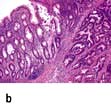
Histology (Fig. 6.28). Histological changes are of variable severity and, though characteristic, are not specific. Villous atrophy can be caused by many other conditions, but coeliac disease is the commonest cause of subtotal villous atrophy.
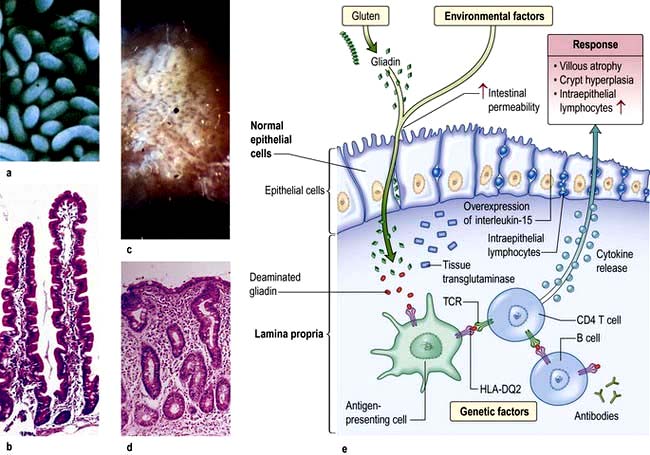
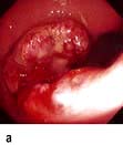
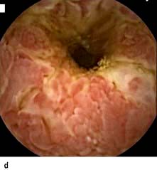
Figure 6.28 Small bowel mucosal appearances – macroscopic and microscopic. (a) Normal mucosa under the dissecting microscope (DM). (b) Normal mucosal histology. (c) Coeliac disease – flattened mucosa. (d) Coeliac disease – showing subtotal villous atrophy. (e) Immune and genetic mechanisms in coeliac disease. TCR, T cell receptor.
(After Green PH, Cellier C. Coeliac disease. New England Journal of Medicine 2007; 357:1731–1743.)
Histological examination shows crypt hyperplasia with chronic inflammatory cells in the lamina propria, and villous atrophy. The enterocytes become cuboidal with an increase in the number of intraepithelial lymphocytes. In the lamina propria there is an increase in lymphocytes and plasma cells. The most severe histological change with mucosal atrophy and hypoplasia is seen in patients who do not respond to a gluten-free diet.
In mild cases, the villous architecture is almost normal but there are increased numbers of intraepithelial lymphocytes.
Serology. Persistent diarrhoea, folate or iron deficiency, a family history of coeliac disease and associated autoimmune disease are indications for serological testing.
The most sensitive tests are for endomysial and anti-tissue transglutaminase antibodies. The sensitivity of these tests is >90% though both are not always positive in the same subject. Titres of either correlate with the severity of mucosal damage so they can be used for dietary monitoring. Standard tests use IgA class antibodies. Selective IgA deficiency occurs in 2.5% of coeliac disease patients but only 0.25% of normals and renders these tests falsely negative. All patients should have concomitant IgA levels tested and if deficient, IgG-based tests should then be used.
HLA typing. HLA-DQ2 is present in 90–95% of CD patients and HLA-DQ8 in about 8%, i.e. most of the rest. The absence of both alleles has a high negative predictive value for coeliac disease. HLA typing may occasionally be useful for risk assessment, e.g. in patients already on a gluten free diet.
Haematology. Mild or moderate anaemia is present in 50% of cases. Folate deficiency is common, often causing macrocytosis. B12 deficiency is rare. Iron deficiency due to malabsorption of iron and increased loss of desquamated cells is common. A blood film may therefore show microcytes and macrocytes as well as hypersegmented polymorphonuclear leucocytes and Howell–Jolly bodies (see p. 406) due to splenic atrophy.
Biochemistry, liver biochemistry and function. In severe cases, biochemical evidence of osteomalacia may be seen (low calcium and high phosphate) and hypoalbuminaemia.
Radiology. A small bowel follow-through may show dilatation of the small bowel with slow transit. Folds become thicker and in severe disease total effacement is seen. Radiology is mainly used when a complication, e.g. lymphoma, is suspected.
Bone densitometry (DXA) should be performed on all patients because of the risk of osteoporosis.
Capsule endoscopy (see p. 233) is used to look for gut abnormalities when a complication is suspected.
Replacement minerals and vitamins, e.g. iron, folic acid, calcium, vitamin D, may be needed initially to replace body stores.
Treatment is with a gluten-free diet for life. Dietary elimination of wheat, barley and rye usually produces a clinical improvement within days or weeks. Morphological improvement often takes months, especially in adults. Oats are tolerated by most coeliacs, but must not be contaminated with flour during their production. Meat, dairy products, fruits and vegetables are naturally gluten free and are all safe.
Gluten-free products can be expensive, unless subsidized by national health services. Patient support organizations such as The Coeliac Society are valuable as information sources and for advice about diet, recipes and gluten-free processed foods. Despite advice, many patients do not keep to a strict diet but maintain good health. The long-term effects of this low gluten intake are uncertain but osteoporosis can occur even in treated cases.
The usual cause for failure to respond to the diet is poor compliance. Dietary adherence can be monitored by serial tests for endomysial antibody (EMA) and tissue transglutaminase (tTG). If clinical progress is suboptimal then a repeat intestinal biopsy should be taken. If the diagnosis is equivocal on the diagnostic mucosal biopsy, or if the patient has already started on a gluten-free diet, then a gluten challenge, i.e. reintroduction of oral gluten, with evidence of jejunal morphological change, can confirm the diagnosis.
Patients should have pneumococcal vaccinations (because of splenic atrophy) once every 5 years (see p. 406).
A few patients do not improve on a strict diet and are said to have non-responsive coeliac disease. Many of these patients are still ingesting gluten (see above). A few of the others may have concomitant problems, e.g. microscopic colitis, IBD, small bowel bacterial overgrowth or lactase deficiency.
A very small percentage will have the rare complication of refractory coeliac disease (RCD). In type 1 RCD, the lymphocytes are normal and the T cell receptors are polyclonal, whilst in type 2 there are abnormal clonal lymphocytes with loss of CD8 and CD3 surface markers. The 5-year survival rates are 93% and 40–60% respectively.
Very rarely, enteropathy-associated T cell lymphoma (EATCL) (8–20% 5-year survival), or ulcerative jejunitis can occur as part of a spectrum of neoplastic T cell disorders. Small bowel adenocarcinoma is also increased in coeliac disease.
Ulcerative jejunitis presents with fever, abdominal pain, perforation and bleeding.
Diagnosis for these conditions is with MRI or barium studies but laparoscopy with full-thickness small bowel biopsies is often required. Steroids and immunosuppressive agents, e.g. azathioprine, are used in ulcerative jejunitis
Carcinoma of the oesophagus as well as extragastrointestinal cancers are also increased in incidence. Malignancy seems to be unrelated to the duration of the disease but the incidence is reduced by a gluten-free diet.
This is an uncommon blistering subepidermal eruption of the skin associated with a gluten-sensitive enteropathy (see also p. 1222). Rarely gross malabsorption occurs, but usually the jejunal morphological abnormalities are not as severe as in coeliac disease. The inheritance and immunological abnormalities are the same as for coeliac disease. The skin condition responds to dapsone but a gluten-free diet improves both the enteropathy and the skin lesion, and is recommended for long-term benefit.
This is a condition presenting with chronic diarrhoea and malabsorption that occurs in residents or visitors to affected tropical areas. The disease is endemic in most of Asia, some Caribbean islands, Puerto Rico and parts of South America. Epidemics occur, lasting up to 2 years, and in some areas repeated epidemics occur at varying intervals of up to 10 years.
The term tropical sprue is reserved for severe malabsorption (of two or more substances) accompanied by diarrhoea and malnutrition. Malabsorption of a mild degree, sometimes following an enteric infection, is quite common in the tropics; it is usually asymptomatic and is sometimes called tropical malabsorption.
The aetiology is unknown, but is likely to be infective because the disease occurs in epidemics and patients improve on antibiotics. A number of agents have been suggested but none has been unequivocally shown to be responsible. Different agents could be involved in different parts of the world.
These vary in intensity and consist of diarrhoea, anorexia, abdominal distension and weight loss. The onset is sometimes acute and occurs either a few days or many years after being in the tropics. Epidemics can break out in villages, affecting thousands of people at the same time. The onset can also be insidious, with chronic diarrhoea and evidence of nutritional deficiency. The clinical features of tropical sprue vary in different parts of the world, particularly as different criteria are used for diagnosis.
Acute infective causes of diarrhoea must be excluded (see p. 293), particularly Giardia, which can produce a syndrome very similar to tropical sprue. Malabsorption should be demonstrated, particularly of fat and B12. The jejunal mucosa is abnormal, showing some villous atrophy (partial villous atrophy). In most cases, the lesion is less severe than that found in coeliac disease, although it affects the whole small bowel. Mild mucosal changes can be seen in asymptomatic individuals in the tropics.
Many patients improve when they leave the sprue area and take folic acid (5 mg daily). Most patients also require an antibiotic to ensure a complete recovery (usually tetracycline 1 g daily for up to 6 months).
Severely ill patients require resuscitation with fluids and electrolytes for dehydration, and nutritional deficiencies should be corrected. Vitamin B12 (1000 µg) is also given to all acute cases.
The prognosis is excellent. Mortality is usually associated with water and electrolyte depletion, particularly in epidemics.
The gut contains many resident bacteria in the terminal ileum and colon. Anaerobic bacteria, e.g. Bacteroides, bifidobacteria, are 100–1000 times more abundant than aerobic (facultative anaerobes), e.g. Escherichia, Enterobacter, Enterococcus. This gut microflora has major functions including metabolic, e.g. fermentation of non-digestible dietary residues into short-chain fatty acids as an energy source in the colon.
The microflora which influences epithelial cell proliferation, is involved in the development and maintenance of the immune system and protects the gut mucosa from colonization by pathogenic bacteria. Bacteria also initiate vitamin K production.
The upper part of the small intestine is almost sterile, containing only a few organisms derived from the mouth. Gastric acid kills some ingested organisms and intestinal motility keeps bacterial counts in the jejunum low. The normal terminal ileum contains faecal-type organisms, mainly Escherichia coli and anaerobes, and the colon has abundant bacteria.
Bacterial overgrowth is normally found associated with a structural abnormality of the small intestine such as a stricture or diverticulum, although it can occur occasionally in the elderly without. E. coli and/or Bacteroides, both in concentrations greater than 106/mL, are found as part of a mixed flora. These bacteria are capable of deconjugating and dehydroxylating bile salts, so that unconjugated and dehydroxylated bile salts can be detected in small bowel aspirates.
The clinical features of overgrowth are chiefly diarrhoea and steatorrhoea. There may also be symptoms due to the underlying small bowel pathology. Steatorrhoea (see p. 263) occurs because of conjugated bile salt deficiency. Some bacteria can metabolize vitamin B12 and interfere with its binding to intrinsic factor, leading to mild B12 deficiency (see Chapter 8) rarely severe enough to produce a neurological deficit. Some bacteria produce folic acid giving a high serum folate. Bacterial overgrowth has only minimal effects on the absorption of other substances. Confirmation of bacterial overgrowth is with the hydrogen breath test (see p. 264).
Small intestinal resection is usually well tolerated, but massive resection leaving <1 m of small bowel in continuity is followed by the short-bowel syndrome. The effects of resection depend on the amount and location of the resection and the presence or absence of the colon. Resection of the jejunum is better tolerated than ileal resection, where there is less adaptation, probably due to low levels of glucagon-like peptide 2 (GLP-2), which is a specific growth hormone for the enterocyte.
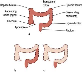
The ileum is the site of specific mechanisms for the absorption of bile salts and vitamin B12. Relatively small resections lead to malabsorption of these substances. Removal of the ileocaecal valve increases the incidence of diarrhoea (Fig. 6.29).
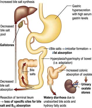
The following occur after ileal resection:
Bile-salt induced diarrhoea: bile salts and fatty acids enter the colon and cause malabsorption of water and electrolytes (see p. 293).
Steatorrhoea and gallstone formation: increased bile salt synthesis can compensate for loss of approximately one-third of the bile salts in the faeces. Greater loss than this results in decreased micelle formation and steatorrhoea, and lithogenic bile and gallstone formation.
Oxaluria and oxalate stones: bile salts in the colon cause increased oxalate absorption with oxaluria leading to urinary stone formation.
B12 deficiency: low serum B12, macrocytosis and other effects of B12 deficiency.
Investigations include a small bowel follow-through, measurement of B12, bile salt retention (SeHCAT) test (see p. 236). A hydrogen breath test may show rapid transit (see p. 264). Many patients require B12 replacement and some need a low-fat diet if there is steatorrhoea. Diarrhoea is often improved by cholestyramine which binds bile salts and reduces the level of diarrhoeogenic bile salts in the colon.
The ileum can compensate for loss of jejunal absorptive function. Jejunal resection may lead to gastric hypersecretion with high gastrin levels; the exact mechanism of this is unclear. Structural and functional intestinal adaptation takes place over the course of a year, with an increase in the absorption per unit length of bowel.
Intestinal failure results from obstruction, dysmotility, surgical resection, congenital defect, or disease-associated loss of absorption and is characterized by the inability to maintain protein-energy, fluid, electrolyte, or micronutrient balance. This most often occurs following resection for Crohn’s disease, mesenteric vessel occlusion (see p. 270), radiation enteritis (see p. 268) or trauma. There are two common situations:
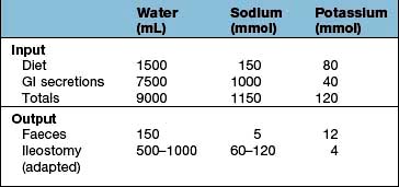
The major problem is of sodium and fluid depletion, and the majority of patients with ≤100 cm of jejunum remaining will require parenteral supplements of fluid and electrolytes, often with nutrients. Sodium losses can be minimized by increasing salt intake, restricting hypotonic fluids between meals and administering oral glucose-electrolyte mixture with a sodium concentration 90 mmol/L. Jejunal transit time can be increased and stomal effluent loss reduced by treatment with the somatostatin analogue octreotide, often used in combination with a proton pump inhibitor, loperamide and codeine phosphate. There is no benefit from a low-fat diet, but fat assimilation can be increased on treatment with cholestyramine and synthetic bile acids.
Because of the absorptive capacity of the colon for fluid and electrolytes, only a small proportion of these patients require parenteral supplementation. Unabsorbed fat results in impairment of colonic fluid and electrolyte absorption so patients should be on a low-fat diet. A high carbohydrate intake is advised as unabsorbed carbohydrate is metabolized anaerobically to short-chain fatty acids (SCFAs) which are absorbed; they also stimulate fluid and electrolyte absorption in the colon and act as an energy source (1.6 kcal/g). Patients are often treated with cholestyramine to reduce diarrhoea and colonic oxalate absorption.
Whipple’s disease is a rare infectious bacterial disease caused by Tropheryma whipplei. About 1000 cases have been described; 87% are males, usually white and middle-aged. It presents with arthritis and arthralgia, progressing over years to weight loss and diarrhoea with abdominal pain, systemic symptoms of fever and weight loss. Peripheral lymphadenopathy and involvement of the heart, lung, joints and brain occur, simulating many neurological conditions.
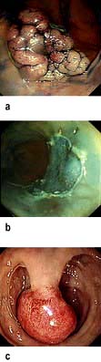
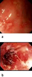
Blood tests show features of chronic inflammation and malabsorption. Endoscopy typically shows pale, shaggy duodenal mucosa with eroded, red, friable patches.
Diagnosis is made by small bowel biopsy. Periodic acid–Schiff (PAS)-positive macrophages are present but are nonspecific. On electron microscopy, the characteristic trilaminar cell wall of T. whipplei can be seen within macrophages. T. whipplei antibodies can be identified by immunohistochemistry. A confirmatory PCR-based assay is available.
Treatment is with antibiotics which cross the blood-brain barrier, such as 160 mg trimethoprim and 800 mg sulphamethoxazole (co-trimoxazole) daily for 1 year. This is preceded by a 2-week course of streptomycin and penicillin or ceftriaxone. Treatment periods of less than a year are associated with relapse in about 40%.
Radiation of >40 Gy will damage the intestine. The chronic effects of radiation are muscle fibre atrophy, ulcerative changes due to ischaemia and obstruction due to radiation-induced fibrotic strictures.
Pelvic irradiation is frequently used for gynaecological and urinary tract malignancies, so the ileum and rectum are the areas most often involved.
At the time of the irradiation, there may be nausea, vomiting, diarrhoea and abdominal pain, usually improving within 6 weeks of completion of therapy.
Chronic radiation enteritis is diagnosed if symptoms persist for ≥3 months. The prevalence is >15%. Abdominal pain due to obstruction is the main symptom. Malabsorption can be due to bacterial overgrowth in dilated segments and mucosal damage.
Many patients suffer from increased bowel frequency.
Treatment is symptomatic, although often unsuccessful in chronic radiation enteritis. Surgery should be avoided if possible, being reserved for obstruction or perforation.
Acute radiation damage to the rectum produces a radiation proctitis with diarrhoea and tenesmus, with or without blood. Local steroids sometimes help initially. When the acute phase heals, mucosal telangiectases form and may cause persistent bleeding. They can be treated with argon plasma coagulation or, under a light anaesthetic, by packing the rectum with a formalin-soaked swab for 2 min, both of which destroy the telangiectases.
Giardia intestinalis (see p. 151) not only produces diarrhoea but can produce malabsorption with steatorrhoea. Minor changes are seen in the jejunal mucosa and the organism can be found in the jejunal fluid or mucosa.
Cryptosporidiosis (see p. 151) can also produce malabsorption.
Patients with HIV infection are particularly prone to parasitic infestation (see Table 6.23).
Drugs that bind bile salts (e.g. cholestyramine) and some antibiotics (e.g. neomycin) produce steatorrhoea.
Orlistat (see p. 220) is used in obesity to reduce fat absorption by inhibiting gastric and pancreatic lipase causing diarrhoea and steatorrhoea.
Thyrotoxicosis: diarrhoea, rarely with steatorrhoea, occurs in thyrotoxicosis owing to increased gastric emptying and increased motility.
Zollinger–Ellison syndrome (see p. 370).
Intestinal lymphangiectasia produces diarrhoea and rarely steatorrhoea (see p. 270).
Lymphoma that has infiltrated the small bowel mucosa causes malabsorption.
Diabetes mellitus: diarrhoea, malabsorption and steatorrhoea occur, sometimes due to bacterial overgrowth from autonomic neuropathy causing small bowel stasis.
Hypogammaglobulinaemia, which is seen in a number of conditions including lymphoid nodular hyperplasia, causes steatorrhoea due either to an abnormal jejunal mucosa or to secondary infestation with Giardia intestinalis.
Protein-losing enteropathy refers to intestinal conditions causing protein loss, usually manifest by hypoalbuminaemia. The causes include Crohn’s disease, tumours, Ménétrier’s disease, coeliac disease and lymphatic disorders (e.g. lymphangiectasia).
Usually protein-losing enteropathy forms a minor part of the generalized disorder, but occasionally hepatic synthesis of albumin cannot compensate for the protein loss, and peripheral oedema dominates the clinical picture. The investigations are described on page 264 and treatment is that of the underlying disorder.
This is the most common congenital abnormality of the GI tract, affecting 2–3% of the population. The diverticulum projects from the wall of the ileum approximately 60 cm from the ileocaecal valve. It is usually symptomless, but 50% contain gastric mucosa that secretes hydrochloric acid. Peptic ulcers can occur and may bleed (see p. 254) or perforate.
Acute inflammation of the diverticulum also occurs and is indistinguishable clinically from acute appendicitis. Obstruction from an associated band rarely occurs.
Tuberculosis (TB) (see also p. 839) can affect the intestine as well as the peritoneum (see p. 302). In developed countries, most patients are from ethnic minority groups, or are immunocompromised due to HIV or drugs. Intestinal tuberculosis is due to reactivation of primary disease caused by Mycobacterium tuberculosis. Bovine TB occurs in areas where milk is unpasteurized and is rare in western countries.
Clinical features are abdominal pain, weight loss, anaemia, fever with night sweats, obstruction, right iliac fossa pain or a palpable mass. The ileocaecal area is most commonly affected, but the colon, and rarely other parts of the gastrointestinal tract, can be involved. One-third of patients present acutely with intestinal obstruction or generalized peritonitis; 50% have X-ray evidence of pulmonary tuberculosis.
Differential diagnosis includes Crohn’s disease and caecal carcinoma.
A small bowel follow-through may show transverse ulceration, diffuse narrowing of the bowel with shortening of the caecal pole.
Ultrasound or CT shows additional mesenteric thickening and lymph node enlargement.
Histology and culture of tissue is desirable, but it is not always possible. Specimens can be obtained by colonoscopy or laparoscopy but laparotomy is required in some cases.
Drug treatment is similar to that for pulmonary TB (see page 842). Treatment should be started if there is a high degree of suspicion.
Systemic amyloidosis may affect any part of the GI tract (see also p. 1042). Rectal biopsy may be diagnostic. Occasionally, amyloid deposits occur as polypoid lesions. The symptoms depend on the site of involvement; amyloidosis in the small intestine gives rise to diarrhoea.
Systemic sclerosis (see p. 538) most commonly affects the oesophagus (see p. 243), although the small bowel and colon are often found to be involved if investigated. There may be no symptoms of this involvement, but diarrhoea and steatorrhoea can occur due to bacterial overgrowth as a result of reduced motility, dilatation and the presence of diverticula.
Intestinal ischaemia results from occlusion of arterial inflow, occlusion of venous outflow or failure of perfusion; these factors may act alone or in combination and usually occur in the elderly.
Arterial inflow occlusion can be caused by atheroma, thrombosis, and embolism (cardiac arrhythmia), including cholesterol emboli (see p. 599), aortic disease (occluding ostia of mesenteric vessels) or vasculitis (see p. 542), thromboangiitis and Takayasu’s syndrome (see p. 745)
Venous outflow occlusion occurs in 5–15% of cases and usually in sick patients with circulatory failure.
Infarction without occlusion can occur due to reduced cardiac output, hypotension and shock causing reduced intestinal blood flow.
An embolus from the heart in a patient with atrial fibrillation is the commonest cause, usually occluding the superior mesenteric artery. Patients present with sudden abdominal pain and vomiting with a distended and tender abdomen, and absent bowel sounds. The patient is hypotensive and ill. Surgery is necessary to resect the gangrenous bowel. Mortality is high (up to 90%) and is related to co-existing disease, the development of multiorgan failure (MOF) (see p. 882) and massive fluid and electrolyte losses in the postoperative period. Survivors may go on to develop nutritionally inadequate short-bowel syndrome (see p. 268).
This is due to atheromatous occlusion or cholesterol emboli of the mesenteric vessels, particularly in the elderly. Good collateral circulation can minimize clinical effects. The characteristic symptom is post-prandial abdominal pain and weight loss. Loud bruits may be heard but, as these are heard in normal subjects, they are of doubtful significance. The diagnosis is made using angiography.
In this condition of unknown aetiology there is eosinophilic infiltration and oedema of any part of the gastrointestinal mucosa. The gastric antrum and proximal small intestine are usually involved either as a localized lesion (eosinophilic granuloma) or diffusely with sheets of eosinophils seen in the serosal and submucosal layers. There is an association with asthma, eczema and urticaria.
The condition occurs mainly in the third decade. The clinical presentation depends on the site of gut involvement. Abdominal pain, nausea and vomiting and upper GI bleeding occur. Peripheral eosinophilia occurs in only 20% of patients. Endoscopic biopsy is useful for making the diagnosis histologically. Radiology may demonstrate mass lesions.
Treatment is with corticosteroids for the widespread infiltration, particularly if peripheral eosinophilia is present.
In some adults, the condition appears to be allergic (allergic gastroenteritis) and is associated with peripheral eosinophilia and high levels of plasma and tissue IgE. Eosinophilic oesophagitis’s relationship to eosinophilic gastroenteritis is unclear.
Dilatation of the lymphatics may be primary or secondary to lymphatic obstruction, such as occurs in malignancy or constrictive pericarditis. Hypoproteinaemia with ankle oedema is the main feature. The rare primary form may be detected incidentally as dilated lacteals on a jejunal biopsy or it can produce steatorrhoea of varying degrees. White-tipped villi are seen on capsule endoscopy. Serum immunoglobulin levels are reduced, with low circulating lymphocytes. Treatment is with a low-fat diet, mid-chain triglycerides and fat-soluble vitamin supplements as required. Octreotide has a dramatic effect in a few primary cases, although the mechanism of action is unknown.
This rare congenital disorder is due to a failure of apo B-100 synthesis in the liver and apo B-48 in the intestinal cell, so that chylomicrons are not formed. This leads to fat accumulation in the intestinal cells, giving a characteristic histological appearance to the jejunal mucosa. Clinical features include acanthocytosis (spiky red cells owing to membrane abnormalities), a form of retinitis pigmentosa, and mental and neurological abnormalities. The latter can be prevented by vitamin E injections.
The small intestine is relatively resistant to the development of neoplasia and only 3–6% of all GI tumours and fewer than 1% of all malignant lesions occur here. The reason for the rarity of tumours is unknown. Explanations include the fluidity and relative sterility of small bowel contents and the rapid transit time, reducing the time of exposure to potential carcinogens. It is also possible that the high population of lymphoid tissue and secretion of IgA in the small intestine protect against malignancy.
Adenocarcinoma of the small intestine is rare and found most frequently in the duodenum (in the periampullary region) and in the jejunum. It is the most common tumour of the small intestine, accounting for up to 50% of primary tumours.
Lymphomas are most frequently found in the ileum. These are of the non-Hodgkin’s type and must be distinguished from peripheral or nodal lymphomas involving the gut secondarily.
In developed countries, the most common type of lymphoma is the B cell type arising from MALT (see p. 468). These lymphomas tend to be annular or polypoid masses in the distal or terminal ileum, whereas most T cell lymphomas are ulcerated plaques or strictures in the proximal small bowel.
A tumour similar to Burkitt’s lymphoma also occurs and commonly affects the terminal ileum of children in North Africa and the Middle East.
There is an increased incidence of lymphoma of the T cell type and adenocarcinoma of the small bowel, as well as an unexplained increase in all malignancies both in the GI tract and elsewhere. The reason for the local development of malignancy is unknown. It is now accepted that coeliac disease is a premalignant condition, but there is no association with the length of the symptoms. Treatment with a gluten-free diet reduces the risk of both lymphoma and carcinoma.
There is a small increase in the incidence of adenocarcinoma of the small bowel in Crohn’s disease.
IPSID is a rare B cell disorder in which there is proliferation of plasma cells in the lamina propria of the upper small bowel producing truncated monoclonal heavy chains, without associated light chains. The α heavy chains are found in the gut mucosa on immunofluorescence and can also be detected in the serum. It occurs usually in countries surrounding the Mediterranean, but it has also been found in other developing countries in South America and the Far East. IPSID predominantly affects people in lower socioeconomic groups in areas with poor hygiene and a high incidence of bacterial and parasitic infection of the gut. IPSID presents as a malabsorptive syndrome associated with diffuse lymphoid infiltration of the small bowel and neighbouring lymph nodes, progressing in some cases to a lymphoma. The condition has also been documented in the developed world.
Clinically, patients present with abdominal pain, diarrhoea, anorexia, weight loss and symptoms of anaemia. There may be a palpable mass, and a small bowel follow-through may detect a mass lesion. Endoscopic biopsy is useful where lesions are within reach. Ultrasound and CT may show bowel wall thickening and the involvement of lymph nodes, which is common with lymphoma. Wireless capsule endoscopy can be used where obstruction by the capsule is not likely, but cannot deliver histology.
Adenocarcinoma. Most patients are treated surgically with a segmental resection. The overall 5-year survival rate is 20–35%; this varies with the histological grade and the presence or absence of lymph node involvement. Radiotherapy and chemotherapy are used in addition.
IPSID. If there is no evidence of lymphoma, antibiotics, e.g. tetracycline, should be tried initially. In the presence of lymphoma, combination chemotherapy is used; in one series the 3–5-year survival rate was 58%.
Lymphoma. Most patients require surgery and radiotherapy with chemotherapy for more extensive disease. The prognosis varies with the type. The 5-year survival rate for T cell lymphomas is 25%, but is better for B cell lymphomas, varying from 50% to 75%, depending on the grade of lymphoma.
These originate from the enterochromaffin cells (APUD cells) of the intestine. They make up 10% of all small bowel neoplasms, the most common sites being in the appendix and terminal ileum. It is often difficult to be certain histologically whether a particular tumour is benign or malignant. A total of 10% of carcinoid tumours in the appendix present as acute appendicitis, secondary to obstruction. Surgical resection of the tumour is usually performed.
Most carcinoids do not secrete hormones or vasoactive compounds, and may present with liver enlargement due to metastases.
Carcinoid syndrome occurs in only 5% of patients with carcinoid tumours and only when there are liver metastases. Patients complain of spontaneous or induced bluish-red flushing, predominantly on the face and neck, sometimes leading to permanent changes with telangiectases.
Gastrointestinal symptoms consist of abdominal pain and recurrent watery diarrhoea. Cardiac abnormalities are found in 50% of patients and consist of pulmonary stenosis or tricuspid incompetence. Examination of the abdomen reveals hepatomegaly. The tumours secrete a variety of biologically active amines and peptides, including serotonin (5-hydroxy-tryptamine – 5-HT), bradykinin, histamine, tachykinins and prostaglandins. The diarrhoea and cardiac complications are probably caused by 5-HT itself, but the cutaneous flushing is thought to be produced by one of the kinins, such as bradykinin. This is known to cause vasodilatation, bronchospasm and increased intestinal motility.
Ultrasound examination confirms the presence of liver secondary deposits.
Urine shows a high concentration of 5-hydroxyindoleacetic acid (5-HIAA) which is the major metabolite of 5-HT.
Serum chromogranin A is raised in nearly all hindgut tumours and 80–90% of patients with symptomatic foregut and midgut tumours.
Treatment is with octreotide and lanreotide; both are octapeptide somatostatin analogues that inhibit the release of many gut hormones. They alleviate the flushing and diarrhoea and can control a carcinoid crisis. Octreotide is given subcutaneously in doses up to 200 µg three times daily initially; a depot preparation 30 mg every 4 weeks can then be used. Lanreotide 30 mg is given every 7–10 days or as a gel 60 mg every 28 days. Long-acting octreotide also sometimes inhibits tumour growth. Interferon and other chemotherapeutic regimens also occasionally reduce tumour growth, but have not been shown to increase survival.
This consists of mucocutaneous pigmentation (circumoral; 95% of patients), hands (70%) and feet (60%) and gastrointestinal polyps. It has an autosomal dominant inheritance. The gene STK11 (also known as LKB1) responsible for Peutz–Jeghers codes for a serine protein kinase and can be used for genetic analysis. The brown buccal pigment is characteristic of the condition. The polyps, which are hamartomas, can occur anywhere in the GI tract but are most frequent in the small bowel. They may bleed or cause small bowel obstruction or intussusception (50% of patients).
Treatment is by endoscopic polypectomy. Balloon enteroscopy may be necessary to reach all the small bowel polyps. Bowel resection should be avoided if possible, but may be necessary in patients presenting with gangrenous bowel due to intussusception. Follow-up is with yearly pan-endoscopy. There is an increased incidence of GI cancers. Non-GI cancers also occur with increased frequency, so yearly screening for uterine, ovarian and cervical cancer should start in the teens, and breast and testicular screening by the age of 20.
Adenomas, lipomas and stromal tumours (see p. 253) are rarely found and are usually asymptomatic and picked up incidentally. They occasionally present with iron deficiency anaemia. In familial adenomatous polyposis (FAP) duodenal adenomas form in one-third of patients and may progress to adenocarcinoma. This is the commonest cause of death in FAP patients who have been treated by prophylactic colectomy.
Two major forms of inflammatory bowel disease are recognized:
There is a degree of overlap between these two conditions in their clinical features, histological and radiological abnormalities; in 10% of cases of IBD causing colitis a definitive diagnosis of either UC or CD is not possible and the diagnosis is termed colitis of undetermined type and etiology (CUTE). It is clinically useful to distinguish between these two conditions because of differences in their management, although in reality they may represent two aspects of the same disease.
Another form of colitis related to microscopic inflammation is termed microscopic colitis; this is subdivided into lymphocytic and collagenous (see p. 281). The distinction between this and IBD is the absence of macroscopic evidence of inflammation.
The incidence of CD varies from country to country but is approximately 4–10 per 100 000 annually, with a prevalence of 25–100/100 000.
The incidence of UC is stable at 6–15/100 000 annually, with a prevalence of 80–150/100 000.
Although both conditions have a worldwide distribution, the highest incidence rates and prevalence have been reported from northern Europe, the UK and North America. Both race and ethnic origin affect the incidence and prevalence of CD and UC. Thus, in North America, prevalence rates of CD are lower in Hispanic and Asian people (4.1, 5.6/100 000, respectively) compared with white individuals (43.6/100 000). Jewish people are more prone to inflammatory bowel disease than any other ethnic group. Prevalence rates also change after migration, thus there is an increasing incidence of Crohn’s disease in the UK-born children of migrants from South-east Asia.
Approximately 25% patients are diagnosed before their 18th birthday and there is increasing evidence that disease commencing in youth is more extensive and more aggressive than that occurring in older patients.
Although the aetiology of IBD is unknown, it is increasingly clear that IBD represents the interaction between several co-factors: genetic susceptibility, the environment, the intestinal microbiota and host immune response (Fig. 6.30a).
Figure 6.30 Inflammatory bowel disease. (a) Schematic diagram showing the aetiopathogenesis. (b) Cellular intestinal processes. Bacterial ligands attach to the epithelium and antigen presenting cells via Toll-like receptors and the NOD protein. This triggers the release of various cytokines.
(Modified from Abraham C, Cho JH. Inflammatory bowel disease. N Engl J Med 2009; 361:2066–2078.)
CD and UC are complex polygenic diseases and having a positive family history is the largest independent risk factor for development of IBD. Up to one in five patients with CD and one in six patients with UC will have a 1st-degree relative with the disease. The monozygotic and dizygotic twin concordance rates for CD are 20–50% and 10%, respectively.
Genome Wide Association Studies have identified multiple susceptibility loci and many of the underlying risk variants have been identified. The major genetic factors are the NOD 2 gene (nucleotide oligomerization domain 2), the autophagy genes and the Th17 pathway (interleukin 23-type 17 helper T cells). The NOD 2 protein on chromosome 16 is an intracellular sensor of bacterial peptidoglycan, present in bacterial cell walls (see below). NOD 2 is expressed in epithelial cells, macrophages, endothelial cells. Individuals who are homozygote or compound heterozygote for one of several mutations in the NOD 2 gene have a significant increased risk of developing ileal Crohn’s disease. Likewise mutations in the autophagy genes ATG16L1 and IRGM (immunity-related GTP-ase M-protein) and IL-23 receptor gene increase CD risk, and mutations in genes associated with the mucosal barrier increase UC risk. However, the presence of IBD associated genes in many unaffected individuals and the failure of the approximately 71 genetic susceptibility loci identified thus far to explain more than around one-fifth of the genetic risk of CD highlights the complexity of the genetic basis of IBD.
Apart from susceptibility, HLA genes on chromosome 6 also appear to have a role in modifying the disease. The DRB*0103 allele, which is uncommon, is linked to a particularly aggressive course of UC and the need for surgery, as well as with colonic CD. DRB*0103 and MICA*010 are associated with perianal disease and DRB*0701 with ileal CD. For the extraintestinal disease complications and HLA links, see p. 275.
Smoking: Patients with CD are more likely to be smokers, and smoking has been shown to exacerbate CD and increase the risk of disease recurrence after surgery. By contrast, there is an increased risk of UC in non- or ex-smokers and nicotine has been shown to be an effective treatment in one small clinical trial.
Non-steroidal anti-inflammatory drugs: NSAID ingestion is associated with both the onset of IBD and flares of disease in patients with an established diagnosis.
Hygiene: Good domestic hygiene has been shown to be a risk factor for CD but not for UC. Poor and large families living in crowded conditions have a lower risk of developing CD. A ‘clean’ environment may not expose the intestinal immune system to pathogenic or non-pathogenic microorganisms such as helminths which seems to alter the balance between effector and regulatory immune responses.
Nutritional factors: Many foods and food components have been suggested to play a role in the aetiopathogenesis of IBD (e.g. high sugar and fat intake) but unfortunately the results of numerous studies designed to define risk have been equivocal. However, breast-feeding may provide protection against inflammatory bowel disease developing in offspring.
Psychological factors such as chronic stress and depression seem to increase relapses in patients with quiescent disease.
Appendicectomy appears ‘protective’ for the development of UC, particularly if performed for appendicitis or for mesenteric lymphadenitis before the age of 20. It also influences the clinical course of UC, with a lower incidence of colectomy and need for immunosuppressive therapy. By contrast, appendicectomy may increase the risk of development of CD.
The gut is colonized by 10 times more bacterial organisms than there are host cells, there being 300–400 distinct bacterial species. The intestinal microbiota play a crucial role in perpetuating intestinal inflammation, both in animal models of disease and patients with IBD. Thus, in transgenic murine models of IBD, animals kept under germ-free conditions do not develop inflammation until bacteria are introduced. Likewise, the number of mucosal adherent bacteria is increased in patients with Crohn’s disease compared to healthy subjects and diversion of the bacterial component of the faecal stream induces clinical remission. However, there is also evidence for an immunoregulatory role for the commensal microbiota, which protect against intestinal inflammation and upregulate epithelial defence mechanisms in animal models of colitis.
Intestinal dysbiosis. There is an alteration in the bacterial flora in patients with Crohn’s disease. Although results vary due to differences in both the patient groups studied and the microbiological method utilized, higher concentrations of Bacteroides and Escherichia coli and lower concentrations of bifidobacteria and F. prausnitzii have been reported in faecal and mucosal samples from patients with CD compared to healthy controls. Lower concentrations of F. prausnitzii have been found in patients with active compared with quiescent disease, and low levels of this organism in CD resection specimens predict subsequent endoscopic disease recurrence.
Specific pathogenic organisms. It has been shown that there is increased E. coli adherence to the ileal-epithelial cells in CD with evidence of invasion into the mucosa. This occurs via E. coli’s type 1 pili to a protein called carcinoma embryonic antigen-related cell adhesion molecule 6 (CEACAM 6). Many authors have also suggested a link between Crohn’s disease and Mycobacterium paratuberculosis, although recent PCR based studies have failed to confirm this and a therapeutic trial of anti Mycobacterium tuberculosis (MTB) therapy was not effective.
Bacterial antigens. Bacteria exert their influence by the interaction of ligands such as peptidoglycan-polysaccharides (PG-PS) and lipopolysaccharides (LPS) interacting with host pattern recognition receptors such as the Toll-like receptor family (cell surface) and the NOD family (intracellular).
Defective chemical barrier or intestinal defensins (see p. 262). Evidence suggests a decrease in human α defensin-1 (HD-1) in both CD and UC and lack of induction of HD-2 and HD-3, HD-5 in CD.
Impaired mucosal barrier function may explain the presence of unusual and potentially pathogenic bacteria, e.g. Mycobacterium paratuberculosis (MAP), Listeria and mucosal adherent E. coli. However, their presence does not necessarily imply causation of the disease.
Butyrate. Sulphate-producing bacteria increase luminal levels of hydrogen sulphide (H2S), which leads to a reduction of butyrate oxidation in colonic mucosa, producing an energy-deficient state and leading to mucosal inflammation. H2S and methanethiol may produce the malodorous flatus that some patients complain of prior to a flare-up.
IBD occurs when the mucosal immune system exerts an inappropriate response to luminal antigens, such as bacteria, which may enter the mucosa via a leaky epithelium (Fig. 6.30b). Bacterial ligands interact with the innate and acquired mucosal immune system via Toll-like receptors expressed on both epithelial and antigen-presenting cells. Recent research has highlighted deficiencies in the clearance of invading bacteria by aspects of the innate immune system such as neutrophils, which may allow inappropriate activation of the acquired immune system. In keeping with the genetic susceptibility loci identified, these findings highlight a deficiency in a component of the inflammasome (an intracellular danger sensor of the innate immune system which can trigger caspase-1-dependent processing of inflammatory mediators, such as IL-1β and IL-18) in patients with IBD. In addition, specific bacterial species have distinct immunological effects mediated by dendritic cells (DC) which sample bacteria from the intestinal lumen and direct the subsequent functional differentiation of naive T cells into effector or regulatory populations. IBD is associated with an imbalance in the relative numbers of intestinal homing effector (Th1 and Th17) and regulatory T cell populations which disturb the normal tolerance to the lumnal antigenic load.
The pro-inflammatory cytokines released by these activated effector T cells stimulate macrophages to secrete pro-inflammatory cytokines such as tumour necrosis factor α (TNF-α), IL-1 and IL-6 in large quantities. These mechanisms result in increased adhesion molecule expression on the intestinal vascular endothelium, which facilitates the recruitment of leucocytes from the circulation and the release of chemokines all of which lead to tissue damage and also attract more inflammatory cells in a vicious circle.
Crohn’s disease is a chronic inflammatory condition that may affect any part of the gastrointestinal tract from the mouth to the anus but has a particular tendency to affect the terminal ileum and ascending colon (ileocolonic disease) (Fig. 6.31). The disease can involve one small area of the gut such as the terminal ileum, or multiple areas with relatively normal bowel in between (skip lesions). It may also involve the whole of the colon (total colitis) sometimes without macroscopic small bowel involvement.
Ulcerative colitis can affect the rectum alone (proctitis), can extend proximally to involve the sigmoid and descending colon (left-sided colitis), or may involve the whole colon (extensive colitis) (Fig. 6.32). In a few of these patients there is also inflammation of the distal terminal ileum (backwash ileitis).
Figure 6.32 Sites of ulcerative colitis (Montreal classification). (a) Extensive colitis = pancolitis, total colitis: entire colon and rectum inflamed. (b) Distal colitis = left-sided colitis: rectum, sigmoid and descending colon inflamed. (c) Proctitis = rectum only inflamed.
In Crohn’s disease, the involved bowel is usually thickened and often is narrowed. Deep ulcers and fissures in the mucosa produce a cobblestone appearance. Fistulae and abscesses may be seen which reflect penetrating disease. An early feature is aphthoid ulceration, usually seen at colonoscopy; later, larger and deeper ulcers appear in a patchy distribution, again producing a cobblestone appearance.
Crohn’s disease. Colonoscopic appearances of (a) aphthoid ulcers typical of Crohn’s disease, (b) deep serpiginous ulceration in colonic Crohn’s disease.
In ulcerative colitis, the mucosa looks reddened, inflamed and bleeds easily (friability). In severe disease there is extensive ulceration with the adjacent mucosa appearing as inflammatory (pseudo) polyps.
In fulminant colonic disease of either type, most of the mucosa is lost, leaving a few islands of oedematous mucosa (mucosal islands), and toxic dilatation occurs. On healing, the mucosa can return to normal, although there is usually some residual scarring.
In Crohn’s disease, the inflammation extends through all layers (transmural) of the bowel, whereas in UC a superficial inflammation is seen. In CD, there is an increase in chronic inflammatory cells and lymphoid hyperplasia, and in 50–60% of patients granulomas are present. These granulomas are non-caseating epithelioid cell aggregates with Langhans’ giant cells.
In ulcerative colitis, the mucosa shows a chronic inflammatory cell infiltrate in the lamina propria. Crypt abscesses and goblet cell depletion are also seen.
The differentiation between these two diseases can usually be made not only on the basis of clinical and radiological data but also on the histological differences seen in the rectal and colonic mucosa obtained by biopsy (Table 6.10).
Table 6.10 Histological differences between Crohn’s disease and ulcerative colitis
Crohn’s disease | Ulcerative colitis | |
---|---|---|
Inflammation |
Deep (transmural) patchy |
Mucosal continuous |
Granulomas |
++ |
Rare |
Goblet cells |
Present |
Depleted |
Crypt abscesses |
+ |
++ |
It is occasionally not possible to distinguish between the two disorders, particularly if biopsies are obtained in the acute phase, and such patients are considered to have a Colitis of Undetermined Type and Aetiology (CUTE). Serological testing for anti-neutrophil cytoplasmic antibodies (ANCA) in UC and anti-Saccharomyces cerevisiae antibodies (ASCA) (CD) may be of value in differentiating the two conditions (see p. 279) although an exact diagnosis can sometimes only be made after examining a surgical colectomy specimen. Occasionally, examination of the colectomy specimen still does not lead to a diagnosis of CD or UC and the patient is labelled as having indeterminate colitis.
These occur with both diseases (Table 6.11) Joint complications are most common, and the peripheral arthropathies are classified as type 1 (pauciarticular) and type 2 (polyarticular).
Table 6.11 Extragastrointestinal manifestations of inflammatory bowel disease
Eyes |
Type 1 attacks are acute, self-limiting (<10 weeks) and occur with IBD relapses; they are associated with other extraintestinal manifestations of IBD activity.
Type 2 arthropathy lasts longer (months to years), is independent of IBD activity and usually associated with uveitis.
The incidence of joint and other extragastrointestinal manifestations is shown in Table 6.11. There is an association of HLA DRB1*0103 with pauciarticular large joint arthritis in UC and CD and small joint symmetrical arthritis with HLAB44. HLA B27 is associated with sacroiliitis.
Alternative causes of diarrhoea should be excluded (see Table 6.21) and stool cultures (including Clostridium difficile toxin assay) must always be performed. Stool microscopy for parasitic diseases such as amoebiasis should be performed in patients with a relevant travel history. Crohn’s disease should be considered in all patients with evidence of vitamin malabsorption, e.g. megaloblastic anaemia, or malnourishment, as well as in children with reduced growth velocity. Ileocolonic tuberculosis (see p. 269) is common in developing countries, e.g. India, which makes a diagnosis of CD difficult. Microscopy and culture for TB of any available tissue is essential in these countries. A therapeutic trial of antituberculosis therapy may be required. Lymphomas can occasionally involve the ileum and caecum although are rare in the patient population at risk from inflammatory bowel disease.
The major symptoms are diarrhoea, abdominal pain and weight loss. Constitutional symptoms of malaise, lethargy, anorexia, nausea, vomiting and low-grade fever may be present and in 15% of these patients there are no gastrointestinal symptoms. Despite the recurrent nature of this condition, some patients have an almost normal lifestyle. However, patients with extensive disease have frequent recurrences and progress from inflammatory to stricturing and penetrating disease. Approximately 50% of patients will require an intestinal resection within 5 years of diagnosis.
The clinical features are very variable and depend partly on the region of the bowel that is affected. The disease may present insidiously or acutely. The abdominal pain can be colicky, suggesting obstruction, but it usually has no special characteristics and sometimes in colonic disease only minimal discomfort is present. Diarrhoea is present in 80% of all cases and in colonic disease it usually contains blood, making it difficult to differentiate from UC. Steatorrhoea can be present in small bowel disease. Diarrhoea can also be due to bile acid malabsorption occurring as a consequence of ileal resection or ileal disease.
Crohn’s disease can also present as an emergency with acute right iliac fossa pain mimicking appendicitis. If laparotomy is undertaken, an oedematous reddened terminal ileum is found. Other causes of an acute ileitis include infections such as Yersinia and tuberculosis.
Crohn’s disease is complicated by anal and perianal disease and this is the presenting feature in 25% of cases, often preceding colonic and small intestinal symptoms (Table 6.12). Enteric fistulae, e.g. to bladder or vagina or abdominal wall, occur in 20–40% of cases.
Table 6.12 Anal and perianal complications of Crohn’s disease
Physical signs are few, apart from loss of weight and signs of malnutrition. Aphthous ulceration of the mouth is often seen. Abdominal examination may be normal although tenderness and/or a right iliac fossa mass are occasionally found. The mass is due either to inflamed loops of bowel that are matted together or to an abscess which may also cause psoas muscle irritation. The anus should always be examined to look for oedematous anal tags, fissures or perianal abscesses.
The presence of extragastrointestinal features of inflammatory bowel disease should be assessed (see Table 6.11).
Anaemia is common and may be the normocytic, normochromic anaemia of chronic disease. However, deficiency of iron and/or folate also occurs. Despite terminal ileal involvement in CD, megaloblastic anaemia due to B12 deficiency is unusual, although serum B12 levels can be below the normal range.
Raised ESR and C-reactive protein (CRP) and a raised white cell count and platelet count.
Hypoalbuminaemia is present in severe disease or as part of an acute phase response to inflammation associated with a raised CRP.
Liver biochemistry may be abnormal.
Blood cultures are required if septicaemia is suspected.
Serological tests. pANCA is negative (p. 279).
Stool cultures including Clostridium difficile toxin assay should always be performed if diarrhoea is present. Microscopy for parasites is essential in patients with a relevant travel history
Faecal calprotectin and lactoferrin are raised in active colonic disease.
Colonoscopy is performed if colonic involvement is suspected except in patients presenting with severe disease (in whom a limited unprepared sigmoidoscopy should be performed). The findings vary from mild patchy superficial (aphthoid) ulceration to more widespread larger and deeper ulcers producing a cobblestone appearance.
Upper GI endoscopy is required to exclude oesophageal and gastroduodenal disease in patients with relevant symptoms and is increasingly being performed in all patients at diagnosis to accurately define the extent of disease as a guide to prognosis.
Small bowel imaging is mandatory in patients with suspected Crohn’s disease. The technique used will depend on availability and local expertise. Techniques include barium follow-through, CT scan with oral contrast, small bowel ultrasound or MRI enteroclysis. The findings include an asymmetrical alteration in the mucosal pattern with deep ulceration, and areas of narrowing or stricturing. Although commonly confined to the terminal ileum (Fig. 6.33), other areas of the small bowel can be involved and skip lesions with normal bowel are seen between affected sites. Axial imaging allows diagnosis of extraintestinal sepsis in patients presenting acutely and is therefore preferred in this situation.
Figure 6.33 Imaging the small bowel in Crohn’s disease. (a) Barium follow-through showing an ulcerated stricture (arrow), pre-stenotic dilatation and separation of the small bowel loops due to fibro-fatty proliferation. (b) A capsule endoscopy picture of an ileal ulcer in a patient with Crohn’s disease. (c) MRI showing a thick-walled segment of ileal inflammation (arrows). (d) A capsule endoscopy showing an ileal Crohn’s disease stricture.
Perianal MRI or endoanal ultrasound are used to evaluate perianal disease.
Capsule endoscopy is used in Crohn’s disease patients who have a normal radiological examination.
Radionuclide scans with indium- or technetium-labelled leucocytes are used in some centres to identify small intestinal and colonic disease inflammation and to localize extraintestinal abscesses.
This can be assessed using simple parameters such as Hb, white cell count, inflammatory markers (raised ESR, CRP and platelet count) and serum albumin. Formal clinical activity indices (e.g. CD Activity Index) are used in research studies. Faecal calprotectin or lactoferrin have the potential to be a simple cheap non-invasive marker of disease activity in IBD and are of value in predicting response to and failure of treatment.
The aim of management is to induce and then maintain clinical remission and achieve mucosal healing to prevent complications. Alternative causes for symptoms such as extraintestinal sepsis, stricture formation, functional GI disease or bile salt malabsorption must be excluded before commencing immunosuppressive therapy. Patients with mild symptoms and no evidence of extensive disease may require only symptomatic treatment. Cigarette smoking should be stopped. In the absence of significant colonic disease diarrhoea can be controlled with loperamide, codeine phosphate or co-phenotrope. Diarrhoea in longstanding inactive disease or after ileal resection may be due to bile acid malabsorption (see p. 293) and should be treated with bile salt sequestrants. Anaemia, if due to vitamin B12, folic acid or iron deficiency, should be treated with the appropriate haematinics. A few patients are intolerant of oral iron and should receive an intravenous iron infusion. Most patients can be treated as outpatients, although severe attacks may require admission and prophylaxis for thrombembolism should be given.
Glucocorticosteroids are commonly used to induce remission in moderate and severe attacks of CD (oral prednisolone 30–60 mg/day). Mild to moderate ileocaecal disease should be treated with controlled release corticosteroids such as budesonide. Budesonide has high topical potency and because of its extensive hepatic inactivation has low systemic availability, which induces less suppression of endogenous cortisol and reduces frequency and intensity of steroidal side-effects. Overall remission/response rates vary from 60% to 90% depending on type, site and extent of disease. Steroids should be avoided in patients with penetrating intestinal or perianal sepsis.
Aminosalicylates have been used but there is little evidence to support their efficacy in CD.
Antibiotics (ciprofloxacin and metronidazole) are used for treating secondary complications of CD (e.g. abscess and perianal disease). Rifaximin has been shown to induce remission in moderately active Crohn’s disease, in a clinical trial.
Exclusive enteral nutrition is the traditional treatment for moderate to severe attacks of Crohn’s disease in paediatric practice, but is underutilized in adults due to issues with compliance to the diet. If enteral diets with low fat (1.3% of total calories) and low linoleic acid content are administered as the sole source of nutrition for 28 days, rates of induction of remission are similar to those obtained with steroids. Relapse rates are high, however, particularly in those with colonic involvement.
Refractory disease. Patients with symptoms that do not respond to conventional therapy should be re-assessed to exclude an alternative diagnosis such as a stricture or penetrating abscess. In patients with disease limited to the terminal ileum, surgical resection is appropriate. In patients with more extensive disease, remission should be induced with an anti-TNF agent either as monotherapy or preferably in combination with an immunosuppressive such as azathioprine (see below).
FURTHER READING
Van Assche G, Dignass A, Reinisch W et al; European Crohn’s and Colitis Organisation (ECCO). The second European evidence-based Consensus on the diagnosis and management of Crohn’s disease: Special situations. J Crohn’s Colitis 2010; 4:63–101.
Van Assche G, Vermeire S, Rutgeerts P. The potential for disease modification in Crohn’s disease. Nat Rev Gastroenterol Hepatol 2010; 7:79–85.
Patients with good prognosis disease (older age at diagnosis, no perianal disease, limited ulceration at index investigations, non-smoker) may not require maintenance therapy. Patients with poor prognosis disease (young age at diagnosis, extensive small bowel disease, deep colonic ulceration, smoker) or those who flare after withdrawing induction therapy require long-term immunosuppression. The goal of maintenance therapy is to prevent disease progression to a stricturing or penetrating phenotype as well as to reduce the need for corticosteroids which are associated with a high burden of side-effects. Therapies that induce mucosal healing result in better outcomes. All maintenance therapies require careful monitoring to ensure optimal disease control and prevent side-effects.
Conventional maintenance therapies include azathioprine (AZA, 2.5 mg/kg per day), mercaptopurine (MP, 1.5 mg/kg per day), methotrexate (25 mg once a week until remission, then reduced to 15 mg per week) (Box 6.6). Long-term treatment with these drugs is necessary as the rate of relapse on discontinuation is high. Patient education regarding side-effects and appropriate monitoring for complications is essential and may increase adherence. Careful monitoring is required as leucopenia can occur. The key enzyme involved in AZA and MP metabolism is thiopurine methyl transferase (TPMT). This enzyme has a significant genetic variation and deficiencies can result in high circulating levels of thioguanine nucleotides with increased risk of bone marrow depression. Assays of TPMT activity are now available and should be performed before treatment. TPMT deficiency is not the only cause of bone marrow depression so 3-monthly blood counts should be performed on all patients.
Anti-TNF agents have clear evidence of benefit in the maintenance of remission in patients with Crohn’s disease. They are indicated in patients with disease refractory to conventional immunosuppressive therapy. Early use of anti-TNF therapy is indicated in selected patients with poor prognosis disease (see above). They are also used to treat complex perianal/rectal disease once sepsis has been drained. Available anti-TNF agents include infliximab (a chimeric anti-NF-α IgG1 monoclonal antibody), adalimumab (a fully humanized anti-TNF IgG1 monoclonal antibody) and certolizumab pegol (a PEGylated Fab’ fragment of a humanized anti-TNF antibody). They neutralize soluble TNF-α, bind to membrane bound TNF-α and induce immune cell apoptosis, although the exact mechanism of action is not defined. In clinical trials they have been shown to exert a steroid sparing effect and result in complete mucosal healing in up to one-third of patients in the long term. This results in reduced need for hospital admission and surgery. They should always be used for a defined maintenance period as they are less effective and induce anti-drug antibodies if used episodically. In patients who are naive to azathioprine, combination therapy increases efficacy and reduces immunogenicity. Their use should be limited to clinicians experienced in the management of Crohn’s disease as they are associated with significant complications including opportunistic infections (including tuberculosis), demyelination and malignancy such as lymphoma.
Novel biological therapies for the treatment of Crohn’s disease that are currently in clinical trials include the anti α4β7 integrin therapy vedolizumab, which acts to reduce leucocyte recruitment to the inflamed intestine. Therapies that target the IL-12/IL-23 pathway such as ursetkinumab are being used in the USA.
Approximately 80% of patients will require an operation at some time during the course of their disease. Nevertheless, surgery should be avoided if possible and only minimal resections undertaken, as recurrence (15% per year) is almost inevitable without prophylactic maintenance therapy. The indications for surgery are:
Failure of medical therapy, with acute or chronic symptoms producing ill-health
Complications (e.g. toxic dilatation, obstruction, perforation, abscesses, enterocutaneous fistula)
Failure to grow in children despite medical treatment.
Presence of perianal sepsis: an examination under anaesthetic is performed, the sepsis is drained and a seton is inserted to ensure ongoing drainage.
In patients with small bowel disease, some strictures can be widened (stricturoplasty), whereas others require resection and anastomosis.
When colonic CD involves the entire colon and the rectum is spared or minimally involved, a subtotal colectomy and ileorectal anastomosis may be performed. An eventual recurrence rate of 60–70% in the ileum, rectum or both is to be expected; however, two-thirds of these patients retain a functional rectum for 10 years. If the whole colon and rectum are involved, a panproctocolectomy with an end ileostomy is the standard operation. In this operation, the colon and rectum are removed and the ileum is brought out through an opening in the right iliac fossa and attached to the skin. The patient wears an ileostomy bag, which is stuck on to the skin over the ileostomy spout. CD patients are not suitable for a pouch operation (see p. 280) as recurrence in the pouch is high.
The major symptom in UC is diarrhoea with blood and mucus, sometimes accompanied by lower abdominal discomfort. General features include malaise, lethargy and anorexia with weight loss, although these features are less than with CD. Aphthous ulceration in the mouth may be seen. The disease can be mild, moderate or severe (Table 6.13), and in most patients runs a course of remissions and exacerbations. Of the patients, 10% have persistent chronic symptoms, while some patients may have only a single attack. Disease extent is defined as limited to the rectum (proctitis), left-sided or extensive (see Fig. 6.32).
Table 6.13 Definition and management of a severe attack of ulcerative colitis
Definition |
|
Stool frequency |
>6 stools/day with blood +++ |
Fever |
>37.5°C |
Tachycardia |
>90/min |
ESR |
>30 mm/h |
Anaemia |
<100 g/L haemoglobin |
Albumin |
<30 g/L |
Management |
|
Admit to hospital |
|
Assess i.v. fluids |
|
Give prophylactic anticoagulation |
|
Monitor daily: |
|
stool frequency |
|
abdominal X-ray |
|
FBC, CRP |
|
albumin |
Proctitis is characterized by the frequent passage of blood and mucus, urgency and tenesmus. There are normally few constitutional symptoms and the stool when passed, may be solid. Patients are nevertheless greatly inconvenienced by the frequency of defecation.
In an acute attack of left-sided or extensive UC, patients have bloody diarrhoea, passing up to 10–20 liquid stools per day. Diarrhoea also occurs at night, with urgency and incontinence that is severely disabling for the patient. Patients with a severe flare of colitis (Table 6.13) require urgent admission for intensive therapy.
Toxic megacolon is a serious complication associated with severe colitis. The plain abdominal X-ray shows a dilated thin-walled colon with a diameter of >6 cm; it is gas filled and contains mucosal islands (Fig. 6.34). It is a particularly dangerous stage of advanced disease with impending perforation and a high mortality (15–25%). Urgent surgery is required in all patients in whom toxic dilatation has not resolved within 48 hours with intensive therapy as above. The differential diagnosis includes an infectious colitis, e.g. C. difficile and CMV.
In general, there are no specific signs in UC. The abdomen may be slightly distended or tender to palpation. Tachycardia and pyrexia are signs of severe colitis and mandate admission. The anus is usually normal. Rectal examination will show the presence of blood. Rigid sigmoidoscopy is usually abnormal, showing an inflamed, bleeding, friable mucosa. Very occasionally, rectal sparing occurs, with normal sigmoidoscopy.
In moderate to severe attacks, iron deficiency anaemia is commonly present and the white cell and platelet counts are raised.
The ESR and CRP are often raised; liver biochemistry may be abnormal, with hypoalbuminaemia occurring in severe disease.
pANCA may be positive. This is contrary to CD, where pANCA is usually negative (see p. 276).
These should always be performed to exclude infective causes of colitis. Stool microscopy to exclude amoebiasis is mandated in patients with a relevant travel history. Faecal calprotectin/lactoferrin will be elevated.
Endoscopy with mucosal biopsy is the ‘gold standard’ investigation for the diagnosis of UC. Colonoscopy also allows assessment of disease activity and extent. In patients with long-term colitis, chromoendoscopy is used to diagnose dysplasia. Full colonoscopy should not be performed in severe attacks of disease for fear of perforation – a limited unprepared sigmoidoscopy should be used to confirm diagnosis.
A plain abdominal X-ray is essential in patients with severe attacks to exclude colonic dilatation. However, the extent of disease is not reliably assessed using this investigation. Other imaging investigations are rarely used in the assessment of patients with UC as endoscopy is preferred. However, inflammation of the colonic wall is detected on ultrasound as is the presence of free fluid within the abdominal cavity. In patients with severe colitis in whom full colonoscopy is contraindicated, disease extent should be assessed by technetium-labelled white cell scan.
Wherever possible, patients with IBD should be managed in patient-focused inflammatory bowel disease clinics with access to a full multidisciplinary team. The mainstay of treatment for mild and moderate disease of any extent is an aminosalicylate which acts topically in the colonic lumen. The active moiety of these drugs is 5-aminosalicylic acid (5-ASA), which is absorbed in the small intestine. Therefore, the various aminosalicylate preparations are designed to deliver the active 5-ASA to the colon. This is achieved by binding of 5-ASA with an azo bond to sulfapyridine (sulfasalazine), 4-aminobenzoyl-β-alanine (balsalazide) or to 5-ASA itself (olsalazine), coating with a pH-sensitive polymer (Asacol), packaging of 5-ASA in microspheres (Pentasa), or a combination of these (Mezavant®). The azo bonds are broken down by colonic bacteria to release 5-ASA within the colon. The pH-dependent forms are designed to release 5-ASA in the proximal colon. Luminal pH profiles in patients with inflammatory bowel disease are abnormal and in some patients capsules of 5-ASA coated with pH-sensitive polymer may pass through into the faeces intact. 5-ASA is released from microspheres throughout the small intestine and colon.
The mode of action of 5-ASA in inflammatory bowel disease is unknown although it may involve the intracellular PPAR-γ signalling pathway. The aminosalicylates have been shown to be effective in inducing remission in mild to moderately active disease and maintaining remission in all forms of disease. There is also evidence that they are chemopreventative for UC-associated colorectal cancer.
Rectal 5-ASA suppositories are the first-line treatment. Topical steroids are less effective than 5-ASA preparations. Oral 5-ASA can be added to increase remission rates. Some cases of proctitis can be ‘resistant’ to 5-ASA treatment and require oral prednisolone.
Topical 5-ASA enemas are the first line treatment. The addition of an oral 5-ASA will increase remission rates. Patients who do not respond to this or have worsening symptoms require oral prednisolone.
Patients with mild to moderate symptoms can be treated with an oral 5-ASA at an adequate dose. The additional of a 5-ASA enema increases remission rates. Patients who do not respond to this or have worsening symptoms require oral prednisolone.
Patients with severe colitis (Table 6.13) or those who do not respond to oral prednisolone should be admitted to hospital and treated initially with hydrocortisone 100 mg i.v. 6-hourly with s.c. low molecular weight heparin to prevent thromboembolism. Investigations to confirm the diagnosis and exclude enteric infection (see above) should be performed and full supportive therapy administered (i.v. fluids, nutritional support via the enteral route if required). The incidence of concomitant C. difficile infection in patients admitted for severe colitis is increasing. This is associated with a significant increase in morbidity and must be excluded. The clinical status of patients should be monitored daily (fever, tachycardia, stool frequency) and daily FBC, CRP, urea and electrolytes should be performed. Repeat abdominal X-rays are required if patients are not improving. Success or failure of medical treatment of a severe attack of UC must be judged by an experienced gastroenterologist and colorectal surgeon. If patients have not responded to i.v. steroids within 3 days either salvage medical therapy or surgery is required. If patients respond to i.v. steroids they should be switched to oral prednisolone which can be weaned over 8–10 weeks. All patients who have been admitted for severe colitis should commence long-term maintenance therapy with a thiopurine (azathioprine/mercaptopurine).
Salvage therapy to avoid colectomy is required for patients with a CRP >45 mg/L or more than eight bowel motions after 3 days of i.v. hydrocortisone. Continuing steroid therapy alone in this situation will delay the inevitable colectomy and increase mortality. Salvage medical therapies with clear evidence of benefit in controlled clinical trials are i.v. ciclosporin 2 mg/kg per day as a continuous infusion or infliximab 5 mg/kg as an infusion. These should only be used by experienced gastroenterologists as part of a multidisciplinary team with colorectal surgeons. Steroids should be weaned rapidly once salvage therapy has commenced to reduce morbidity. Patients who respond should be treated with oral ciclosporin or further infliximab infusions respectively, while being commenced on maintenance thiopurine therapy.
While the treatment of UC remains primarily medical, surgery continues to have a central role because it may be life-saving, is curative and eliminates the long-term risk of cancer. The main indication for surgery is for a severe attack which fails to respond to medical therapy. Other indications are listed in Box 6.7. In expert centres laparoscopic surgery is often used to improve postoperative pain, recovery time and cosmesis.
In acute disease, subtotal colectomy with end ileostomy and preservation of the rectum is the operation of choice. At a later date, a number of surgical options are available and are best carried out in a specialist colorectal centre. These include proctectomy with a permanent ileostomy, or to avoid a permanent ileostomy an ileo-anal anastomosis can be formed (Fig. 6.35). The ileoanal pouch is anastomosed to the anus at the dentate line following excision of the remaining rectum. A third of patients, however, will experience ‘pouchitis’, in which there is inflammation of the pouch mucosa with clinical symptoms of diarrhoea, bleeding, fever and at times exacerbation of extracolonic manifestations (Fig. 6.36). The incidence of pouchitis is twice as high in patients with primary sclerosing cholangitis and is also raised in patients with a positive ANCA and backwash ileitis prior to colectomy. Two-thirds of pouchitis cases will recur either as acute relapsing or chronic unremitting forms. The mainstay of treatment is antibiotics (metronidazole ± ciprofloxacin). Treatment is not always satisfactory and steroids may be required. The probiotic VSL#3 has been shown to be effective to prevent the onset of pouchitis and to maintain remission in pouchitis patients with antibiotic induced mucosal healing (Box 6.8).
Figure 6.36 Pouchitis. Fibreoptic sigmoidoscopic appearances. (a) Mild changes. (b) Severe haemorrhagic ulceration.
Box 6.8
Probiotic use in inflammatory bowel disease
Probiotics are live microorganisms which when ingested can modify the composition of enteric microflora. Commonly used probiotics are lactobacilli, bifidobacteria, non-pathogenic E. coli.
Evidence of dysbiosis (dysbacteriosis) in patients with IBD
Evidence that specific bacteria are pro-inflammatory and others immunoregulatory
In vitro isolates of normal bacteria inhibit growth of pathogenic bacteria
Regulatory signals between bacterial flora and intestinal epithelial cells maintain mucosal integrity.
Pouchitis (see p. 280) – VSL#3 can prevent onset and maintain a remission
E. coli Nissle 1914 may be useful in maintaining remissions in ulcerative colitis.
One-third of patients with distal inflammatory proctitis due to UC will develop more proximal disease, with 5–10% developing total colitis. One-third of patients with UC will have a single attack and the others will have a relapsing course. One-third of patients with UC will undergo colectomy within 20 years of diagnosis.
Patients with UC and extensive Crohn’s colitis have an increased incidence of developing dysplasia and subsequent colon cancer. The risk of dysplasia is related to the extent and duration of disease as well as the presence of untreated mucosal inflammation. A family history of colorectal cancer and the presence of primary sclerosing cholangitis also increase the risk. Appropriate colonoscopic screening strategies according to guidelines are used by many, although evidence for overall benefit is lacking. Patients with CD of the small intestine have a small increase in the incidence of small bowel carcinoma.
Women with inactive IBD have normal fertility. Fertility, however, may be reduced in those with active disease, and patients with active disease are twice more likely to suffer spontaneous abortion than those with inactive disease.
The rate of relapse of UC in pregnant patients is similar to non-pregnant patients and is often due to inappropriate discontinuation of maintenance therapy. The risk of a flare-up in the puerperal period is enhanced in patients who have a flare-up in the 1st trimester. Patients with CD, like those with UC, do not have an increased risk of flare-up during pregnancy. Relapse (if it does occur) is, however, more likely during the 1st trimester.
Aminosalicylates, steroids and azathioprine are safe at the time of conception and during pregnancy. Methotrexate is teratogenic and is contraindicated. The sulfapyridine moiety of sulfasalazine impairs spermatogenesis, so the partners of women trying to conceive should be treated with an alternative aminosalicylate. There is no good evidence that male patients with IBD should stop either AZA or 6MP. Infliximab and adalimumab cross the placenta in the 3rd trimester of pregnancy; patients who become pregnant while on an anti-TNF agent should be managed on a case-by-case basis by an expert in this scenario.
Population-based studies demonstrate mortality in UC is similar to that in the general population. The two exceptions are patients with severe colitis who have a slightly higher mortality in the first year after diagnosis and patients aged over 60 at the time of diagnosis. Although currently it is unclear whether there is a slightly higher overall mortality in patients with CD, those with extensive jejunal and ileal disease and those with gastric and duodenal disease have been shown to have a relatively higher mortality.
Patients with this group of disorders present with chronic or fluctuating watery diarrhoea. Although the macroscopic features on colonoscopy are normal, the histopathological findings on biopsy are abnormal. There are three distinct forms of microscopic inflammatory colitis:
Microscopic ulcerative colitis. There is a chronic inflammatory cell infiltrate in the lamina propria, with deformed crypt architecture, and goblet cell depletion with or without crypt abscesses. Treatment is as for UC; many patients respond to treatment with aminosalicylates alone.
Microscopic lymphocytic colitis. There is surface epithelial injury, prominent lymphocytic infiltration in the surface epithelium and increased lamina propria mononuclear cells.
Microscopic collagenous colitis. There is a thickened subepithelial collagen layer (>10 µm) adjacent to the basal membrane with increased infiltration of the lamina propria with lymphocytes and plasma cells and surface epithelial cell damage. It is predominantly a disorder of middle-aged or elderly females, and is associated with a variety of autoimmune disorders (arthritis, thyroid disease, limited cutaneous scleroderma (see p. 539) and primary biliary cirrhosis). The incidence of both microscopic lymphocytic and collagenous colitis is increased in patients with coeliac disease and this must be excluded in these patients. Treatment of microscopic and collagenous colitis is usually with budesonide. There is also evidence of benefit for aminosalicylates, bismuth-containing preparations, and if refractory, prednisolone and azathioprine. A small number of patients with microscopic lymphocytic and collagenous colitis have co-existing bile acid malabsorption and as such can respond to cholestyramine. Prognosis is good.
FURTHER READING
Melmed GY, Targan SR. Future biologic targets for IBD: potentials and pitfalls. Nat Rev Gastroenterol Hepatol 2010; 7:110–117.
Mowat C, Cole A, Windsor A et al; IBD Section of the British Society of Gastroenterology. Guidelines for the management of inflammatory bowel disease in adults. Gut 2011; 60:571–607.
Noomen CG, Hommes DW, Fidder HH. Update on genetics in inflammatory disease. Best Practice and Research. Clin Gastroenterol 2009; 23:233–243.
Rutgeerts P, Vermeire S, Van Assche G. Biological therapies for inflammatory bowel diseases. Gastroenterology 2009; 136:1182–1197.
The large intestine starts at the caecum, on the posterior medial wall of which is the appendix.
The colon is made up of ascending, transverse, descending and sigmoid parts, which join the rectum at the rectosigmoid junction (Fig. 6.37).
The muscle wall consists of an inner circular layer and an outer longitudinal layer. The outer layer is incomplete, coming together to form the taenia coli, which produce the haustral pattern seen in the normal colon.
The mucosa of the colon is lined with epithelial cells with crypts but no villi, so that the surface is flat. The mucosa is full of goblet cells. A variety of cells, mainly lymphocytes and macrophages, are found in the lamina propria.
The blood supply to the colon is from the superior and inferior mesenteric vessels. Generally there are good anastomotic channels, but the caecum and splenic flexure are areas where ischaemia can occur. The colon is innervated mainly by the enteric nervous system with input from the parasympathetic and sympathetic pathways. Spinal afferent neurones from the dorsal root ganglia innervate the entire colon.
The rectum is about 12 cm long. Its interior is divided by three crescentic circular muscles producing shelf-like folds. These are the rectal valves that can be seen at sigmoidoscopy. The anal canal has an internal and an external sphincter.
The main roles of the colon are the absorption of water and electrolytes (Table 6.14) and the propulsion of contents from the caecum to the anorectal region. Approximately 1.5–2 L of fluid pass the ileocaecal valve each day. Absorption is stimulated by short-chain fatty acids which are produced predominantly in the right colon by the anaerobic metabolism of dietary fibre by bacterial polysaccharidase enzyme systems. Colonic contents are mixed, aiding absorption by non-propagative segmenting muscular contractions. High-amplitude propagative colonic contractions cause propulsion. Peristalsis is induced by the release of serotonin (5-HT) from neuroendocrine cells in response to luminal distension. Serotonin activates the HT4 receptors, which in turn results in the activation of sensory (calcitonin gene-related peptide, CGRP) neurones. Normal colonic transit time is 24–48 h with normal stool weights of up to 250 g/day.
The role of the rectum and anus in defecation is complex. The rectum is normally empty. Stool is propelled into the rectum by propagated colonic contractions. Sensation of fullness, a desire to defecate and urgency to defecate are experienced with increasing volumes of rectal content (threshold 100 mL). The sensations are associated with rectal contraction and a relaxation of the internal anal sphincter, both of which serve to push the stool down into the proximal anal canal. This increases the defecatory urge, which can only be suppressed by vigorous contraction of the external sphincter and puborectalis. If conditions are appropriate for defecation the subject sits or squats, contracts the diaphragm and abdominal muscles and relaxes the pelvic floor muscles, including the puborectalis, and the anal sphincter muscles with the result that stool is expelled.
‘Constipation’ is a very common symptom, particularly in women and the elderly. A consensus definition used in research (the Rome III criteria) defines constipation as having two or more of the following for at least 12 weeks: infrequent passage of stools (<3/week), straining >25% of time, passage of hard stools, incomplete evacuation and sensation of anorectal blockage. According to these definitions ‘constipation’ affects more than one in five of the population.
Many symptoms are attributed by patients to constipation and include headaches, malaise, nausea and a bad taste in the mouth. Other symptoms include abdominal bloating and/or discomfort (undistinguishable from the irritable bowel syndrome) as well as local and perianal pain. The causes of constipation are shown in Table 6.15.
Table 6.15 Causes of constipation
General
|
This relies on the history. When there has been a recent change in bowel habit in association with other significant symptoms (e.g. rectal bleeding), a colonoscopy or CT pneumocolon is indicated. By these means, gastrointestinal causes such as colorectal cancer and narrowed segments due to diverticular disease (Table 6.15) can be excluded.
Constipation can be classified into three broad categories but there is much overlap:
Defecatory disorders with slow transit can occur together (3%).
In normal-transit constipation, stool traverses the colon at a normal rate, the stool frequency is normal and yet patients believe they are constipated. This is likely to be due to perceived difficulties of evacuation or the passage of hard stools. Patients may complain of abdominal pain or bloating. Normal-transit constipation can be distinguished from slow-transit constipation by undertaking marker studies of colonic transit. Capsules containing 20 radio-opaque shapes are swallowed on days 1, 2 and 3 and an abdominal X-ray obtained 120 hours after ingestion of the first capsule. Each capsule contains shapes of different configuration and the presence of more than four shapes from the first capsule, six from the second and 12 from the third denotes moderate to severe slow transit (Fig. 6.38).
A ‘paradoxical’ contraction rather than the normal relaxation of the puborectalis and external anal sphincter and associated muscles during straining may prevent evacuation (pelvic floor dyssynergia, anismus). These are mainly due to dysfunction of the anal sphincter and pelvic floor. An anterior rectocele is a common problem where there is a weakness of the rectovaginal septum, resulting in protuberance of the anterior wall of the rectum with trapping of stool if the diameter is >3 cm. In some patients the mucosa of the anterior rectal wall prolapses downwards during straining (see p. 287) impeding the passage of stool, while in others there may be a higher mucosal intussusception.
In some patients, the rectum can become unduly sensitive to the presence of small volumes of stool, resulting in the urge to pass frequent amounts of small-volume stool and the sensation of incomplete evacuation.
The defecatory disorders can often be characterized by performing evacuation proctography and tests of anorectal physiology.
Slow-transit constipation occurs predominantly in young women who have infrequent bowel movements (usually less than once a week). The condition often starts at puberty and the symptoms include an infrequent urge to defecate, bloating, abdominal pain and discomfort. Some patients with severe slow-transit constipation have delayed emptying of the proximal colon and others a failure of ‘meal-stimulated’ colonic motility. Histopathological abnormalities have been demonstrated in the colons of some patients with severe slow-transit constipation, and some patients have co-existing disorders of small intestinal motility, consistent with a diagnosis of chronic idiopathic intestinal pseudo-obstruction (see p. 301).
Any underlying cause should be treated. In patients with normal and slow-transit constipation the main focus should be directed to increasing the fibre content of the diet in conjunction with increasing fluid intake.
The use of laxatives should be restricted to severe cases. Types of laxatives available are listed in Box 6.9. Osmotic laxatives act by increasing colonic inflow of fluid and electrolytes; this acts not only to soften the stool but to stimulate colonic contractility. The polyethylene glycols (macrogols) have the advantage over the synthetic disaccharide lactulose in that they are not fermented anaerobically in the colon to gas which can distend the colon to cause pain. The osmotic laxatives are preferred to the stimulatory laxatives, which act by stimulating colonic contractility and by causing intestinal secretion. Prucalopride is a high affinity 5HT4 agonist which increases colonic transit and is an effective therapy for refractory constipation.
Box 6.9
Laxatives and enemas
Linaclotide, a minimally absorbed peptide agonist of guanylate cyclase-C receptor, is being used in chronic constipation.
Patients with defecatory disorders should be referred to a specialist centre as surgery may be indicated, for example, for anterior rectocele or internal anal mucosal intussusception. Anterior mucosal prolapse can be treated by injection, and those with pelvic floor dyssynergia (anismus) can benefit from biofeedback therapy.
The term ‘megacolon’ is used to describe a number of congenital and acquired conditions in which the colon is dilated. In many instances, it is secondary to chronic constipation and in some parts of the world Chagas’ disease is a common cause.
All young patients with megacolon should have Hirschsprung’s disease excluded. In this disease, which presents in the first years of life, an aganglionic segment of the rectum (megarectum) gives rise to constipation and subacute obstruction. Occasionally, Hirschsprung’s disease affecting only a short segment of the rectum can be missed in childhood. A preliminary rectal biopsy is performed and stained with special stains for ganglion cells in the submucosal plexus. In doubtful cases full-thickness biopsy, under should be obtained. A frozen section is stained for acetylcholinesterase, which is elevated in Hirschsprung’s disease. Manometric studies show failure of relaxation of the internal sphincter, which is diagnostic of Hirschsprung’s disease. This disease can be successfully treated surgically.
Treatment of other causes of a megacolon is similar to that of slow-transit constipation, but saline washouts and manual removal of faeces are sometimes required.
Of the healthy population over the age of 65, 7% experience a degree of incontinence. Incontinence occurs when the intrarectal pressure exceeds the intra-anal pressure and is classified as minor (inability to control flatus or liquid stool, causing soiling) or major (frequent and inadvertent evacuation of stool of normal consistency). The common causes of incontinence are shown in Table 6.16. Obstetric injury is a common cause and sphincter defects have been found in up to 30% of primiparous women. Endoanal ultrasonography or pelvic MRI are the investigations of choice in the assessment of anal sphincter damage (Fig. 6.39). Neurophysiological investigation of pudendal nerve function, anal sensation and anal sphincter function may be required to elicit the cause of the problem.
Table 6.16 Aetiology of faecal incontinence
Congenital |
Figure 6.39 Endoanal ultrasound scan, axial mid canal image, showing a large tear between 10 and 1 o’clock (arrows) following vaginal delivery, involving the external (EAS) and internal anal sphincters (IAS) and resulting in faecal incontinence.
(Courtesy of Professor Clive Bartram, Princess Grace Hospital, London.)
Initial management of minor incontinence is bowel habit regulation. Loperamide is the most potent antidiarrhoeal agent which also increases internal sphincter tone.
Biofeedback is effective in some people with faecal incontinence associated with impaired function of the puborectalis muscle and the external anal sphincter. Sacral spinal nerve stimulation has been shown to be effective in the treatment of patients with a functionally deficient but morphologically intact external anal sphincter. Treatment with bulking agent injection is being evaluated.
Surgery may be required for anal sphincter trauma and should only be carried out in specialist centres.
SIGNIFICANT WEBSITE
NICE. Clinical guidelines: Faecal incontinence: http://www.nice.org.uk/guidance/CG49
Occlusion of branches of the superior mesenteric artery (SMA) or inferior mesenteric artery (IMA), often in the older age group, commonly presents with sudden onset of abdominal pain and the passage of bright red blood per rectum, with or without diarrhoea. There may be signs of shock and evidence of underlying cardiovascular disease. The anatomy of the vascular supply to the colon results in a watershed area at the splenic flexure which is therefore the most common affected site. This condition has also been described in women taking the contraceptive pill, patients on nicorandil and in patients with thrombophilia (see p. 424) and small- or medium-vessel vasculitis (see p. 542).
On examination the abdomen may be distended and tender. A straight abdominal X-ray often shows thumb-printing (a characteristic sign of ischaemic disease) at the splenic flexure.
The differential diagnosis includes other causes of acute colitis. An unprepared flexible sigmoidoscopy is the investigation of choice: biopsies showing epithelial cell apoptosis and lamina propria fibrosis are characteristic. A colonoscopy should be performed when the patient has fully recovered to exclude the formation of a stricture at the site of disease and confirm mucosal healing. Patients without evidence of underlying cardiovascular disease should be screened for thrombophilia and vasculitis.
This is a rare condition in which multiple gas-filled cysts are found in the submucosa of the intestine, chiefly the colon. The cause is unknown but some cases are associated with chronic obstructive pulmonary disease. Patients are usually asymptomatic, but abdominal pain and diarrhoea do occur and occasionally the cysts rupture to produce a pneumoperitoneum. This condition is diagnosed on X-ray of the abdomen, barium enema or sigmoidoscopy when cysts are seen.
Treatment is often unnecessary but continuous oxygen therapy will help to disperse the largely nitrogen-containing cysts.
Diverticula are frequently found in the colon and occur in 50% of patients over the age of 50 years. They are most frequent in the sigmoid, but can be present throughout the whole colon.
The term diverticulosis indicates the presence of diverticula; diverticulitis implies that these diverticula are inflamed; diverticular colitis refers to crescenteric inflammation on the folds in areas of diverticulosis. It is perhaps better to use the more general term diverticular disease, as it is often difficult to be sure whether the diverticula are inflamed. The precise mechanism of diverticula formation is not known. There is thickening of the muscle layer and, because of high intraluminal pressures, pouches of mucosa extrude through the muscular wall through weakened areas near blood vessels to form diverticula. An alternative explanation is cholinergic denervation with increasing age which leads to hypersensitivity and increased uncoordinated muscular contraction. Diverticular disease seems to be related to the low-fibre diet eaten in developed countries and is rare in rural Africa.
Diverticulitis occurs when faeces obstruct the neck of the diverticulum causing stagnation and allowing bacteria to multiply and produce inflammation. This can then lead to bowel perforation (peridiverticulitis), abscess formation, fistulae into adjacent organs, or even generalized peritonitis.
Diverticular disease is asymptomatic in 95% of cases and is usually discovered incidentally on a colonoscopy or barium enema examination. No treatment other than advice to increase dietary fibre is required in those patients. In symptomatic patients intermittent left iliac fossa pain or discomfort and an erratic bowel habit commonly occur. In severe disease, luminal narrowing can occur in the sigmoid colon, giving rise to severe pain and constipation. In the absence of clinical signs of acute diverticulitis a colonoscopy or ‘virtual colonoscopy’ (see p. 233) is the investigation of choice. Barium enema (Fig. 6.40) combined with flexible sigmoidoscopy is also used. Treatment of uncomplicated symptomatic disease is with a well-balanced (soluble and insoluble) fibre diet (20 g/day) with smooth muscle relaxants if required.
This most commonly affects diverticula in the sigmoid colon. It presents with severe pain in the left iliac fossa, often accompanied by fever and constipation. These symptoms and signs are similar to appendicitis but on the left side. On examination, the patient is often febrile with a tachycardia. Abdominal examination shows tenderness, guarding and rigidity on the left side of the abdomen. A palpable tender mass is sometimes felt in the left iliac fossa.
Blood tests. A polymorphonuclear leucocytosis is often present. The ESR and CRP are raised.
CT colonography (Fig. 6.41) will show colonic wall thickening, diverticula and often pericolic collections and abscesses. There is usually a streaky increased density extending into the immediate pericolic fat with thickening of the pelvic fascial planes. These findings are diagnostic of acute diverticulitis (95% sensitivity and specificity) and differ from those of malignant disease. Sigmoidoscopy and colonoscopy are not performed during an acute attack.
Figure 6.41 CT of lower abdomen, showing acute diverticulitis (arrow). The bowel wall is thickened and there is loss of clarity of the pericolic fat. A narrow segment of bowel is seen to the left of the diseased segment.
Ultrasound examination is often more readily available and is cheaper. It can demonstrate thickened bowel and large pericolic collections, but is less sensitive than CT.
Mild attacks can be treated on an outpatient basis using oral antibiotics such as ciprofloxacin and metronidazole. Patients with sign of systemic upset (fevers/tachycardia), significant abdominal pain or co-morbidity require admission for bowel rest, i.v. fluids and i.v. antibiotic therapy.
Perforation, which usually occurs in association with acute diverticulitis, can lead to formation of a paracolic or pelvic abscess or generalized peritonitis. Surgery may be required.
Fistula formation into the bladder, causing dysuria or pneumaturia, or into the vagina, causing discharge.
Intestinal obstruction (see p. 301) usually after repeated episodes of acute diverticulitis.
Bleeding is sometimes massive. In most cases, the bleeding stops and the cause of the bleeding can be established by colonoscopy and sometimes angiography. In rare cases, emergency segmental colectomy is required.
Mucosal inflammation in areas of diverticula occurs, giving the appearance of a segmental colitis at endoscopy which may resemble Crohn’s disease. This may cause diarrhoea which responds to 5-ASA therapy.
Pruritus ani, or an itchy bottom, is common. Perianal excoriation results from scratching. Usually the condition results from haemorrhoids or overactivity of sweat glands. Treatment consists of enhanced toilet hygiene, keeping the area dry; and avoiding the use of perfumed moisturizing creams. Secondary causes include threadworm (Enterobius vermicularis) infestation, fungal infections (e.g. candidiasis) and perianal eczema, which should be treated appropriately.
Haemorrhoids (primary – internal; second degree – prolapsing; third degree – prolapsed) usually cause rectal bleeding, discomfort and pruritus ani. Patients may notice red blood on their toilet paper and blood on the outside of their stools. They are the most common cause of rectal bleeding (Fig. 6.22). Diagnosis is made by inspection, rectal examination and proctoscopy. If symptoms are minor no treatment is required; depending on severity of symptoms, treatment is with rubber band ligation or surgery. Injection of sclerosant is also used, but may be associated with significant complications.
An anal fissure is a tear in the sensitive skin-lined lower anal canal distal to the dentate line which produces pain on defecation. It can be an isolated primary problem in young to middle-aged adults or occur in association with Crohn’s disease or ulcerative colitis, in which case perianal abscesses and anal fistulae can complicate the fissure. Diagnosis can usually be made on the history alone and confirmed on peri-anal inspection. Rectal examination is often not possible because of pain and sphincter spasm. The spasm not only causes pain but impairs wound healing. In severe cases, proctoscopy and sigmoidoscopy should be performed under anaesthesia to exclude other anorectal disease. Initial treatment is with local anaesthetic gel and stool softeners. Use of 0.4% glyceryl trinitrate and 2% diltiazem ointments are of benefit. Botulinum toxin is used in chronic fissures but lateral subcutaneous internal sphincterotomy is also used for severe cases.
The anatomy of perianal fistulae may be simple or complex (Fig. 6.42). The fistulae usually present as abscesses and heal after the abscess is incised. In other cases a small discharging pilonidal sinus may be noted by the patient. Endoanal ultrasonography, magnetic resonance and/or examination under anaesthetic is usually required to define the primary and any secondary tracks, exclude sepsis and detect any associated disease such as Crohn’s disease and tuberculosis. Treatment is with surgical incision and drainage with antibiotics.
All these conditions are thought to be related, with rectal prolapse being the unifying pathology. Some patients with SRUS do not have prolapse but strain excessively and ulcerate the anterior rectal wall, which is forced into the anus during attempts at defecation. Constipation and chronic straining may be precipitating causes. Patients commonly present with slight bleeding and mucus on defecation, tenesmus and sensation of anal obstruction.
SRUS is commonly on the anterior wall of the rectum within 13 cm of the anal verge and this is sometimes difficult to distinguish from cancer and Crohn’s disease during endoscopic examination. SRUS has typical histological features of nonspecific inflammatory changes with bands of smooth muscle extending into the lamina propria.
Asymptomatic SRUS should not be treated. Symptomatic patients should be advised to stop straining and measures taken to soften the stool. If rectal prolapse can be demonstrated during defecation, this should be repaired; in severe cases surgical treatment by resection rectopexy may be indicated. Surgical treatment for complete rectal prolapse is also required.
A colonic polyp is an abnormal growth of tissue projecting from the colonic mucosa. They range from a few millimetres to several centimetres in diameter and are single or multiple, pedunculated, sessile or ‘flat’.
Colonoscopic appearance of (a) a sessile flat polyp with Indigo carmine; (b) colon after resection of a sessile polyp; (c) a large pedunculated polyp.
Many histological types of polyps are found in the colon (Table 6.17). However, adenomas are the precursor lesions in most cases of colon cancer.
Sporadic adenomas. An adenoma is a benign, dysplastic tumour of columnar cells or glandular tissue. They have tubular, tubulovillous or villous morphology. The vast majority of adenomas are not inherited and are termed ‘sporadic’. Although many sporadic adenomas do not become malignant in the patient’s lifetime, they have a tendency to progress to cancer via increasing grades of dysplasia due to progressive accumulation of genetic changes (adenoma – carcinoma sequence). Factors favouring malignant transformation in colorectal polyps and the relation between adenoma size and likelihood of cancer are shown in Box 6.10.
Box 6.10
Factors affecting risk of malignant change in an adenoma
Higher risk | Lower risk | |
---|---|---|
Size |
>1.5 cm |
<1 cm |
Type |
Sessile or flat |
Pedunculated |
Histology |
Severe dysplasia |
Mild dysplasia |
Villous architecture |
Tubular architecture |
|
Squamous metaplasia |
|
|
Number |
Multiple polyps |
Single polyp |
The progression from benign polyp to cancer is shown in Figure 9.1.
The likelihood of an adenoma being present increases with age; they are rare before the age of 30 years. By the age of 60–70, 5% of asymptomatic subjects will have a polyp of ≥1 cm, or cancer with no symptoms, and up to 50% will have at least one small <1 cm adenoma. Removal of polyps at colonoscopy and subsequent surveillance reduces the risk of development of colon cancer by approximately 80%. It is thought that the remaining 20% are either newly formed, missed, or difficult to detect, e.g. a flat adenoma. Techniques such as chromoscopy using dye spray or narrow band imaging are now being used to assist in their detection (flat adenomas account for approximately 12% of all adenomas).
Polyps in the rectum and sigmoid often present with rectal bleeding. More proximal lesions rarely produce symptoms and most are diagnosed on barium enema, CT colonography or on colonoscopy performed for screening or for other reasons. Large villous adenomas can present with profuse diarrhoea with mucus and hypokalaemia.
Once a polyp has been found, it is almost always possible to remove it endoscopically. Surveillance guidelines dictate the frequency of repeat investigations:
At 5 years, if 1 or 2 adenomas <1 cm are found
At 3 years if there are 3–4 small adenomas or at least one >1 cm
At 1 year if there are ≥5 small adenomas or there are ≥3, at least one of which is >1 cm.
If any doubt exists about the completeness of excision of any polyp, then an earlier repeat examination is suggested.
About 5% of colorectal cancers have a well-defined single gene basis.
Familial adenomatous polyposis (FAP) is an autosomal dominant condition arising from germline mutations of the APC gene located on chromosome 5q21-q22. More than 825 different mutations have been identified. Penetrance is virtually 100%. It is characterized by the presence of hundreds to thousands of colorectal and duodenal adenomas. The mean age of adenoma development is 16 years; the average age for developing colorectal cancer is 39 years. Tracing and screening of relatives is essential, usually after 12 years of age, and affected individuals should be offered a prophylactic colectomy, often before the age of 20. Surgical options include colectomy and ileorectal anastomosis which requires lifelong surveillance of the rectal stump, or a restorative proctocolectomy or pouch procedure with complete removal of rectal mucosa.
Cystic gland polyps, predominantly in the proximal stomach, and duodenal adenomas are frequently found in FAP, as well as other extraintestinal lesions such as osteomas, epidermoid cysts and desmoid tumours. The duodenal adenomas may progress to cancer and are the commonest cause of death in colectomized patients with FAP. Congenital hypertrophy of the retinal pigment epithelium (CHRPE) occurs in many families with FAP. Other cancers in FAP include thyroid, pancreatic and hepatoblastomas.
APC gene mutations can be found in about 80% of families with FAP. Once the mutation has been identified in an index case, other family members can be tested for the mutation and screening can then be directed at mutation carriers. If a mutation cannot be found in a known FAP case, all family members should undergo clinical screening with regular colonoscopy.
Attenuated FAP may be missed as it presents later (44 years average age) and has fewer polyps (<100), which tend to occur more on the right side of the colon than on the left. It may be indistinguishable from sporadic cases but the gene mutation is in the APC germline.
MYH-associated polyposis. MYH (MUT Y Homolog-associated) polyposis is an autosomal recessive inherited syndrome of multiple colorectal adenomas and cancer. MYH is a base-excision-repair gene that corrects oxidative DNA damage. MYH-AP may account for 7–8% of families with the FAP phenotype in whom APC mutations cannot be found. Subjects with multiple adenomas or an FAP phenotype without APC mutations and with a family history compatible with a recessive pattern of inheritance should be tested for MYH-AP.
Hereditary non-polyposis colon cancer (HNPCC; Lynch syndrome). HNPCC is called ‘non-polyposis’ to distinguish it from FAP, though polyps are formed in the colon and may progress rapidly to colon cancer. It affects 1 : 5000 people, causing 3–10% of colorectal cancer cases.
The disease is caused by a mutation in one of the DNA mismatch repair genes, usually hMSH2 or hMLH1 but others (hMSH6, PMS1 and PMS) have been reported. Mismatch repair genes are responsible for maintaining the stability of DNA during replication. Inheritance is autosomal dominant. The defect in function of the mismatch repair mechanism causes naturally occurring highly repeated short DNA sequences known as microsatellites to be shorter or longer than normal, a phenomenon called microsatellite instability (MSI).
Onset of cancer is earlier than in sporadic cases, at age 40–50 or younger. Tumours have a predilection for the right colon, in contrast to sporadic cases. In contrast to FAP, the lifetime risk of colon cancer (penetrance of the gene) in mutation carriers is 70–80%. Other cancers are also more common in HNPCC: stomach, small intestine, bladder, skin, brain and hepatobiliary system. Female patients are at risk for endometrial and ovarian cancer.
The diagnosis is made from the family history of colon cancer at a young age and the presence of associated cancers in the family. These are formalized in the various editions of the Amsterdam and the Bethesda criteria (Table 6.18).
Table 6.18 Diagnostic criteria for hereditary non-polyposis colon cancer (HNPCC)
Modified Amsterdam Criteria |
Bethesda Guidelines |
MSI-H, microsatellite instability – high.
Turcot’s syndrome consists of FAP or hereditary non-polyposis colon cancer with brain tumours.
Gardner’s syndrome has in addition to FAP desmoid tumours, osteomas of the skull and other lesions.
Hamartomatous polyps are commonly large and stalked. The inherited syndromes show autosomal dominant inheritance and include:
Juvenile polyps, which occur mainly in children and teenagers and are found mainly in the colon and histologically show mucus retention cysts. Most are sporadic, but a syndrome of juvenile polyposis is defined as: >3–5 juvenile colonic polyps, juvenile polyps throughout the GI tract, or any number of polyps with a family history. It is an autosomal dominant condition and the relevant gene has been identified (Table 6.17). The polyps are a cause of bleeding and intussusception in the 1st decade of life. There is also an increased risk of colonic cancer (relative risk (RR) of 34), and surveillance and removal of polyps must be undertaken.
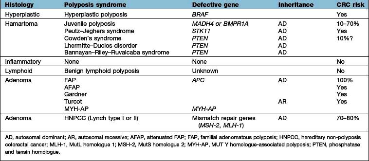
Peutz–Jeghers syndrome (see p. 271).
PTEN hamartoma–tumour syndrome (PHTS), which includes Cowden’s syndrome, Bannayan–Riley–Ruvalcaba syndrome and all syndromes caused by germline Phosphatase and Tensin Homolog (PTEN) mutations. Cowden (multiple hamartoma) syndrome is associated with characteristic skin stigmata and intestinal polyps regarded as hamartomas but with a mixture of cell types. These patients have an increased risk of various extraintestinal malignancies (thyroid, breast, uterine and ovarian). These syndromes are uncommon and together account for <1% of colon cancer cases.
Hyperplastic (metaplastic) polyps. These are frequently found in the rectum and sigmoid colon. These pale, sessile mucosal nodules usually measure <5 mm and are normally without significant malignant potential. However, metaplastic polyposis is defined as the presence of more than 10 colonic metaplastic polyps, some of which are large. These phenotypes are rare but appear to exhibit an increased risk of colon cancer.
Colorectal cancer (CRC) is the third most common cancer worldwide and the second most common cause of cancer death in the UK.
Each year approximately 40 000 new cases are diagnosed in England and Wales (68% colon, 32% rectal cancer) and it is registered as the cause of death in about half this number. The prevalence rate per 100 000 (at all ages) is 53.5 for men and 36.7 for women. The incidence increases with age; the average age at diagnosis is 60–65 years. Approximately 20% of patients in the UK have distant metastases at diagnosis. The disease is much more common in westernized countries than in Asia or Africa.
Factors related to risk of colorectal cancer are shown in Table 6.19.
Table 6.19 Risk factors in colorectal cancer
|
Most colorectal cancers develop as a result of a stepwise progression from normal mucosa to adenoma to invasive cancer. This progression is controlled by the accumulation of abnormalities in a number of critical growth-regulating genes. These include APC mutation and loss, K-ras mutation, Smad2/4loss, and TP53 mutation and loss, and altered DNA methylation with progression to carcinoma. CDK 8 has recently been found to regulate gene expression in the proliferation of colorectal cancer, and it also regulates the WNT/beta-catenin signalling pathway (p. 26) involved in many colon cancers. Microsatellite instability (MSI, see p. 288) and chromosomal instability (CIN) are frequently detected in colon cancers. A third group, MACS (Microsatellite And Chromosomal Stable), is also recognized. CIN indicates loss of heterozygosity (LOH) in a number of cancer-related genes, though the underlying mechanisms are not well understood. About 15% of sporadic colorectal cancers show MSI and 50% exhibit LOH.
A family history of CRC confers an increased risk to relatives. Family history is, next to age, the most common risk factor for CRC. FAP (Fig. 6.43) is the best-recognized syndrome predisposing to colorectal cancer but represents less than 1% of all colorectal cancers. Hereditary non-polyposis colorectal cancer (HNPCC) accounts for 3–10% of familial cancer (see p. 288).
Figure 6.43 Percentages of colon cancer according to family risk. HNPCC, hereditary non-polyposis colorectal cancer; FAP, familial adenomatous polyposis.
Additionally, some colon cancers arise, at least in part, from an inherited predisposition, so-called familial risk (Table 6.20). Estimates of their frequency range from 10% to 30% of all CRC but the genes involved have yet to be identified. The risk of CRC can be estimated from a family history matched with empirical risk tables so that appropriate advice regarding screening can be offered.
Table 6.20 Lifetime risk of colorectal cancer in 1st-degree relatives of a patient with colorectal cancer
Population risk |
1 in 50 |
One 1st-degree relative affected (any age) |
1 in 17 |
One 1st-degree and one 2nd-degree relative affected |
1 in 12 |
One 1st-degree relative affected (age <45) |
1 in 10 |
Two 1st-degree relatives affected |
1 in 6 |
Autosomal dominant pedigree |
1 in 2 |
Houlston RS, Murday V, Harocopos C et al. Screening and genetic counselling for relatives of patients with colorectal cancer in a family cancer clinic. British Medical Journal 1990; 301:366–368.
Most colorectal cancers are, however, sporadic and occur in individuals without a strong family history. Their distribution is shown in Figure 6.44.
CRC, which is usually a polypoid mass with ulceration, spreads by direct infiltration through the bowel wall. It involves lymphatics and blood vessels with subsequent spread, most commonly to the liver and lung. Synchronous cancers are present in 2% of cases. Histology is adenocarcinoma with variably differentiated glandular epithelium with mucin production. ‘Signet ring’ cells in which mucin displaces the nucleus to the side of the cell are relatively uncommon and generally have a poor prognosis.
Symptoms suggestive of colorectal cancer include change in bowel habit with looser and more frequent stools, rectal bleeding, tenesmus and symptoms of anaemia. Looser and more frequent stools, with or without abdominal pain, are common symptoms of left-sided colonic lesions. Rectal and sigmoid cancers often bleed, blood being mixed in with the stool. Presentation with constipation with hard stools is not a risk factor for colon cancer. A rectal or abdominal mass may be palpable. Cancers arising in the caecum and right colon are often asymptomatic until they present as an iron deficiency anaemia. Cancer may present with intestinal obstruction.
Patients aged over 35–40 years presenting with new large bowel symptoms should be investigated. Digital examination of the rectum is essential and examination of the colon should be performed in all cases. Hepatomegaly is present if there are large liver metastases.
Carcinoma in the ascending colon. (a) Colonoscopic appearance of a large irregular ulcer. (b) Histopathology.
Colonoscopy is the ‘gold standard’ for investigation and allows biopsy for histology. Biopsy of the tumour is mandatory, usually at endoscopy.
Double-contrast barium enema can visualize the large bowel but is now superseded by CT colonography.
Endoanal ultrasound and pelvic MRI are used for staging rectal cancer.
Chest, abdominal and pelvic CT scanning to evaluate tumour size, local spread and liver and lung metastases – this contributes to the tumour staging.
PET scanning is useful for detecting occult metastases and for evaluation of suspicious lesions found on CT or MR.
MR is also useful for evaluating suspicious lesions found on CT or US, especially in the liver.
Serum carcinoembryonic antigen (CEA) is of little use for primary diagnosis and should not be performed as a screening test. It is useful for follow-up; rising levels suggest recurrence.
Faecal occult blood tests are used for mass population screening and are of value in hospital or general practice.
Treatment should be undertaken by multidisciplinary teams working in specialist units. About 80% of patients with colorectal cancer undergo surgery (often laparoscopically), though fewer than half of these survive more than 5 years. The operative procedure depends on the cancer site. Long-term survival relates to the stage of the primary tumour and the presence of metastatic disease. There has been a gradual move from using Dukes’ classification to using the TNM classification system (see p. 477). Long-term survival is only likely when the cancer is completely removed by surgery with adequate clearance margins and regional lymph node clearance.
Total mesorectal excision (TME) is required for rectal cancers and removes the entire package of mesorectal tissue surrounding the cancer. A low rectal anastomosis is then performed. Abdomino-perineal excision which requires a permanent colostomy is reserved for very low tumours within 5 cm of the anal margin. TME combined with preoperative radiotherapy reduces local recurrence rates in rectal cancer to around 8% and improves survival. Pre- or postoperative chemotherapy reduces local recurrence rates but had no effect on survival in a recent study.
A segmental resection and restorative anastomosis with removal of the draining lymph nodes as far as the root of the mesentery is used for cancer elsewhere in the colon. Surgery in patients with obstruction carries greater morbidity and mortality. Where technically possible, preoperative decompression by endoscopic stenting with a mesh-metal stent relieves obstruction so surgery can be elective rather than emergency, and is probably associated with a decrease in morbidity and mortality.
Local transanal surgery is very occasionally used for early superficial rectal cancers.
Surgical or ablative treatment of liver and lung metastases prolongs life where treatment is technically feasible and the patient is fit enough to undergo the treatment.
Radiotherapy is not helpful for colonic cancers proximal to the rectum because of difficulties delivering a sufficient dose to the tumour without excess toxicity to adjacent structures, particularly the small bowel.
Adjuvant postoperative chemotherapy improves disease-free survival and overall survival in stage III (Dukes’ C) colon cancer (see p. 478). Those with Dukes’ B tumours with advanced features such as vascular invasion may also benefit.
Treatment of advanced colorectal cancer is discussed on page 477.
All patients who have surgery should have a total colonoscopy performed before surgery to look for additional lesions. If total colonoscopy cannot be achieved before surgery, a second ‘clearance’ colonoscopy within 6 months of surgery is essential. Patients with stage II or III disease should be followed up with regular colonoscopy and CEA measurements; rising levels of CEA suggest recurrence. Annual CT scanning of the chest and abdomen to detect operable liver metastases should be performed for up to 3 years post-surgery.
Faecal occult blood (FOB) tests have been studied as a screening test for colorectal cancer. Several large randomized studies have demonstrated a reduction in cancer-related mortality of 15–33%. Immunological based FOB tests are superior to the conventional guaiac based systems. The disadvantage of screening with FOB is its relatively low sensitivity, which means many negative colonoscopies. In FOBT screen-positive patients in the UK National Bowel Cancer Screening Programme (NHS BCSP), about 10% have cancer, 40% have adenomas and the colon is normal in 50%.
Flexi-sigmoidoscopy screening has been shown to reduce the mortality from CRC but not overall mortality.
Colonoscopy is the ‘gold standard’ technique for the examination of the colon and rectum and is the investigation of choice for high-risk patients. Universal screening strategies have been recommended in the USA, but the shortage of skilled endoscopists, the expense, the need for full bowel preparation and the small risk of perforation make colonoscopy impractical as a population screening tool at present.
CT colonography (‘virtual colonoscopy’) (see Fig. 6.5) is being increasingly used.
Genetic testing and stool DNA tests also contribute to screening programmes.
FURTHER READING
Atkin WS, Edwards R, Kralj-Hans I et al; UK Flexible Sigmoidoscopy Trial Investigators. Once-only flexible sigmoidoscopy screening in prevention of colorectal cancer: a multicentre randomised controlled trial. Lancet 2010; 375:1624–1633.
Cunningham D, Atkin W, Lenz HJ et al. Colorectal cancer. Lancet 2010; 375:1030.
Markowitz SD, Bertagnolli MM. molecular origins of cancer: molecular basis of colorectal cancer. N Engl J Med 2009; 361:2449–2460.