Rewarming is the major objective of therapy. For mild hypothermia (30° to 35° C [86° to 95° F]), only external application of heat lamps or immersion in water (38° to 42° C [100.4° to 107.6° F]) is necessary to restore core temperature with little risk of complications. Lower temperatures require core rewarming by any of several modalities: delivery of warm humidified oxygen, intravenous infusion of fluids, rectal lavage or peritoneal lavage (dialysis) with warm fluids, hemodialysis, application of external warmth to core circulation areas (groin, axilla, posterior neck region), and/or extracorporeal blood rewarming.
Supportive therapy includes maintenance of ventilation, cardiac monitoring, monitoring of renal function, and correction of fluid and acid-base imbalances. The prognosis is directly correlated with the degree of hypothermia, method of rewarming, and presence of underlying medical conditions.
Nursing care consists of monitoring vital functions and assisting with therapies. Obtaining a history from the family or other observers, including outside environmental temperature, length of exposure to elements, location of exposure site (e.g., outside or inside a vehicle or structure), and any care that may have been given, is essential. If trauma is associated with the hypothermia, ascertain the mechanism and circumstances of injury.
Prevention: Anticipation of cold conditions and knowledge of cold survival techniques are the basis of prevention. Children living in cold climates should have adequate protection when outdoors. Multiple layers of warm clothing are more effective than a single heavy layer for reducing the rate of heat loss, although they do not prevent it. Families living in cold climates should take precautions against unexpected prolonged exposure to cold (e.g., store extra blankets, food rations, and other equipment in their vehicles in the event of an unexpected mechanical breakdown).
Loss of central core heat can be reduced by 50% when an individual assumes the fetal position or when a person in water remains still. A person suspected of having hypothermia should be moved to a sheltered area, and wet clothing should be removed and replaced with dry, warm garments. Administration of warm, high-calorie liquids is important if the person is conscious.
Adverse reactions to drugs are seen more often in the skin than in any other organ, although drugs can affect any organ of the body. The reaction may be a result of toxicity related to drug concentration, individual intolerance to the average dosage of the drug, or an allergic or idiosyncratic response. The manifestations may be associated with side effects or secondary effects of a drug, either of which are unrelated to its primary pharmacologic actions.
Although any drug is capable of producing almost any form of reaction in the susceptible individual, some have a tendency to produce a particular reaction consistently, and some are more likely than others to produce an untoward effect. Many such effects are allergenic responses following a prior administration of the drug, even a topical application. Other factors influence response to a drug in a particular individual. For example, the incidence of reaction increases with the amount and the number of drugs given.
Manifestations of drug reactions may be delayed or immediate. A period of 7 days is usually required for a child to develop sensitivity to a drug that has never been administered previously. With prior sensitivity the manifestations appear almost immediately. Rashes—exanthematous, urticarial, or eczematoid—are the most common manifestation of adverse drug reactions in children. However, individual drug reactions may vary from a single lesion to extensive, generalized epidermal necrosis. Cutaneous manifestations can resemble almost any skin disease and can be seen in almost any degree of severity. With few exceptions, the distribution of a drug eruption is widespread (because it results from a circulating agent); appears as an inflammatory response with itching; is sudden in onset; and may be associated with constitutional symptoms such as fever, malaise, gastrointestinal upset, anemia, and liver and kidney damage.
Another common response is a fixed eruption (i.e., a recurrent eruption at the same site with each readministration of the drug). The lesion, a purplish red round or oval plaque with a sharp border seen most frequently on the extremities, disappears slowly, and the pigmentation deepens with each episode.
In most cases treatment for simple cutaneous reactions consists of discontinuation of the drug. Sometimes a decision is made to continue the drug (such as an antibiotic in an infant or small child) until the cause of the rash is clearly indicated. In urticarial-type eruptions, antihistamines may be ordered, and for widespread and severe lesions corticosteroids are beneficial. Severe anaphylactic reactions are a medical emergency. (See Anaphylaxis, Chapter 29.)
The most effective means of management is prevention. Parents always remember a severe response. A careful history taking will elicit evidence of a previous drug reaction. The history should include the name of the drug, nature of the reaction, drug dose, and how soon after administration the reaction occurred. (See History Taking, Chapter 6.)
Nurses who suspect that medication is the cause of the rash should withhold any further dose and report the eruption to the practitioner. Frequent offenders in drug reactions are penicillin and sulfonamides, and nurses must be alert to this possibility. However, even commonplace drugs such as aspirin and barbiturates, chemical agents in some foods, flavoring agents, and preservatives are capable of producing an undesired response. Persons who have severe reactions should wear a medical identification bracelet or necklace in case of emergency or inadvertent administration of the offending agent.
Erythema multiforme is an acute cutaneous disorder that may be associated with infections (usually viral) or ingestions of drugs. The characteristic lesion consists of an urticarial plaque with a dusky or vesicular center, which appears primarily on the palms, soles, and extensor surfaces.
Treatment involves discontinuing the drug, applying wet compresses for erosive lesions, and administering analgesics for discomfort. Antihistamines may be prescribed for pruritus.
Stevens-Johnson syndrome is the severe form of erythema multiforme characterized by lesions of the skin and mucous membranes, fever, and multiple systemic symptoms. The disease is presumed to be a hypersensitivity reaction to certain drugs, although the reaction may follow an upper respiratory tract infection. The disorder is relatively rare, occurs at any age, and is more common in males than in females.
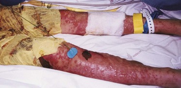
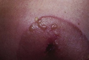
The syndrome begins with flulike symptoms: malaise, sore throat, fever, and severe headache. Inflammation of the glans penis (balanitis), eyes (conjunctivitis), or mouth and pharynx (stomatitis) appears next, followed in a few days by an erythematous papular rash. The lesions can involve any cutaneous surface, including the palms and soles, but usually spare the scalp. They can be scattered or confluent. The initial lesions enlarge by peripheral expansion with a vesicular center that often becomes bullous (Fig. 18-12). Mucous membrane ulceration often becomes severe enough to interfere with eating, and many patients have pulmonary involvement.
Mild disease requires only symptomatic treatment. However, severe disease necessitates hospitalization. Fluid and nutritional requirements are high, and patients often respond to topical lidocaine to relieve oral pain and a liquid diet. Intravenous opioids, such as morphine, and parenteral feedings may be needed for extensive oral involvement. Meticulous mouth care is important, and skin care frequently requires management in a burn unit. (See Stomatitis, Chapter 16, and Burns, Chapter 29.) Daily ophthalmologic examination is advised. Dry eyes are a problem, as is risk of chronic mild symblepharon (adhesion of lids to the eyeball). Eye lubricants promote moisture and comfort. Antibiotics are administered to patients with positive culture results, but the use of corticosteroids is controversial.
The mortality is estimated at 10% to 15% during the acute phase, especially in patients with pulmonary involvement. The disease is self-limiting, and the skin lesions gradually disappear without scarring in 2 to 3 weeks but may recur on reexposure to an offending drug. Wound care usually consists of cleansing with normal saline and providing a moist environment to promote wound healing. Hydrogel dressings (gauze impregnated with gel) are usually soothing and do not stick to the wound with removal (see Wound Care section, p. 691). The family needs emotional support to cope with the life-threatening nature of the disease.
Toxic epidermal necrolysis is a drug-induced injury to the skin characterized by a generalized erythematous rash that rapidly evolves into bullae and peeling. It appears to be a hypersensitivity reaction with precipitating factors similar to those responsible for erythema multiforme. The more common offending drugs are phenobarbital, phenytoin, allopurinol, sulfonamides, and penicillin. In children the clinical appearance is the same as that seen in staphylococcal scalded skin syndrome.
The disease begins with a prodromal period of fever and malaise. Symptoms include a generalized erythematous rash that rapidly evolves into bullae and extensive epidermal peeling, and oral lesions similar to those observed in Stevens-Johnson syndrome. Treatment consists of withdrawal of the offending drug, fluid and electrolyte replacement, and skin management as for severe burns. The disease can be protracted, and mortality can range from 25% to 50%. It is essential that families of children receiving antiepileptics or sulfonamides be informed of the significance of a rash and the importance of promptly reporting it to their health professional.
A number of other skin disorders also occur in children (Table 18-9). Psoriasis can occur in children younger than 16 years of age, and photosensitivity eruptions associated with inherited diseases appear early in childhood. Ichthyoses are a heterogeneous group of disorders characterized by scaling that creates challenging treatment problems.
TABLE 18-9
*National Alopecia Areata Foundation, 14 Mitchell Blvd., San Rafael, CA 94903; 415-472-3780; fax: 415-472-5343; e-mail: info@naaf.org; www.naaf.org.
†Children’s Tumor Foundation (see p. 711 for contact information).
Neurofibromatosis 1 (NF1), or von Recklinghausen disease, is a relatively common disorder with an autosomal dominant inheritance pattern. It occurs in 1 in 3000 persons and has a high mutation rate. The manifestations are highly variable and appear to result from a defect that alters peripheral nerve differentiation and growth.
Initial clinical presentation involves small, discrete, flat, pigmented skin lesions with smooth edges (café-au-lait spots, pigmented nevi) (Fig. 18-13) and/or axillary or inguinal freckling that develops in early infancy or childhood. Slow-growing cutaneous and subcutaneous neurofibromas that grow along the course of a peripheral nerve appear in later childhood or adolescence and increase in number with age. Lisch nodules, dome-shaped clear to yellow or brown elevations on the iris surface, develop before puberty in most affected individuals. Elephantiasis (thickening and enfolding of the skin) may also occur.
Fig. 18-13 Café-au-lait patches. (From Seidel HM, Ball JW, Daines JE, et al: Mosby’s guide to physical examination, ed 6, St Louis, 2006, Mosby.)
Other characteristics include developmental delay, cognitive impairment, seizures, scoliosis or kyphosis, short stature, macrocephaly, speech defects, learning disabilities, and problems with fine and gross motor skills (Johnson, Saal, Lovell, et al, 1999). Severity varies within the same family: one family member may have only café-au-lait spots or axillary freckling, whereas another may have more severe manifestations.
Diagnosis is established by physical findings based on National Institutes of Neurofibromatosis Fact Sheet (Box 18-1). A family history is elicited to determine whether the specific case is inherited or represents a new mutation. Risk for transmitting the disorder to offspring is 50%. Therapy is limited to excision of tumors that produce pain or impair function and symptomatic management of manifestations.
Nursing care involves recognition of signs that indicate a possibility of the disease, referral for diagnosis, and family counseling and support. It is important that a diagnosis be made, even when the only manifestations are a few café-au-lait spots. The family needs to know the genetic implications and be alert for signs that indicate the child is developing more serious characteristics of the disease. Cancer occurs in patients with the disorder, although the rates vary widely. Other members of the family should be assessed for evidence of the disorder.
In addition, families need to know that children with NF1 have a significantly increased risk for learning, social, and emotional problems. Parents need to receive written materials about these problems that they can share with teachers and other adults who interact with the child. Children with NF1 should be screened using systematic standardized tests, and more aggressive and intensive treatment should be provided for speech, motor, and cognitive deficits that may contribute to social and emotional problems (Johnson, Saal, Lovell, et al, 1999).
The Children’s Tumor Foundation* is an organization dedicated to increasing public awareness of NF1, providing help and support to families affected by the disorder, and stimulating research.
Arthropods include insects and arachnids (mites, ticks, spiders, and scorpions). Manifestations and management of skin lesions caused by arthropods are given in Table 18-10.
Fig. 18-14 Brown recluse spider bite; note central necrosis surrounded by purplish area and blisters. (From Weston WL, Lane AT: Color textbook of pediatric dermatology, St Louis, 1991, Mosby.)
Some proteins in insect venom are species specific; others are common to a number of species, and crossover reactivity is common. The usual local response to a sting is sharp pain; a local wheal (<5 cm [2 inches] in diameter); and erythema accompanied by intense itching at the site, lasting less than 24 hours. The reaction is produced by enzymes; cytotoxic proteins; and vasoactive compounds, primarily histamine and kinins.
Systemic reactions can occur and, in some instances, can be life threatening. More benign systemic reactions begin several minutes to several hours after the sting and consist of simple urticaria, erythema, pruritus, and angioedema. Serious, life-threatening reactions usually begin within 5 to 10 minutes after the sting and include airway obstruction secondary to laryngeal edema, bronchospasm, hypotension, and cardiovascular collapse.
To prevent contact with stinging and biting insects, teach children behaviors that reduce the likelihood of injury. In addition, topical insect repellents generally provide safe and effective protection for several hours. The best all-purpose repellents contain the active ingredient DEET, which is effective for a variety of insects and arachnids, including mosquitoes, chiggers, ticks, fleas, deerflies, and sand flies. Protection may last from 1 to several hours, but effectiveness is influenced by the concentration of active ingredients. The product must be reapplied after sweating, swimming, wiping, or exposure to rain. Because adverse effects have been reported in young children and because long-term effects of DEET are unknown, parents are advised against excessive application or prolonged use, especially of products with high concentrations of DEET. The insect repellent should not be applied to children’s hands because it may be rubbed into the eyes. It should be removed with soap and water when the child is brought inside.
Most bites are managed by simple symptomatic measures such as use of cool compresses, application of calamine lotion, and prevention of secondary infection. Often treatment consists of application of a substance that relieves the swelling and discomfort and can be made from common household products.
When a hymenopteran (a bee, in particular) stings, its barbed stinger penetrates into the skin. The stinger also contains a nerve ganglion, muscles, and a venom sac. As long as the stinger remains in the skin, the muscles push the stinger deeper, and venom is pumped into the wound. A study of different methods of extracting the stinger revealed that the method of removal did not influence the amount of venom injected into the wound. There was no difference in the amount of venom injected when the stinger was removed by external compression with forceps and when the stinger was flicked or scraped off the skin. The influencing factor was the amount of time from the bee sting to removal of the stinger; the longer the time interval, the greater the amount of venom injected (Visscher, Vetter, and Camazine, 1996). The best approach is to remove the bee stinger as quickly as possible and to get away from the vicinity of other bees to prevent further injury.
Children need to know how to avoid contact with bees and to recognize the insect (e.g., it is not part of the flower). For those who have become sensitized to hymenoptera stings and demonstrate a severe life-threatening systemic response, intramuscular administration of epinephrine provides immediate relief, and the drug must be available for emergency use. For hypersensitive children, a kit must be available that contains epinephrine, a hypodermic syringe, and perhaps ephedrine and an antihistamine preparation. Hypersensitive children should wear medical identification, such as a bracelet. Nurses should remind parents to frequently check the expiration date on the kit and replace an outdated one. Families should find out whether a nurse is available at the school; if not, someone at the school should be designated to inject the epinephrine in case of an emergency.
Children with a history of generalized reactivity to an insect sting should undergo skin testing with the radioallergosorbent test and possibly immunotherapy with venous extract (desensitization) to prevent serious or fatal reactions. In the United States venous extracts are available for the honeybee, yellow jacket, yellow hornet, and wasp.
Most arachnids in the United States, including tarantulas, are relatively harmless. All spiders produce venom that is injected via fangs. Some are unable to pierce the skin; others produce a venom that is insufficiently toxic to be harmful. A local tissue reaction is relieved by cool compresses or the methods described for hymenoptera stings.
Only scorpions and two spiders—the brown recluse and the black widow—inject venom deadly enough to require immediate attention. Children bitten by these arachnids must receive medical attention as soon as possible.
Ticks are troublesome because they become partially imbedded in the skin as they feed. The recommended method for removal is to grasp the tick with curved forceps as close as possible to the point of attachment and pull straight up with a steady, even pressure. If a portion of the body (e.g., the head) remains, it can be removed with a sterile needle in the same manner as a sliver. The bite is cleansed with soap and a disinfectant after removal.
To avoid ticks, children should wear long pants tucked into the socks and a long-sleeved shirt when walking in infested areas, especially in the spring and summer. Ticks can also be picked up by dogs and other household pets. Parents are advised to check their children carefully for the organisms when their children have been in areas where ticks might be acquired.
Nonhuman mammal bites are common injuries and are inflicted by both wild and domestic animals. Wild animal bites are discussed in relation to rabies in Chapter 37, and these wounds are treated in the same way as the bites of domestic animals such as dogs, cats, hamsters, and mice. The present discussion is directed primarily toward dog bites.
Approximately half the victims of dog bites are younger than 5 years of age; boys are bitten more frequently than girls (Bernardo, Gardner, O’Connor, et al, 2000). Stray dogs are seldom involved in attacks; most dogs involved are owned by the family of the victim or by a neighbor. Most dog attacks occur in or adjacent to the owners’ yards, and the attack is usually preceded by verbal or physical contact with the animal (Bernardo, Gardner, O’Connor, et al, 2000). This problem is increasing because of a growing trend of acquiring large, aggressive guard dogs. Most animal bites are caused by dogs. Cat bites are less frequent, although cat scratches are extremely common (see Cat Scratch Disease, p. 703). Most injuries caused by dogs and cats are to the upper extremities. Small children are more likely to receive bites or scratches to the head, face, and neck because they tend to put their heads near the animal’s head and flail their arms rather than protecting their heads.
Animal bites are potentially serious because of the likelihood of significant infection. Injuries vary in intensity from small puncture wounds to complete avulsion of tissue that is associated with significant crush injury. Dog bites are seen as lacerations or evulsions; cats exert less biting force, but their sharp teeth penetrate more deeply, inoculating organisms deep into tissues.
The location of a bite influences the incidence of infection. Injuries to the arm and hand tend to become infected more often than those on the legs, scalp, and face. Redness, swelling, and tenderness develop around the site of injury, often accompanied by purulent or serosanguineous drainage. It may be difficult to assess hand infection because most lymphatic drainage is contained in the dorsal subcutaneous space, and swelling occurs in this area when the injury may be elsewhere.
Therapeutic Management: General wound care consists of rinsing the wound with copious amounts of saline or Ringer’s lactate delivered under pressure via a large syringe and washing the surrounding skin with mild soap. A clean pressure dressing is applied, and the extremity is elevated if the wound is bleeding. Medical evaluation is advised because of the danger of tetanus and rabies, although dogs in most urban areas are required to be immunized against rabies. Bites from wild animals, such as raccoons, skunks, foxes, and bats, are potentially dangerous. (See Rabies, Chapter 37.)
Prophylactic antibiotics are indicated for puncture wounds and wounds in areas where infection could result in cosmetic or functional impairment. Extensive lacerations are débrided and loosely sutured to allow for drainage in the event of infection. Primary closure of jagged, irregular wounds with associated crush injury and devitalized tissue is contraindicated, except for facial wounds because of cosmetic reasons. Tetanus toxoid is administered according to standard guidelines (see Chapter 12), and rabies protocol is followed. Injuries to poorly vascularized areas such as the hands are more likely to become infected than those in more vascularized areas such as the face. Puncture wounds are more likely to become infected than are lacerations.
Nursing Care Management: The most important aspect related to animal bites is prevention. Children should understand animal behavior and develop respect for animals. It is vital that they learn how to treat animals and how to react to them. Parents should monitor their children’s behavior with dogs and instruct them not to tease or surprise a dog, invade its territory, interfere with its feeding or sleeping, take its toy, or interact with a sick or injured dog or a dog with pups (Humane Society of the United States, 1998).
Parents who are considering getting a pet, especially a dog, for themselves or their children should select a dog that is least likely to be a danger to their children. The level of sociability with children is the key to a selection, and dogs range from dangerous and unsuitable to tolerant of children and well behaved.
Parents should obtain dogs from a reputable source that breeds dogs for good temperament. After the dog is purchased, obedience training and socialization should begin to prevent the dog from developing behaviors that are undesirable to the family and neighborhood. Such training is often provided through veterinary services or animal shelters (Bernardo, Gardner, O’Connor, et al, 2000). Prevention programs should educate families about preventing bites and responsible ownership (i.e. training, socializing, neutering).
Children often acquire lacerations from the teeth of other humans in rough play, during fights, or as victims of child abuse. Many preschool children bite others out of frustration or anger. Because human dental plaque and gingiva harbor pathogenic bacteria, all human bite wounds should receive attention. Delaying treatment increases the risk of infection.
If the laceration is less than 6 mm (0.25 inch) long, the wound can be treated at home. The wound is washed vigorously with soap and water, and a pressure dressing is applied to stop bleeding. Ice applications minimize discomfort and swelling. Increased pain or redness at the wound site is an indication that the child should receive medical attention for antibiotic therapy. Tetanus toxoid is needed if the child is insufficiently immunized. Wounds larger than 6 mm should receive medical attention.
In spring snakes emerge from winter hibernation hungry for food and water. The best prevention for snake bites is to avoid snakes, since most people are bitten when trying to handle them. Approximately 1600 venomous bites from domestic snakes were reported to the American Association of Poison Control Centers in 1998 (Litovitz, Klein-Schwartz, Caravati, et al, 1999). Asian and African snakes are far more dangerous than those in the United States and Europe. The major families in the United States are the Crotalidae (pit vipers), which include rattlesnakes, copperheads, and cottonmouths, and the Elapidae, which include coral snakes and cobras. Most bites are attributed to the Crotalidae species.
The manifestations and morbidity are highly variable and depend on the species and size of the snake, the amount of venom injected, the time of year, the child’s age and size, and the location of the bite. Not every bite from a poisonous snake injects venom (Schexnayder and Schexnayder, 2000).
The initial action after snakebite is to move the victim away from the area, attempt to calm the child, and place the child at rest. A loose tourniquet applied proximal to the bite delays the flow of lymph, which can carry the venom to the systemic circulation. It should not be tight enough to occlude circulation; a pulse should be palpable distal to the bite. Remove any constricting items of clothing or jewelry from the affected limb. Apply a splint to immobilize the limb, and transport the victim to the nearest medical facility.
If the child has been bitten by a large snake, if less than 30 minutes (some authorities say 5 minutes) has elapsed since the child was bitten, and if medical help is more than 30 minutes away, suction may be beneficial. Suction should be applied by a suction device such as the Sawyer extractor, which is very effective if used within 3 minutes.
If possible, the dead snake should be transported with the patient for identification. If there is any possibility that a child has been bitten by a coral snake, aggressive use of antivenom is indicated; once symptoms occur, it is difficult to stop the respiratory paralysis and death.
Dental caries (cavities) is one of the most common chronic diseases that affect individuals at all ages; it is the principal oral problem in children and adolescents. Although the overall incidence of dental caries in children has decreased since the introduction of fluoridation, reducing the incidence and consequences of the disorder remains important. Dental caries, if untreated, results in total destruction of the involved teeth. The ages of greatest vulnerability are 4 to 8 years for the primary dentition and 12 to 18 years for the secondary or permanent dentition. (See Figs. 12-13 and 17-15 for sequence of tooth eruption.)
Dental caries is a multifactorial disease. It involves a number of elements: (1) the host, (2) microorganisms, (3) substrate, and (4) time.
Host: The prevalence of caries is directly related to the tooth size and morphologic characteristics and to the consistency, composition, and amount of saliva. The incidence of caries is increased in teeth that are improperly developed, crowded, or deeply fissured. The areas most subject to attack by bacteria are grooves and fissures, interdermal areas, gum margins, and other smooth surfaces. Newly erupted teeth that have not yet acquired sufficient surface minerals are more susceptible to decay than those that have been erupted for 2 years or longer. Hereditary factors influence resistance and susceptibility, and similar patterns and anatomic characteristics often appear in successive generations. Salivary flow can mechanically clean away bacteria and food debris. It also contains buffering systems, lysozymes, peroxidases, and immunoglobulins that influence the development of caries.
Microorganisms: Certain types of microflora that produce different effects contribute to the formation of dental caries. Acidogenic bacteria act on fermentable carbohydrates in dental plaque to produce organic acids that decalcify hard surface tooth enamel. With the inner organic matrix exposed, proteolytic organisms and acids digest and destroy the inner tooth structure. These destructive organisms are harbored and protected in a gelatinous plaque formed on the tooth surface by another group of bacteria that are thought to play no primary role in production of decay.
Substrate: Caries formation is strongly influenced by the two concurrent processes that continually operate on enamel surfaces: acid production and acid neutralization by saliva. The material on which the acid-forming bacteria act consists essentially of carbohydrates. Among the fermentable carbohydrates, sucrose has been consistently implicated as the most cariogenic. Sucrose-containing substances, especially in forms that cling (such as chewy candy) or that promote prolonged contact with the teeth (such as hard candy and lollipops), when ingested between meals, contribute markedly to the development of dental caries. Saliva, some foods, and chewing gum after a meal tend to help neutralize much of the acid formed from sucrose.
Time and Other Factors: Bacterial enzymes act on salivary glycoproteins to produce a tenacious protein matrix on the tooth surface. This substance, along with the microorganisms, forms dental plaque. If plaque removal is inadequate or nonexistent for a significant length of time (a few days), the plaque is metabolized by the bacteria to form acid, which initiates the demineralization of enamel.
Other factors that contribute to caries formation are heredity, the amount of fluoride in drinking water, and the child’s general state of health. Hereditary factors influence both resistance and susceptibility to dental caries. For example, structural defects, such as deep fissures on occlusive surfaces, predispose the teeth to decay, and individuals in whom acid formation exceeds neutralization are prone to caries. The effectiveness of the buffering action of saliva is highly variable among individuals.
Poor oral hygiene that permits the accumulation of food debris on tooth surfaces allows acid-forming bacteria to thrive and proliferate. Removal of food particles and bacteria-laden plaque inhibits destructive acid formation.
The susceptibility to dental decay is also influenced by the child’s general health. Children who suffer from chronic debilitating disease show increased caries activity, as do children with systemic conditions that alter the quality and quantity of saliva produced.
Because the permanent teeth erupt during middle childhood, children are more susceptible to the development of dental caries during this time than at any other age. Caries penetrates the vulnerable teeth rapidly at this age, whereas carious activity is slower and more irregular at later ages.
Caries on visible surfaces are easily detected by oral inspection. Large, extensive caries are apparent to the untrained eye, but small, beginning lesions are best identified by trained professionals. Caries between the teeth may not be located without x-ray examination. A common site of decay is the fissures of the molars.
Well-informed health care professionals can provide dental information and make periodic dental assessments. However, dentists are prepared to provide both of these services and are the only ones qualified to treat most dental problems. Prophylaxis, including hygiene and fluoride treatment, is the major thrust of dental therapy. (See Chapter 14.) Plasticized sealant, applied to the deep fissures and grooves of healthy teeth, is effective in blocking cavity formation. Treatment of dental caries involves removal of all carious portions of the tooth as soon as decay is detected, preparation of a retentive cavity, and replacement of the lost portion of the tooth with a material that is durable in the mouth environment. This restoration of involved teeth not only prevents progression of established caries but also reduces the number of bacteria in the oral cavity to decrease the danger to uninvolved teeth.
Oral inspection is an integral part of the nursing assessment of the child. If there is evidence of dental caries or another unhealthy state, the child is referred for dental services. Many families have a family dentist or a pedodontist who can provide needed care. However, an alarming number of children do not receive regular dental supervision, and a significant number reach adulthood without being examined or treated by a dentist. Nurses can be active members of preventive educational programs and serve as counselors to families regarding the importance of regular dental care, oral hygiene, and dietary management.
Nurses should encourage good oral hygiene and teach correct tooth cleaning to both children and their parents. The random brushing allowed during the early childhood years should be replaced by more careful and methodic cleaning techniques. Children should brush their teeth and use dental floss according to the method recommended by their dentists. (See Chapter 14.) Regular administration of fluoride is also important. (See Chapter 14.) Families should be aware of the fluoride content of their drinking water, including bottled water if it is used. School-age children can usually manage the chewable tablets, which have both a topical and a systemic effect.
Restriction of cariogenic foods is important to prevent dental caries, but this should be viewed as an activity in which all family members are involved and not simply a directive for the child to obey. It should not be communicated in such a way that the child interprets the withholding of sweets as a punishment.
Concern has been generated about the sugar content of children’s pharmaceutical products, especially because children with chronic conditions such as seizure disorders, asthma, and recurrent urinary tract or ear infections must take medications over a period of years. Children with chronic illness who regularly take medications containing sugar are cautioned to brush their teeth after taking the medication, just as they would after eating any carbohydrate substance. Children taking tricyclic antidepressants are also prone to develop dental caries.
The greatest task for nurses is counseling children and families to develop sound dental hygiene and nutritional practices. School nurses have an excellent opportunity to participate in community detection of dental needs, to educate children in dental hygiene, to make referrals, and to motivate children to comply with prophylaxis and treatment.
Children should be prepared for dental services in such a way that visits to the dentist are a positive experience. Keeping appointments and following through on recommended treatments and practices are habits that extend beyond childhood.
Periodontal disease, an inflammatory and degenerative condition involving the gums and tissues supporting the teeth, often begins in childhood and accounts for a significant amount of tooth loss in adulthood. The more common periodontal problems are gingivitis (simple inflammation of the gums) and periodontitis (inflammation of the gums and loss of connective tissue and bone in the supporting structures of the teeth). An uncommon condition is acute necrotizing ulcerative gingivitis (“trench mouth”).
The most prevalent periodontal disease, gingivitis, is a reversible inflammatory disease that begins very early in many children and is most often associated with the buildup of plaque on the teeth. Changes take place in the plaque bacteria, in both the type and number of organisms, that cause them to release a variety of destructive exotoxins, enzymes, and other noxious agents. These agents produce an inflammatory reaction in the gingival tissues, which causes the gums to become red, edematous, tender, and subject to bleeding at slight irritation.
Management is directed toward prevention by conscientious brushing and flossing and by depriving the bacteria of the substrates required to produce the disease. The implementation and maintenance of preventive dental practice, including the use of fluoride, and conscientious brushing and flossing are effective in preventing both caries and periodontal disease.
Nursing care of the child with periodontal disease is primarily supportive; it includes education regarding dental hygiene and regular inspection of the gingival tissues for signs of early inflammation. Advise the child to see the dentist at any sign of inflammation or irritation.
Nurses caring for teenagers should observe for the use of chewing tobacco. The easily detectable clinical lesions appear as tooth erosion, periodontal destruction, and red or white mucosal alterations. The primary site of lesions is the anterior mandibular mucobuccal fold region.
When teeth of the upper and lower dental arches do not approximate in the proper relationships, the physiologic function of mastication is less effective, and the cosmetic effect is less pleasing. Teeth that are uneven, crowded, or overlapping or are otherwise unable to meet their opponents in the opposite jaw in the appropriate relationships may be predisposed to dental disease. More than half of children 12 to 17 years of age suffer from malocclusions that could be corrected.
The most common cause of malocclusion is hereditary factors, but abnormal growth and habits such as thumb sucking and tongue thrusting also contribute to the disordered alignment and occlusion of the teeth. Treatment of malocclusion includes eliminating habits that aggravate the deformity and initiating corrective therapy at the optimum time. Orthodontic treatment is usually most successful when it is started in the later school-age years or the early adolescent years, after the last primary teeth have been shed and before growth ceases. However, because some deformities can be corrected at an earlier age, referral should be made as soon as malocclusion is evident. For example, removal of extra teeth or impacted teeth or prosthetic replacement of missing teeth can prevent problems from developing.
The nurse who detects malocclusion is obligated to recommend that the teeth be examined by a dentist. The sooner the child is evaluated, the sooner treatment can begin.
Although orofacial appearance is a subjective phenomenon, it can have an adverse effect on a child’s self-esteem and body image. Poorly aligned teeth can be a source of both psychologic and physical stress to affected children. Many children with severe malocclusion are teased by their peers or siblings.
If fixed appliances or braces are applied, the child is advised that there will be some discomfort for a few days. During the period of orthodontic treatments, which averages 18 to 30 months, proper oral hygiene is vital. Although the bands or brackets protect the teeth they cover, plaque can collect on the unprotected surfaces or under loose-fitting bands. The teeth should be brushed with a fluoride toothpaste after every meal and snack and at bedtime, using the method recommended by the dentist. Some orthodontists recommend using an oral irrigating device to remove food from between the teeth and around the braces. However, the device does not remove plaque and is not a substitute for thorough brushing. Some foods can damage the braces; others may be difficult to remove from the teeth during cleaning. Forbidden foods include chewing gum, ice, nuts, toffee and hard candy, corn on the cob, uncut apples, hard taco shells, nachos, and popcorn.
Occasionally, tooth movement or poking at the braces with a pencil or other object may cause an arch wire to break or protrude. If this happens, instruct the child to cover the broken portion with a special wax provided by the dentist and schedule an appointment as soon as possible. Regular visits are usually scheduled every 3 to 6 weeks. After the braces are taken off, a removable or permanent retainer must be used to maintain the desired position of the teeth. Placement of a permanent wire behind the front teeth requires no compliance from the youngster.
Sometimes children need considerable reinforcement for compliance with orthodontic treatment. It may be difficult for some to relate the present barriers of discomfort, inconvenience, and embarrassment to the future reinforcers of improved appearance and dental health. Teenagers with a heightened awareness of body image and physical attractiveness are especially at risk for noncompliance. (See Chapter 27 for a discussion of compliance.)
Dental injury is not uncommon in childhood. Most injuries occur after bicycle and playground mishaps and include fractures of varying degrees of severity, chipping, dislocation, or avulsion. All tooth injuries require prompt treatment by a competent dentist to prevent permanent displacement or loss. Delayed examination and diagnosis of tooth damage can result in infection or pulp involvement. Because it can affect the remaining teeth, loss of a permanent tooth requires professional attention to maintain normal alignment and position of teeth.
Trauma usually involves the maxillary incisors, and children with protruding teeth, craniofacial abnormalities, or neuromuscular disorders are more likely to sustain dental injuries.
A permanent tooth that is avulsed (evulsed, exarticulated, or “knocked out”) should be reimplanted by the child, parent, or nurse and stabilized as soon as possible so that the blood supply to the tooth can be reestablished and the tooth kept alive (American Academy of Pediatric Dentistry, 2009) (see Emergency Treatment box). If the tooth is replaced within 15 minutes, there is a better chance that it will become reattached and the roots will not resorb or the crown exfoliate. Avulsed primary teeth are usually not reimplanted.
Before reimplantation it is important to carefully rinse a dirty tooth in milk or saline solution or under running water to avoid disturbing the adhering periodontal membrane, which is essential to the success of the reimplantation. The tooth is held by the crown, not the root, while rinsing, with the drain plugged. The tooth is then fit back into its socket as atraumatically as possible (Troupe, 1995). If the tooth is reimplanted almost immediately, excessive pressure is not needed; however, it becomes extremely difficult after clot formation (in approximately 10 minutes). The tooth is held in place by the child during transportation to a dentist. If the child or parents are reluctant to reimplant the tooth, the next best alternative is to place the tooth in Viaspan, Hank’s Balanced Salt Solution, cold milk, saliva, contact lens solution, saline, or water for transport to the dentist (American Academy of Pediatric Dentistry, 2009). Cold milk has precisely the osmolality needed to maintain fluid balance within the tissues surrounding the tooth. Water is the least desirable storage medium because the hypotonic environment causes rapid cell lysis.
After reimplantation, the tooth usually becomes firmly attached, although endodontic therapy is often required. If reimplantation is not permanent, the tooth may be retained anywhere from 6 months to 12 years and facilitates normal development and occlusion, since loss of teeth during the period of permanent tooth eruption may adversely affect such development.
As with all mouth trauma, tooth avulsion causes a large amount of bleeding, which is frightening to children and their families. The nurse or anyone faced with dental trauma should be prepared to cope with the emotionality that accompanies tooth avulsion. Using a calm approach and providing gentle reassurance to the child are often successful in reducing anxiety.
Enuresis (bed-wetting) is a common and troublesome disorder that is defined as intentional or involuntary passage of urine into the bed (usually at night) or into clothes during the day in children who are beyond the age when voluntary bladder control should normally have been acquired. The inappropriate voiding of urine must occur at least twice a week for a minimum of 3 months, and the chronologic or developmental age of the child must be at least 5 years (see Cultural Competence box). In addition, the urinary incontinence must not be related to the direct physiologic effects of a substance (e.g., diuretics) or a general medical condition (e.g., diabetes mellitus or diabetes insipidus, spina bifida, seizure disorder, or sickle cell disease).
Enuresis can also be defined as primary (bed-wetting in children who have never been dry for extended periods) or secondary (the onset of wetting after a period of established urinary continence). The passage of urine may occur only during nighttime sleep (nocturnal), only during the waking hours (diurnal), or during both times of the day. The nocturnal type is most common.
Enuresis is more common in boys, and nocturnal bed-wetting usually ceases between 6 and 8 years of age (American Academy of Pediatrics, 2003). Although most children with enuresis do not have coexisting psychopathology, some children do have other developmental disorders, learning problems, or behavior difficulties, such as increased motor activity and aggression.
Enuresis can cause serious psychologic problems. The degree of impairment is related to the effect on the child’s social life, such as not being able to attend overnight camps, and the effect on others, who may ostracize or ridicule the youngster. Adolescents with enuresis have described themselves as being tense, having difficulty sleeping, and having bad dreams. Children state that they are embarrassed about the disorder and are often hesitant to sleep at other children’s homes. Avoiding overnight excursions can impede normal socialization or self-esteem. Enuresis can influence self-esteem if parental response to the disorder is harsh or punitive. In some instances, enuresis may serve as a trigger for child abuse. Although behavior problems can be associated with these psychologic effects, research suggests that adults treated for enuresis as children have normal psychologic profiles.
No clear cause for enuresis has been determined. However, predictive factors have been noted, including longer duration of sleep in infancy, a positive family history, and a slower rate of physical development in children up to 3 years of age. There is a high concordance rate of enuresis in monozygotic (identical) twins and an even higher one in dizygotic (nonidentical) twins, which suggests more than a pure genetic link in the disorder. Approximately 75% of all children with functional enuresis have a first-degree relative who has, or has had, the disorder.
Enuresis is primarily an alteration of neuromuscular bladder functioning and as such is benign and self-limiting. Emotional factors may influence the symptom. Some children exhibit temporary regressive behavior resulting in enuresis after the birth of a sibling or other trauma. Other children, such as those with attention deficit hyperactivity disorder (ADHD), may have occasional “accidents” when they become so involved in play that they are unaware of a full bladder or “forget” to empty the bladder. In other children enuresis may be related to problems with toilet training, such as the age at which training began; the emotional atmosphere surrounding the training situation; or an excessive amount of emotional dependence on the parent, usually the mother. Occasionally enuresis can be a behavioral manifestation of a personality disorder.
Although several theories have been proposed, no one theory thoroughly explains enuresis. The sleep theory stems from parental reports that these children sleep more soundly and are difficult to arouse from sleep.
Another theory relates to functional bladder capacity, the volume of urine voided after maximum delay of micturition. Although there is evidence that some children with enuresis have a smaller bladder capacity than unaffected children, other evidence suggests that this is not the cause. For example, children without enuresis but with a smaller bladder capacity awaken during the night to void, whereas children with enuresis do not awaken.
The nocturnal polyuria theory suggests that the kidneys of these children fail to concentrate urine during sleep because of insufficient secretion of antidiuretic hormone (ADH). The ADH circadian rhythm may thus be a significant biologic marker in enuresis, but additional research must be conducted to clarify its role.
The dysfunctional detrusor activity theory suggests that an unstable bladder detrusor muscle spontaneously contracts to produce bed-wetting, either because of abnormal innervation or as a result of other, unknown reasons. Studies to explore this theory have yielded contradictory and inconclusive results; more research is needed to clarify these contradictions and to determine whether there is a relationship between ADH production and detrusor activity.
The predominant symptom of enuresis is urgency that is immediate and accompanied by acute discomfort, restlessness, and sometimes urinary frequency. With nocturnal enuresis, the child may or may not feel urgency. If awareness of the urgency is present, the child often reports difficulty awakening to urinate. Spontaneous voiding during sleep occurs, which usually results in multiple nightly incidents. Spontaneous remission of nocturnal enuresis occurs in approximately 15% of cases. However, in some cases nocturnal enuresis continues into adolescence and adulthood.
During the initial phases of evaluation, a routine physical examination is performed to rule out physical causes, such as urinary tract infection, structural disorders, major neurologic deficits, nocturnal epilepsy, disorders that increase the normal output of urine (e.g., diabetes mellitus and diabetes insipidus), and disorders that impair the concentrating ability of the kidneys (e.g., chronic renal failure or sickle cell disease). The examination may include diagnostic evaluation of functional bladder capacity. Normal bladder capacity (in ounces) is the child’s age plus 2; therefore normal bladder capacity for 6-year-old is 8 oz (237 ml). Functional bladder capacity is determined by having the child hold off voiding until the strongest urgency is felt, at which time the child voids into a measurement container. A bladder volume of 300 to 350 ml (10 to 12 oz) is sufficient for retention of a night’s urine.
If psychologic difficulties are evident or a personality disorder is suspected, seek a routine psychiatric evaluation.
A history of wetting behavior is obtained, including information about the toilet-training process. Assess parental attitudes by listening and asking parents how they have attempted to cope with the wetting. An important feature of assessment is a baseline count of enuretic incidents and the time of day when each occurs. This is necessary not only to establish diagnostic reliability but also to confirm outcome success after treatment. The baseline information is gathered for 1 to 2 weeks by the child and family. It usually consists of a chart or calendar given to the family on which they indicate the date of the incident, the time of the incident, and the approximate volume of the urinary output.
Enuresis not resulting from known organic causes has been treated in several ways. No single method has achieved universal endorsement. Frequently, more than one technique is employed. In some cases, a spontaneous decrease in bed-wetting occurs with age and irrespective of the treatments used (American Academy of Pediatrics, 2003). Successful treatment is defined as a specified period of dry nights, varying from 7 to 28 consecutive dry nights.
Conditioning therapy involves training the child to awaken to urinate after a stimulus is given, especially with a urine alarm. The device consists of a moisture-sensitive wire pad that is placed inside the underpants and is attached to a bell or buzzer. When the system detects moisture, the bell or buzzer sounds, which fully awakens the child. The child is thus conditioned to awaken at the initiation of micturition or to the stimulus of the bell or buzzer and eventually learns to continue voiding in the toilet. The urine alarm can be very effective, but children may relapse once they stop using it. Relapse is addressed by reinstituting the alarm during sleep. This method is inexpensive compared with drug therapy and has no side effects.
Retention control training was developed after the observation of reduced functional bladder capacity in children who were bed wetters. The child drinks fluids and delays urination as long as can be tolerated to stretch the bladder to accommodate increasingly larger volumes of urine. The use of Kegel, or pelvic muscle, exercises may be helpful in children with daytime enuresis.
In the waking schedule treatment, the child is awakened during the night at intervals to void. This method has been successful in reducing, but not eliminating, wetting incidents.
Drug therapy is increasingly being prescribed to treat enuresis. Three types of drugs are used: tricyclic antidepressants, antidiuretics, and antispasmodics. The selection depends on the interpretation of the cause. The drug used most frequently is the tricyclic antidepressant imipramine (Tofranil), which exerts an anticholinergic action in the bladder to inhibit urination. The dosage and time of administration are individualized, and the drug is given in amounts sufficient to lighten sleep but not to cause wakefulness. Some practitioners prescribe low doses, which reduces bed-wetting in two thirds of children. However, it is important to note that almost all children relapse when the medication is stopped. The suggested length of treatment is 6 to 8 weeks, followed by gradual withdrawal over 4 weeks. Because this drug is dangerous in overdosage, caution parents about safe use and the need to keep supplies of the drug from the reach of younger siblings.
Anticholinergic drugs, especially oxybutynin, reduce uninhibited bladder contractions and may be helpful for children with daytime urinary frequency. Success has also been achieved with desmopressin acetate (DDAVP) nasal spray, an analog of vasopressin, which reduces nighttime urinary output to a volume less than functional bladder capacity. Typically, the child receives two sprays before bedtime. The medication is generally well tolerated but may cause nasal irritation or, rarely, headache or nausea. A preparation of desmopressin acetate is also available in tablet form. This preparation is as effective and safe as the nasal spray but avoids the problem of nasal irritation.
One of the challenges of using the drug therapeutically is the difficulty in simulating the normal circadian rhythm of ADH secretion. Another concern is the expense of the treatment. Although DDAVP is effective in reducing the number of wet nights, only about 25% of children become completely dry, and the relapse rate is about 80% to 90% (Bosson and Lynn, 2001).
Other therapies and treatment options include stream interruption, paired associations, overlearning, reinforcement systems, and self-monitoring (motivation therapy). Frequently these therapies are coupled with other treatment modalities. In conclusion, alarms are the most effective treatment for enuresis, but desmopressin may provide temporary relief (Glazener, Evans, and Petro, 2004a, 2004b). Counseling may be beneficial in helping the child, and sometimes the family, adjust to the bed-wetting.
It is imperative that punishment not be used to correct enuresis. Supportive therapy such as teaching the child to change pajamas and bed linens, as well as restriction of fluids before bedtime, should be used in place of punishment. Behavior modification techniques, such as placing stars or stickers on a chart for each dry night and providing rewards for achieving a certain goal, are also helpful. Token or social reinforcement can also be used to enhance the rewards for success.
No matter what techniques are used, the nurse can help both children and parents understand the problem of enuresis, the treatment plan, and the difficulties they may encounter in the process. Essential to the success of any method is the supportive management of parents and their children. Both need encouragement and patience. The problem is discussed with both the parent and the child, since any treatment involves and requires the child’s active participation. In some treatment interventions the child is in charge of the intervention; therefore parents must learn to support the child rather than intervene themselves. For example, children can strip their wet covers, limit fluids, and use the toilet before bedtime (American Academy of Pediatrics, 2006).
Parents should also be taught to observe for side effects of any medications used. All children with primary enuresis should be encouraged to void before bedtime. Diapering should be avoided. Positive reinforcement in the form of keeping diaries to record dry nights has been effective in fostering motivation in children.
The most important predictor for the outcome of treatment is family difficulties. Family disturbances influence the initial arrest of the enuresis, the relapse rate, and the long-term success rate.
Many parents believe that enuresis is caused by an emotional disturbance and fear that they have somehow produced the situation by improper childrearing practices. They need reassurance that the bed-wetting is not a manifestation of emotional disturbance and does not represent willful misbehavior. Parents need to understand that punishment such as scolding, shaming, and threatening is contraindicated because of their negative emotional impact and limited success in reducing the behavior. Encourage parents to be patient, to be understanding, and to communicate love and support to the child.
Communication with children is directed toward eliminating the emotional impact of the problem; relieving feelings of shame, guilt, and the burden of parental disapproval; building self-confidence; and motivating them toward independent control. More important, the nurse can provide consistent support and encouragement to help children through the inconsistent and unpredictable treatment process. Children need to believe that they are helping themselves and to maintain feelings of confidence and hope.
Encopresis is repeated voluntary or involuntary passage of feces of normal or near-normal consistency in places not appropriate for that purpose according to the individual’s own sociocultural setting. The event must occur at least once a month for a minimum of 3 months, and the child’s chronologic or developmental age must be at least 4 years. The fecal incontinence must not be caused by physiologic effects of a substance (e.g., laxatives) or a general medical condition except through a mechanism involving constipation. The consistency of the stool may vary from normal or near-normal to liquid, with a more liquid stool seen especially in individuals who have overflow incontinence secondary to fecal retention.
A child 4 years of age or older who has never achieved fecal continence is said to have primary encopresis. This type is more frequently observed as a result of neglect, lax training methods, mental subnormalities, and familial causes. Secondary encopresis is fecal incontinence occurring in a child over 4 years of age after a period of established fecal continence. The disorder is more common in males than in females.
One of the most common causes of encopresis is constipation, which may be precipitated by environmental change, such as having a new sibling, moving to a new house, changing schools, or even having to use new or unfamiliar toilet facilities. Chronic, severe constipation has a tendency to impair the usual movement and contractions of the colon, which can lead to fecal obstruction. Abnormalities in the digestive tract (e.g., Hirschsprung disease, anorectal lesions, malformations, and rectal prolapse) and medical conditions such as hypothyroidism, hypokalemia, hypercalcemia, lead intoxication, myelomeningocele, cerebral palsy, muscular dystrophy, and irritable bowel syndrome are also associated with constipation, which can lead to encopresis (Coughlin, 2003). Voluntary retention of stool may also follow an incident of painful defecation (e.g., in a child with anal fissures). Involuntary retention may be produced by emotional problems caused by the encopresis, which sets up a fear-pain cycle and results in learned abnormal defecation patterns. Psychogenic encopresis, in which the soiling is caused by emotional problems, is often related to a disturbed mother-child relationship.
Normally, children and adolescents have one or two soft-formed stools per day. Children with soiling problems tend to form large-bore stools, which are painful to excrete. Therefore they tend to avoid defecation and withhold stooling. Stool held in the rectum and sigmoid colon loses water and progressively hardens, which causes successively more painful bowel movements and a stretched rectal vault. Over time the child will lose the urge to defecate on his or her own (Montgomery, 2008). A pain-retention-pain cycle is established. Many children have diarrhea or loose leakage in their clothing and pass small amounts of hard stool, which suggests leakage around an impaction.
Children may experience exacerbations with transitions in the school setting. Some reasons for developing retentive tendencies at this time are fear of using school bathrooms, a busy schedule, and the interruption of an established time schedule for bowel evacuation. Children may also react to stress with bowel dysfunction.
The manifestation of simple constipation is painful expulsion of hard, pelletlike stools. Voluntary retention is usually temporary, with a history of a painful precipitating episode and blood-streaked stools. Involuntary retention is associated with a history of abdominal pain, distention, moodiness, poor appetite, and accumulation of stools with periodic passage of voluminous stools. Children display a characteristic posturing during suppression of colonic signals to defecate—stiffening, standing in a corner with straight legs and a bright red face, “doing a little dance,” “crawling,” or hiding behind furniture or behind a tree when playing outdoors. They typically hide soiled underwear. It is not unusual for soiling to take place after bathing because of reflex stimulation.
The child with encores often feels ashamed and may wish to avoid situations (e.g., camp or school) that might lead to embarrassment. School performance and attendance are affected as the child’s offensive odor becomes a target for scorn and derision by classmates. The child is not well liked by peers and may be severely rejected by the parents as a result of the symptom. Rejection by peers and parents causes further withdrawal and other behavioral manifestations.
Direct treatment toward the cause of the soiling. To determine the cause, perform a complete physical examination, including a rectal examination. An abdominal x-ray film may be obtained to determine the severity of impaction. Diet, lubricants, and a toilet ritual that encourages the child to establish normal defecation are used. Fecal impaction is relieved by lubricants such as mineral oil; osmotic laxatives such as lactulose, sorbitol, or polyethylene glycol (PEG or MiraLax); and magnesium hydroxide. Customary dosages are usually insufficient. Mineral oil should be avoided in children who have dysphagia or vomiting to prevent risk of aspiration. Dietary changes may be helpful, including elimination of milk and dairy products and consumption of increased amounts of high-fiber foods, such as fruits, vegetables, and cereals, as well as increased fluids. Behavior therapy may be indicated to eliminate any fear that has developed as a result of painful defecation. Psychotherapeutic intervention with the child and the family may become necessary.
A thorough history of the soiling is essential—when soiling began, how often it occurs and under what circumstances, and whether the child uses the toilet successfully at all. Because the parents and child are reluctant to volunteer information, direct questioning about the soiling is more successful.
Education regarding the physiology of normal defecation, toilet training as a developmental process, and the treatment outlined for the particular family is a prerequisite to a successful outcome. The regimen prescribed for stimulating elimination is explained to parents. Bowel retraining with mineral oil, a high-fiber diet, and a regular toileting routine is essential in treating encopresis or chronic constipation.
Encourage the child to sit on the toilet 10 to 15 minutes after meals for intervals of 10 minutes. Placing a footstool below the feet may relax the abdomen and make the child more comfortable. Enemas may be needed for impactions, but long-term use prevents the child from assuming responsibility for defecation. Initially lubricants are given liberally, but stimulant cathartics often cause abdominal cramps that can frighten the child. Positive reinforcement such as giving stickers, praising the child, and awarding special activities may encourage the child to participate in the bowel regimen.
Family counseling is directed toward reassurance that most problems resolve successfully, although the child may have relapses during periods of stress, such as vacations or illness (see Family-Centered Care box). If encopresis persists beyond occasional relapses, the condition needs to be reevaluated. Behavior modification techniques are explained, and the family is assisted with a plan suited to the particular situation.
Attention Deficit Hyperactivity Disorder
ADHD refers to developmentally inappropriate degrees of inattention, impulsiveness, and hyperactivity. Some hyperactive-impulsive or inattentive symptoms must have been present before age 7 years and must be present in at least two settings. Prevalence rates for ADHD vary but range from approximately 4% to 12%, depending on whether they are based on school samples or community samples (American Academy of Pediatrics, 2000). ADHD is seen more frequently in boys than in girls. The symptoms of ADHD were first recognized in the early 1900s. Several different names have been applied to the disorder. ADHD was originally called minimal brain damage, then minimal brain dysfunction, and in the mid-1900s the term hyperkinetic reaction of childhood was given to the symptoms. Currently the term attention deficit hyperactivity disorder has been adopted by the American Psychiatric Association (2000).
Case Study—Attention Deficit Hyperactivity Disorder
Difficulties associated with ADHD are most often school related or academic. Family and social relationships can also be affected if aggressive behavior and mood lability interfere with peer relationships, cause difficulties in social interactions, or make discipline difficult. Children with ADHD are at greater risk for conduct disorders, oppositional defiant disorders, depression, anxiety disorders, and developmental disorders such as speech and language delays and learning disabilities than are children without ADHD (American Academy of Pediatrics, 2000).
Early identification of affected children is important because the characteristics of ADHD significantly interfere with the normal course of emotional and psychologic development. Many children develop maladaptive behavior patterns that hinder psychosocial adjustment. Their behavior evokes negative responses from others, and repeated exposure to negative feedback adversely affects the child’s self-concept (see Research Focus box).
The exact cause of ADHD is unknown. A combination of organic, genetic, and environmental factors is probably involved. A variety of factors put a child at risk for symptoms of ADHD. ADHD is seen more often in children who have family members with ADHD, especially the father, a brother, or an uncle. There is also an increased incidence of substance abuse, conduct disorders, learning disabilities, depression, and antisocial personality disorder in families of children with ADHD. Chromosomal or genetic abnormalities such as fragile X syndrome have been implicated in ADHD. Girls with Turner syndrome have a high incidence of impaired spatial abilities and right-left directional sense, and a large number of boys with Klinefelter syndrome have learning, behavior, or peer problems. A sex-linked factor may be operating because the disorder is much more common in boys than in girls.
Other risk factors include exposure to toxins or medications, perinatal complications, chronic otitis media, head trauma, meningitis, neurologic infections, and mental disorders such as the affective disorders.
Another popular theory is the concept of a developmental lag. Distractibility, short attention span, and impulsiveness are normal characteristics of children at a much younger developmental level. However, research indicates that symptoms of ADHD do not diminish with age. Symptoms such as inattentiveness and impulsivity last into adolescence and young adulthood in many affected individuals (King, 2000). In addition, hyperactivity may be a normal variant of innate temperament in some children who represent the extreme end of the normal distribution curve for activity.
Support for a neurochemical etiology is suggested by the fact that many children with ADHD respond to medications that affect the central nervous system. Some children may have an absence or insufficiency of norepinephrine, dopamine, and serotonin. These neurotransmitters normally occur in high concentrations in the brain and affect activity level, mood, and awareness. It is hypothesized that children who lack these neurotransmitters experience learning difficulties in reading, math, and language and are prone to impulsivity. Many of these children respond to treatment with psychostimulants such as methylphenidate hydrochloride, which increases dopamine and norepinephrine levels (American Academy of Pediatrics, 2001). The fact that some children with ADHD manifest decreased symptoms in stressful situations (such as in the physician’s or principal’s office) provides additional support for this theory, since stress increases the level of norepinephrine.
Another neurochemical theory suggests that symptoms result from an excess of norepinephrine and/or an alteration in the reticular activating system of the midbrain, an area that controls consciousness and attention. This excess or abnormality interferes with the function of filtering out extraneous stimuli. Consequently, children are unable to focus on one stimulus and are compelled to respond to every stimulus in the environment. They demonstrate hyperactive behaviors that result from cognitive “flooding” and exaggerated arousal that overwhelms the attention filters and overrides inhibitory processes. Other theories maintain that symptoms of ADHD result from dysfunction in the brain circuits of the behavioral inhibition system; structural abnormalities in the prefrontal cortex, caudate, and thalamus; and a gene variant known to code for a receptor for dopamine.
Interest in diet as a factor in hyperactivity continues to generate controversy. Some believe that the observed behavior patterns are related to an innate sensitivity to food items such as sucrose or food additives such as aspartame (NutraSweet). This theory does not have widespread support and has not been validated by empirical studies. Nevertheless, some children do show improvement when certain foods are eliminated from the diet, particularly those that cause hyperallergic reactions, such as chocolate, cow’s milk, and eggs. A recent study by Konofal, Lecendreux, Arnulf, and colleagues (2004) suggests that low iron stores may contribute to ADHD because iron is a coenzyme of dopamine synthesis and that these children may benefit from iron supplementation.
The behaviors exhibited by the child with ADHD are not unusual aspects of child behavior. The difference lies in the quality of motor activity and the developmentally inappropriate inattention, impulsivity, and hyperactivity that the child displays. The manifestations may be numerous or few, mild or severe, and vary with the child’s developmental level. Any given child will not have every manifestation that is characteristic of the syndrome, and the degree of severity is highly variable. Mild manifestations of the symptoms may not be apparent in some educational and family environments, whereas severe symptomatology will be recognizable in most environments. Every child with ADHD is different from all other children with ADHD (Box 18-2).
Most behavioral manifestations are apparent at an early age, but the learning disabilities may not become evident until the child enters school. The disorder is unpredictable; it may remit spontaneously at any age, and the number of years that a child will require treatment is unknown.
A major clinical manifestation is distractibility. The stimuli may come from external sources or internal sources. Children frequently demonstrate immaturity relative to chronologic age. Selective attention is often seen, in which the child has difficulty attending to “nonpreferred” tasks, such as completing chores or finishing homework. The child may not consider the consequences of behavior, may take excessive physical risks (often beginning early in life), and may demonstrate inappropriate social skills.
Children with ADHD demonstrate one of three subtypes (American Psychiatric Association, 2000).
1. Combined type—Six (or more) symptoms of inattention and six (or more) symptoms of hyperactivity-impulsivity have persisted for at least 6 months. Most children and adolescents with the disorder have the combined type.
2. Predominantly inattentive type—Six (or more) symptoms of inattention (but fewer than six symptoms of hyperactivity-impulsivity) have persisted for at least 6 months.
3. Predominantly hyperactive-impulsive type—Six (or more) symptoms of hyperactivity-impulsivity (but fewer than six symptoms of inattention) have persisted for at least 6 months. Inattention may often still be a significant clinical feature in such cases.
Course of ADHD: ADHD is relatively stable throughout early adolescence for most children. Some children experience decreased symptoms during adolescence and adulthood, but a significant number of these children carry symptoms into adulthood. The goal for children with learning disabilities is to help them identify areas of weakness and learn to compensate for them.
The basic characteristics outlined in Box 18-2 are used to establish a clinical diagnosis of ADHD. It is important to emphasize the need for a complete and thorough multidisciplinary evaluation of the child, incorporating the efforts of the pediatrician (often a developmental pediatrician or pediatric neurologist), psychologist, pediatric nurse, classroom teacher, reading and math specialist, special education teacher, possibly a speech therapist, and the child’s parents. The clinicians and professionals must first determine whether the child’s behavior is age appropriate or truly problematic.
A history, both medical and developmental, and a description of the child’s behavior should be obtained from as many observers of the child as possible, especially the parents and teachers, along with the health professionals involved. Descriptions of the child’s behavior in home and school situations should be included. In obtaining descriptive material, the interviewer must question the observers carefully, because some persons, especially parents, may be so concerned with gross behaviors that they overlook less distressing but equally important symptoms. For example, parents may report a “colicky” infant, a child who began to run soon after walking, a toddler who was compelled to touch everything in sight, and a child who resisted sleep until exhausted. A pregnancy and birth history may provide clues to a situation that might have produced an episode of hypoxia.
A physical examination, including vision and hearing screening and a detailed neurologic evaluation, will help rule out any severe neurologic disorders. Psychologic testing, especially projective tests, is valuable in identifying visual-perceptual difficulties, problems with spatial organization, and other phenomena that suggest cortical or diencephalic involvement, and it helps to identify the child’s intelligence and achievement levels.
Behavioral checklists and adaptive scales are also helpful in measuring social adaptive functioning in children with ADHD. Psychiatric disorders, medical problems, and traumatic experiences are ruled out, including lead poisoning, seizures, partial hearing loss, psychosis, and witnessing of sexual activity and/or violence.
Management of the child with ADHD involves many approaches, including family education and counseling, medication, proper classroom placement, environmental manipulation, and behavior therapy and/or psychotherapy for the child (see Research Focus box).
Behavior Therapy and Psychotherapy: Behavior therapy focuses on the prevention of undesired behavior. Families are helped to identify new appropriate contingencies and reward systems to meet the child’s developing needs. They may also receive instruction in effective parenting skills, such as delivering positive reinforcement, rewarding small increments of desired behaviors, and providing age-appropriate consequences (e.g., time-out, response cost). Through collaborative teamwork, parents learn techniques to help the child become more successful at home and in school.
Use of organizational charts for completing self-care activities and use of a word processor instead of manually writing out assignments are emphasized.
Pharmacologic Therapy: The most frequently used medications are the psychostimulants: methylphenidate hydrochloride (Ritalin) and dextroamphetamine sulfate (Dexedrine) (American Academy of Pediatrics, 2001). The majority of ADHD cases are treated with the psychostimulant methylphenidate. Psychostimulants cause an increase in dopamine and norepinephrine levels that leads to stimulation of the inhibitory system of the central nervous system. Children are given a small dosage initially, and the dosage is gradually increased until the desired response is achieved. Children who receive stimulants should be monitored carefully for the development of tics during initial treatment, and stimulants should be avoided in children who have a history of ticlike behaviors, a family history of Tourette syndrome (TS), or ADHD combined with TS.
Other medications used in the treatment of ADHD include the tricyclic antidepressants, primarily imipramine, desipramine (Norpramin), and nortriptyline (Pamelor). The tricyclic antidepressants block norepinephrine and serotonin at the nerve endings and increase the action of these substances in nerve cells. Clonidine, used occasionally in the treatment of ADHD, has been recommended primarily for children with ADHD and coexisting conditions such as sleep disturbances (American Academy of Pediatrics, 2001).
Regularly scheduled reevaluation of the child is essential with all of these medications to determine medication effectiveness, detect and evaluate any side effects, monitor development and health status (especially growth and blood pressure), and assess family interaction.
Nurses, especially school nurses, are active participants in all aspects of management of the child with ADHD. Nurses in the community setting work with families in the home on a long-term basis to help plan and implement therapeutic regimens and to evaluate the effectiveness of therapy. They coordinate services and serve as a liaison between health and education professionals directly involved in the child’s therapy program. School nurses understand the child’s special needs and work with teachers. Nurses in any setting (community, school, hospital, practitioner’s office) provide support and guidance to children and families during the difficult period of the child’s growing up with a disabling condition.
Management begins with an explanation to the parents and the child about the diagnosis, including the nature of the problem and the practitioner’s concept of the underlying central nervous system basis for the disorder. Most parents are confused and feel some measure of guilt. To some parents, a diagnosis of ADHD is confirmation of the fear that their child has some irreversible, serious disease; to others it is a relief. All need the opportunity to vent their feelings and suspicions. A common complaint of parents is that health professionals do not listen to what they have to say about their child. The health professional should focus on building self-esteem by encouraging the family to focus on developing their child’s strengths (e.g., sport, hobbies, and talents) rather than just weaknesses (Jellinek, 2008).
Parents need information about the prognosis and an understanding of the treatment plan. The greater their understanding of the disorder and its effects, the more likely they will be to carry out the recommended program of therapy. It is important that they understand that the therapy is not necessarily a panacea and that it will extend over a long period. This has particular significance for changes they need to make in environmental management. Reading material to help the child and family is available from a variety of sources.
Medication: Psychostimulants are prescribed for administration on a variety of schedules, but the most common schedule is twice daily, usually in the morning at breakfast and at noon. Many school-age children take their medication at home in the morning before going to school and at lunchtime in the school health suite. Both parents and school nurses should be sensitive to the issue of peer stigma and the feelings that children have in relation to taking these medications at school (see Family-Centered Care box).
Some medications are begun at low dosage and increased until the desired effect is attained. When the child’s response to the medication is evaluated, it is helpful to obtain reports from the teacher, as well as from the parents, because the parents may see the child when the effects of the drug are wearing off. Observing the child’s behavior through visits to the home and school is useful for assessing attention span, interactional patterns with others at school, and performance on academic tasks. The nurse can consult with the teacher and analyze data needed to regulate dosage based on recorded, systematic observations of the child’s behaviors.
Parents need to be informed of the possible side effects of medications. The psychostimulants have similar side effects that include weight loss, abdominal pain, headaches, decreased appetite, sleeplessness, increased crying and irritability, nervous stimulation, and cardiovascular stimulation (see Critical Thinking Exercise). The use of caffeine decreases the efficacy of these drugs, and insulin requirements may also be altered. If decreased appetite is a concern, giving the psychostimulants with or after meals rather than before, encouraging consumption of nutritious snacks in the evening when the effects of the medication are decreasing, and serving frequent small meals with healthy “on the go” snacks are helpful interventions. Sleeplessness is reduced by administering medication early in the day.
Children taking tricyclic antidepressants display a dramatic increase in the incidence of dental caries. The marked anticholinergic action of the drugs increases saliva viscosity and produces a dry mouth. Emphasis on rigorous dental hygiene, conscientious home fluoride treatments, regular visits to the dentist, limited intake of refined carbohydrates, and use of artificial saliva is an important nursing function. The child should drink plenty of fluids and be well hydrated.
The issue of continuous administration of psychostimulants and their relationship to growth suppression is another area of concern for parents. Long-term use of dextroamphetamine may result in suppression of growth. Although some practitioners have suggested “drug holidays” on the weekends and during summer vacations, children who respond well to medication often benefit from continuous therapy. For many children, the symptoms of ADHD do not disappear on the weekends or during vacations. For these children, continuous medication may provide an enhancement that allows them not only to succeed in school but also to function successfully in other social situations and to develop a positive self-image.
Parents often express concern that their child will become addicted to the psychostimulants or the antidepressant drugs. Both types of drugs have the potential for abuse, and all children taking these drugs should be monitored closely for psychologic dependence, tolerance, depression, and other adverse behavior changes or idiosyncratic effects. Most children with ADHD are not interested in abusing their drugs because the effect of the drugs in these children is opposite that produced in normal individuals. However, caution parents to keep these drugs safely stored away from young children who may inadvertently ingest them and adolescents who may abuse these drugs.
Environmental Manipulation: Encourage families to learn how to modify the environment to allow the child to be more successful. Consistency is especially important for children with ADHD. Consistency between families and teachers in terms of reinforcing the same goals is essential. Fostering improved organizational skills requires a more highly structured environment than most children need. The child should be encouraged to make more appropriate choices and to take responsibility for his or her actions.
Other helpful interventions include teaching parents how to make organizational charts (e.g., listing all activities that must be performed before leaving for school) and decrease distractions in the environment while the child is completing homework (e.g., turning the television off, having a consistent study area equipped with needed supplies) and helping parents to understand ways to model positive behaviors and problem solving. The focus is on strategies to help the child succeed and cope with deficits while emphasizing strengths.
Appropriate Classroom Placement: Children with ADHD need an orderly, predictable, and consistent classroom environment with clear and consistent rules. Homework and classroom assignments may need to be reduced, and more time may need to be allotted to allow the child to complete tests. Verbal instructions should be accompanied by visual references such as written instructions on the blackboard. Schedules may need to be arranged so that academic subjects are taught in the morning when the child is experiencing the effects of the morning dose of medication. Low-interest and high-interest classroom activities should be intermingled to maintain the child’s attention and interest. Regular and frequent breaks in activity are helpful because sitting in one place for an extended time may be difficult. Computers are helpful for children who have difficulty with written assignments and fine motor skills.
If learning disabilities exist, special training activities may be accomplished in self-contained classes limited to six to eight children, in special resource rooms with equipment and teaching teams, by mobile consultants who move from room to room to provide assistance to teachers and children, and in special first-grade programs in which high-risk children receive special attention to prevent or reduce the need for services as they progress. The purpose of programs for children with special learning disabilities is to assist them toward more successful achievement, personal adjustment, and retention in the regular classroom.
Psychiatric, Psychologic, and Social Therapies: Counseling or therapy can be helpful for children who demonstrate signs of anxiety or depression. Therapy can help the child develop a healthier self-esteem and practice problem-solving strategies. The adolescent may benefit from group work focusing on social skill development. Parents of children with ADHD can face a lot of stress, and therapy may be indicated for parents and other family members.
Learning disorders exist when the individual’s achievement on individually administered, standardized tests in reading, mathematics, or written expression is substantially below that expected for age, schooling, and level of intelligence. The learning problems significantly interfere with academic achievement or activities of daily living that require reading, mathematics, or writing skills (Kelly, 2005). Not included are learning problems that result primarily from visual, hearing, or motor disabilities; cognitive impairment; emotional disturbances; or environmental disadvantage. The types of disabilities include dyslexia (difficulty with reading, letter reversal), dysgraphia (difficulty with writing), dyscalculia (difficulty with calculation), right-left confusion, and short attention span.
A comprehensive battery of tests is needed to confirm a learning disability. These include intelligence tests (these children tend to have normal or above-average intelligence); hand-eye coordination tests; and measurements of auditory and visual perception, comprehension, and memory. Often a wide gap exists between verbal and performance scores on intelligence tests.
Special training activities in the schools can assist in areas of deficit such as visual and auditory perception and other areas involving integration and coordination. Programs for children with special learning disabilities assist them in successful achievement, personal adjustment, and eventual retention in the regular classroom (Lambros and Leslie, 2005). According to Public Law 94-142, the Education for All Handicapped Children Act, children with learning disorders must receive free public education in the least restrictive environment possible. (See Chapters 1 and 22.)
Nurses must understand which type of learning disability a child has to best provide direction for the child, parents, and teachers. Children with an auditory perceptual deficit appear unable to follow directions or to comprehend large amounts of verbal teaching. These children need to learn with diagrams, pictures, demonstration, and written lists. Children with a visual perceptual deficit may have difficulty reading, lining up numbers for mathematical operations, or judging distance. These children may have dyslexia and may do better with demonstration and a verbal approach. Children with an integrative deficit may have difficulty sequencing data or storing and retrieving sensory data. Multisensory techniques should be used, and comprehension should be checked frequently throughout instruction. Children with motor deficits may need to use computers or typewriters in the classroom because their handwriting will not improve. They may need to find alternatives to physical competition that requires coordination of movement (Selekman and Snyder, 2000). The Learning Disabilities Association of America* provides information and support to families who have a child with a learning disability. An online interactive website (www.ldonline.org) is also available for parents, teachers, and children with learning disabilities.
Children with learning disorders grow up to be adults with learning disorders. The goal is to help them identify their area of weakness and to compensate for it.
A tic is an involuntary, recurrent, random, rapid, highly stereotyped movement or vocalization. Tics occur in 10% to 35% of all children (Table 18-11). Tics can be simple or complex and can involve eye movements, other motor movements, or vocalizations (Box 18-3). Tics decrease during concentration, are markedly diminished during sleep, and become more exaggerated when the affected children are experiencing stress or excitement. Obsessive-compulsive behaviors, in the form of ritualistic activities, may also be present and can occur in individuals free of tics. A number of medications can precipitate or exacerbate tics.
Almost all mild, transient tic disorders of childhood are self-limiting and disappear within a few months, usually less than a year. The most common tics involve the eyes, head, and face, and treatment does not affect recovery. Tic disorders can begin at any time during childhood. Boys are affected at least three times more often than girls, and transient tic disorders are observed in other family members (see Research Focus box).
Motor or vocal tics are considered chronic if they persist for longer than 1 year (Shapiro, 2002). The most severe of the chronic tic disorders is Gilles de la Tourette syndrome, more commonly referred to as just Tourette syndrome. Diagnosis of a tic disorder is based on clinical observations.
Most tic disorders resolve by late childhood or adolescence without treatment and cause no physical harm to the child. Therapeutic management consists primarily of support to the child and family, reassurance about the prognosis, and education regarding expectations (of the child) for control. Although the child is able to suppress the manifestations to some degree, persistent pressure for control constitutes an additional stress to an affected child. Medications may provide some relief of symptoms of chronic tics. Genetic counseling is advisable for families of children with chronic tics.
TS is the most complex and severe of the tic disorders. It begins between ages 2 and 16 years, persists throughout life, and is characterized by rapidly repetitive multiple motor and vocal movements. The cause is uncertain; most theories implicate abnormalities of various neurotransmitters or a dysregulation in brain circuits that connect the basal ganglia to the motor cortex. TS is an inheritable disorder that is three times more likely to occur in boys than in girls (Centers for Disease Control and Prevention, 2009b).
The manifestations of TS wax and wane in intensity and exhibit a continuing pattern of change in which old tics disappear and new tics develop (Box 18-4). The onset is usually mild, and the initial tic is of brief duration. The minor tics then come and go, becoming more intense and lasting longer. Some tics may be severe from the onset, often with no symptom-free periods. A high percentage of children with TS have associated obsessive-compulsive symptoms (e.g., recurring thoughts or the need to arrange and rearrange objects, repeatedly turn the light switch off and on, tie and retie their shoes, and so on). Other problems associated with TS include ADHD, disruptive behavior, and learning disabilities (Shapiro, 2002). For some children, these associated symptoms may be more disturbing than the tics. Diagnosis is based on clinical observations, especially if other family members are affected. The tics do not lead to physical deterioration or affect the child’s life expectancy.
Treatment of TS is primarily symptomatic and consists of child and family education and support. Children with more severe tics sometimes obtain symptomatic relief from medications. Various α-adrenergic agonists, neuroleptics, SSRIs, tricyclic antidepressants, and stimulants may be prescribed depending on the most significant symptoms to be alleviated. Psychostimulants and tricyclic antidepressants have been used to treat the coexisting symptoms of ADHD. Antidepressant medications such as clomipramine (Anafranil), fluoxetine (Prozac), and sertraline (Zoloft), which block the reuptake of serotonin in the brain, have also been used to treat obsessive-compulsive symptoms associated with TS. The goal is to use the lowest dose of medication that reduces symptoms to an acceptable level while enhancing the child’s development (Shapiro, 2002; Zinner 2004). Genetic counseling is also advised.
Education of children, families, teachers, and others involved in children’s everyday life is a major aspect of therapy. Punishment for the behaviors is inappropriate because they are involuntary. Affected children are often quick to anger, have a low tolerance of frustration, and may engage in temper tantrums. These children need to be guided toward acceptable substitute behaviors to develop normally, both socially and emotionally. For example, suggesting that a child retire to a quiet area to gain control of emotions or providing a pillow, stuffed toy, or punching bag on which to vent feelings is often helpful.
Influential persons in the children’s lives must help foster feelings of self-esteem. Children with TS are in a constant, ongoing battle to control their impulses and need positive relationships with their parents to become well adjusted. A child’s self-concept can be damaged if parents react to the disability with controlling behaviors, guilt, anger, or hostility.
School nurses can help children with TS cope with teasing by their classmates, can advocate to ensure they are not barred from extracurricular activities, and can educate teachers and classmates about the effects of TS and which behaviors the child can and cannot control in the classroom. Many children with TS experience difficulty writing and benefit from using a tape recorder or computer in the classroom. They may also need extra time when taking standardized tests.
Nurses can assist families in long-term monitoring of symptoms, which includes establishing the waxing and waning pattern and determining whether symptoms interfere with development and adaptation or require more intensive therapy. Families of children taking medication need to be alert to possible side effects, including lethargy, personality change, increased appetite and overweight, depression, parkinsonian symptoms (tremor; muscle rigidity; shuffling gait; hypokinesia; and difficulty chewing, swallowing, and speaking), and anticholinergic symptoms (confusion, excitement, dilated pupils, blurred vision, dry mouth, and dysphagia).
The family may benefit from referral to health agencies such as the local health departments, social services, and parent groups. The Tourette Syndrome Association* is active in research and education and provides services to affected children and their families.
Posttraumatic stress disorder (PTSD) refers to the development of characteristic symptoms after exposure to an extremely traumatic experience or catastrophic event. The traumatic experience or catastrophic event is typically life threatening to self or a significant other and may involve grotesque mutilation or death, serious injury, or physical coercion. An accident; an assault or victimization; a natural disaster (earthquake, flooding, train wreck, plane crash); sexual abuse; or witnessing of a suicide, homicide, beating, shooting, or other act of violence can lead to PTSD. It is important to note that PTSD is not limited to children who have lived in war-torn countries. Events such as automobile, school, and recreational accidents and bullying have been identified as causes of PTSD (Sundelin-Wahlsten, Ahmad, and von Knorring, 2001).
The response to the event must involve intense fear, helplessness, or horror. In children the response must involve disorganized or agitated behavior. The characteristic symptoms include persistent reexperiencing of the traumatic event, persistent avoidance of stimuli associated with the trauma, numbing of general responsiveness, and persistent symptoms of increased arousal. The response to the event occurs in three stages. The initial response to the stressor is intense arousal, which usually lasts from a few minutes to 1 or 2 hours, depending on the stressor and the individual. The stress hormones are at the maximum as the individual prepares for “fight or flight.” A prolonged arousal phase may indicate psychosis.
The second phase, which lasts approximately 2 weeks, is one in which defense mechanisms are mobilized. It is a period of calm in which the event appears to have produced no impression. The victim feels numb, and stress hormone secretion is absent. The reaction is outside the individual’s awareness, is not well controlled, and involves some type of behavior pattern. Defense mechanisms are less adaptive to specific situations and may not be what the situation demands. Denial that anything is wrong is a frequently observed defense mechanism.
The third phase is one of coping, which normally extends over 2 to 3 months. This is a phase of consciously directed inquiry. The victim wants to know what happened and appears to be getting worse, when actually he or she is getting better. Numerous psychologic symptoms such as depression, repetitive phenomena, phobic symptoms, anxiety symptoms, and conversion reactions may be apparent. Children frequently display repetitive actions. They play out the situation over and over again in an attempt to come to terms with their fear. Flashbacks are common. This phase can be self-perpetuating, and a prolonged reaction can develop into an obsession with the traumatic event. Some traumatic effects remain indefinitely.
Children need to deal with any traumatic event; much hinges on the intensity of the event and their reactions to it. Children’s reactions depend heavily on their social environment and the way in which their caretaking adults react to the event. Children usually react in the same manner as their caregivers (contagious pathology); therefore it is important to be aware of these reactions. In the second, or defense, phase of the PTSD the appropriateness of the defense mechanism must be assessed, and children must be assisted in the application of their defense. If children do not engage in some catharsis or if their defense phase is prolonged, they may need referral for special psychologic help.
Coping is a learned response, and children in the third phase can be helped to use their coping strategies to deal with their fears. Children usually are willing to accept reasoning. Those who are assisted in their catharsis and allowed expression will survive without serious lasting effects. Encourage them to play out the stress and/or discuss their feelings about the event. If they are unable to do this, they may become obsessed with the traumatic event and need professional help. Conversion reactions are common obsessive behaviors in children suffering from PTSD.
Children need professional help if any of the phases of PTSD are prolonged. Boys tend to have a prolonged defense phase more often than do girls. Occasionally the precipitating event will go unrecognized, and the affected child will engage in what is considered to be unusual behavior. Children exhibiting any sudden change in behavior need to be assessed for exposure to a traumatic event. When the change in behavior is determined to be caused by a traumatic event, treatment can be implemented.
Children other than beginning students who resist going to school or who demonstrate extreme reluctance to attend school for a sustained period as a result of severe anxiety or a fear of school-related experiences are said to have school phobia. The terms school refusal and school avoidance are also used to describe this behavior. School phobia occurs in children of all ages, but it is more common in children 10 years of age and older. School avoidance behaviors occur in both boys and girls and in children from all socioeconomic levels.
Anxiety that frequently verges on panic is a constant manifestation, and children can develop symptoms as a protective mechanism to keep them from facing the situation that distresses them. Physical symptoms are prominent and may affect any part of the body—anorexia, nausea, vomiting, diarrhea, dizziness, headache, leg pains, or abdominal pains. The children may even develop a low-grade fever. A striking feature of school phobia is the prompt subsiding of symptoms when it is evident that the child can remain at home. Another significant observation is absence of symptoms on weekends and holidays, unless they are related to other places such as Sunday school or parties. Occasional mild reluctance to attend school is not uncommon among schoolchildren, but if the fear continues for longer than a few days, it must be considered a serious problem.
The onset is usually sudden and precipitated by a school-related incident. By taking a careful history, nurses find out whether a poor attendance record is due to trivial reasons.
A number of factors can cause school phobia. Sometimes the complaints are related to a transient, specific cause, such as fear of a mismatched or overcritical teacher; fear of failing an examination or giving an oral recitation for a painfully shy child; or discrimination based on race, dress, or physical defect. Sometimes it may be related to a school bully or threatening gang. An insecure home situation in which children fear that they may be deserted by a parent may be the basis of anxiety, especially if the parent has previously threatened to leave.
A frequent source of fear is separation anxiety growing out of a strong, dependent relationship between the mother and child in which the child is reluctant to leave the mother and she is equally reluctant to have the child leave her (although this feeling may be unconscious on the mother’s part). The intense need for closeness between mother and child is normal in infancy, but the persistence of this type of relationship into childhood is inappropriate.
Characteristically, these children are not afraid to go to school; rather, they are afraid to leave home. They fear that something dreadful might happen while they are separated from their families. No event is required to trigger the associated behaviors. However, symptoms may be precipitated by a situation that intensifies the mutual dependency between the mother and the child, such as illness, arrival of a new baby, a move to a strange neighborhood or a new school, or parental discord.
In some instances children have an unrealistic, exaggerated view of their abilities and achievements. When they feel threatened by incidents that challenge their estimates of themselves, such as a minor episode that leads to embarrassment, return to school after an absence, transfer to another class, or even imagined social or academic failure, they become anxious and withdraw, frequently seeking proximity to the mother. Sometimes the step-up in expectations at school or change of important personnel at school (e.g., teacher or principal) is a contributing factor. Occasionally the child may be suffering from an undiagnosed learning disability.
The treatment for school phobia depends on the cause. If the reason for the problem is an examination, a relationship with a bully, or a mismatch between teacher and child, it can be dealt with accordingly. When the child is helped to understand and cope with the fear, the symptoms usually disappear. In severe cases when returning to school is unsuccessful, professional psychiatric consultation is usually desirable to help identify possible distorted family relationships or a personality disturbance in the child and to help both the child and the family understand the sources of the problem.
Some children with a moderately severe separation anxiety disorder and school refusal may be treated with a tricyclic antidepressant. However, psychiatric evaluation is almost always required before anxiolytic agents are prescribed.
Treatment of school phobia depends on the cause. The primary goal is to return the child to school. The longer the child is permitted to stay out of school, the more difficult it is to reenter. Parents must be convinced gently but firmly that immediate return is essential and that it is their responsibility to insist on school attendance.
A school reentry protocol may be necessary for a child with severe symptoms. In reentry programs, the child role-plays routines that are involved in getting ready for school and that occur at school. Relaxation techniques are also useful. The child usually goes to school for a half-day initially and then progresses to a full day. Children may be rewarded with points for each period during the day that they are able to remain in school. These points are then redeemed for rewards (e.g., playing with favorite toys or social rewards). Often the school nurse can provide both the teacher and the parents with support in carrying out this plan.
Prevention: School phobia and other dependency problems can be avoided by encouraging independence at appropriate times during infancy and early childhood. For example, by 6 months of age children can be left with a baby-sitter during a parents’ night out. Two-year-olds can be left at home (while awake) with a sitter. By 3 years of age children should experience being left somewhere other than their home (e.g., grandparents’ home). As soon as they are able, they should be able to feed, dress, and wash themselves. By 3 to 4 years of age children can be allowed to play in the yard by themselves, and later they should be allowed to play in the neighborhood by themselves.
Specific clues indicate that a child may be experiencing first-time fear of school and may need help to cope. Extra preparation may be needed for children who are fearful, have trouble adjusting to new situations, or are clinging. Many individuals continue to manifest some form of fear throughout the school years. When the problem is identified early and treated effectively, negative emotions surrounding school are minimized, and the child is less likely to carry residual fears throughout life.
For most first-time school fears, simple reassurances and a little advance preparation are all that is necessary. Direct contact with the school and teachers is an excellent way to allay anticipatory anxiety. Parents can take the child to visit the school about a month before school starts, introduce the child to the teacher, and let the child experience the classroom firsthand.
Bedtime is also an excellent time to help children resolve first-day jitters. Bedtime stories and books suited to the occasion are available from bookstores and libraries. Several videotapes and tape recordings are also available to help children cope with a variety of common fears (dark, nightmares, baby-sitters, doctors, dentists, monsters).
Parents who suspect that their child may be especially frightened may want to accompany the child to school and wait outside the classroom the first day. A gradual breakaway over succeeding days should relieve their child’s and their own anxiety. If the distress extends over a long period, professional help may be necessary.
Recurrent abdominal pain (RAP) is a complaint of childhood that is often attributed to psychogenic causes, although it can be a symptom of either psychosomatic or organic disease. RAP is traditionally defined as three or more separate episodes of abdominal pain at least 3 months before diagnosis that interferes with functioning. This condition has subclassified functional abdominal pain (FAP), and a list of criteria has been developed to assist in the diagnosis of FAP (Yacob and Di Lorenzo, 2009). The disorder affects school-age children 4 to 18 years of age but is more common in children over the age of 8 and occurs in girls more often than in boys (Scholl and Allen, 2007).
Only a minority of youngsters with RAP have an organic basis for their pain. Organic causes include inflammatory bowel disease, peptic ulcer disease, lactose intolerance, pelvic inflammatory disease, urinary bladder infection, and pancreatitis. Psychogenic causes of abdominal pain, such as school phobia, depression, acute reactive anxiety, and conversion reaction, account for a small number of cases. Most children with RAP suffer from FAP.
In cases in which no organic disorder is identifiable, the abdominal pain of RAP has been attributed to dysfunction (Smith, 2001). Dysfunctional conditions causing RAP include constipation, chronic stool retention, overeating, irritable colon, and intestinal gas with heightened awareness of intestinal motility or dysmotility. Normally, intestinal contents arrive at the distal portion of the intestine with a relatively high fluid content, and fluid is extracted in the distal colon and rectum. If the normally relaxed distal intestine fails to relax and prevents the flow of its contents toward the rectum, the resulting excessive distention and spasms of the distal intestinal musculature produce pressure on nerve endings, causing pain.
The symptoms of RAP may result from multiple causes, and it is important to assess a number of factors that could place a child at risk for this condition. These include (1) somatic predisposition, dysfunction, or disorder; (2) lifestyle and habit, including routines, diet, and life tempo; (3) temperament and learned response patterns, such as the child’s behavior style, personality, and learned coping skills; and (4) milieu and critical events (i.e., the child’s intimate surroundings [familial, social, and cultural norms] and unexpected sources of stress or gratification).
Children at risk for RAP tend to be high achievers who have extensive personal goals or whose parents have unusually high expectations. They are described as being more mature and sensitive than others or as worriers. At risk are children who are overly concerned about what others think about them but have difficulty meeting the expectations of parents, teachers, and others. They are uncomfortable with expressions of anger or argument, especially directed at those persons who are significant in their lives. School attendance is adversely affected, and these children generally exhibit poor learning performance. It is not uncommon for symptoms to be aggravated during school days.
Children with RAP have real pain that is usually located in the periumbilical and/or epigastric area. On palpation the pain is more likely to be experienced in the epigastric area or in the lower right or left quadrant and is accompanied by vague tenderness without muscle guarding. The pain is irregular in time, duration, and intensity and is associated with either loose or pellet-formed stools. Other symptoms that may accompany the abdominal pain are headache, flushing, pallor, dizziness, and fatigue. Nausea, vomiting, diarrhea, and dysuria are sometimes part of the syndrome. The symptoms reflect the heightened intensity of response to stimulation of the autonomic bowel sites. Loose stools are a result of the exaggerated propulsive motility, and the pain is caused by the sharply increased mechanical tension in the gut.
Diagnosis is based on a complete family history, the child’s health history, physical examination, and laboratory tests. The family history may provide evidence of a hereditary disorder or mimicry of adult symptoms. The child is evaluated for evidence of an organic basis for symptoms, such as pain that radiates to the back, pain that awakens the child from sleep, persistent right upper or right lower quadrant pain, unexplained or recurrent fever, weight loss, gastrointestinal blood loss, significant vomiting, chronic severe diarrhea, or family history of inflammatory bowel disease (American Academy of Pediatrics, 2005). Pain is assessed for location, quality, frequency, duration, any associated symptoms, alleviating factors, and exacerbating factors (Smith, 2001).
Treatment involves providing reassurance and reducing or eliminating symptoms. Hospitalization may be necessary, and the child frequently shows improvement in the hospital environment. Initial efforts are directed toward ruling out organic causes of the pain, relieving discomfort, and attempting to determine the situations that precipitate attacks.
Emphasize a high-fiber diet, psyllium bulk agents, lubricants such as mineral oil, and bowel training for pain associated with bowel patterns. Treatment may also include acid-reduction therapy for pain associated with dyspepsia; antispasmodic agents, smooth muscle relaxants, or low doses of psychotropic agents for pain. Dietary modifications may include removal of dairy products, fructose, and gluten for 2 to 3 weeks to rule out lactose intolerance, sensitivity to high sugar content, and celiac disease. Other treatments include cognitive-behavior therapy and biofeedback.
The nurse can be instrumental in assessment and management of RAP in children. Many techniques used in a routine assessment elicit information that might help identify factors that contribute to the child’s symptoms. Evaluate the child’s social and psychologic adjustment, and obtain the details of the pain directly from the child. Questions that provide clues to parent-child relationships and the way the family deals with angry feelings provide information for diagnosis and management. Relationships with peers, school problems, and other concerns of the child need to be explored. Note any evidence of depression.
Once the diagnosis has been established, the parents and the child need an explanation of the pain, which can be compared to a skeletal muscle cramp, “charley horse,” or headache for easier comprehension. Reassurance that the symptoms are not unique to their child and that the pain is rarely associated with a severe disease can help relieve parental fears and anxieties.
Discuss a high-fiber diet with the child and family (see Chapter 33), and emphasize bowel training. The child is encouraged to establish a pattern of sitting on the toilet for 10 to 15 minutes immediately after breakfast to take advantage of the increased colonic activity following meals. If necessary, have the child use stimulatory suppositories to induce early morning defecation.
Once parents are reassured that there is no organic cause for the pain, they need guidance on what to do during a pain episode. Often they feel helpless and anxious, which tends to compound the child’s distress. The simple measure of having the child rest in a peaceful, quiet environment and providing comfort will often relieve the symptoms in a short time. Application of a heating pad may also ease the discomfort. (See Nonpharmacologic [Pain] Management, Chapter 7.) If pain is not relieved by these simple measures, teach parents how to administer antispasmodics, if prescribed. For example, if pain is precipitated by meals, having the child take the medication 20 to 30 minutes before mealtime may prevent an episode.
The most valuable assistance that the nurse can provide is support and reassurance to the family. When open communication is established and families are able to see a relationship between stress-provoking situations and the child’s symptoms, the chance for remedial action is enhanced. Follow-up care and continued support are essential because the symptoms tend to remit and exacerbate; therefore the availability of a supportive health professional can be a source of comfort to the child and family.
Conversion reaction, also known as hysteria, hysterical conversion reaction, and childhood hysteria, is a psychophysiologic disorder with a sudden onset that can usually be traced to a precipitating environmental event. The disorder is observed with equal frequency in both sexes in childhood, but affected girls outnumber affected boys during adolescence. The manifestations involve primarily the voluntary musculature and special senses and include abdominal pain, fainting, pseudoseizures, paralysis, headaches, and visual field restriction. Once considered rare in childhood, the disorder occurs more frequently than has generally been acknowledged. The most commonly observed symptom is seizure activity, which can be differentiated from symptoms of neurogenic origin by formal tests, the most useful of which is the finding of a normal electroencephalogram.
Many children with conversion reaction have experienced a major family crisis before the onset of symptoms, such as loss of a parent or other significant person through death, divorce, or moving. The families of children with conversion reaction characteristically display problems in communication and depression or hypochondriasis in a parent.
Educating the child and family regarding the cause of emotional stresses or feelings and alternative approaches to coping with stress may alleviate the child’s symptoms. If deep personality problems are evident, psychiatric consultation is indicated. Nursing care is similar to that for the child with RAP.
Depression in childhood is often difficult to detect because children may be unable to express their feelings and tend to act out their problems and concerns. Authorities agree that childhood depression exists, but the manifestations may differ from those in depressed adults. The characteristics of depression are largely determined by parallel developments in symbolism, language, and cognitive development. Younger children demonstrate a more cause-and-effect relationship between the stressors and the depressive manifestations. In older children the relationships between stressful events and depression are less clear. Their reactions are less physiologic and more cognitively complex, and the observed behaviors tend to be age specific. Depressed children exhibit a distinctive style of thinking characterized by low self-esteem, hopelessness, and a tendency to explain negative events in terms of personal shortcomings.
Critical Thinking Exercise—Depression
Some states of depression are temporary (e.g., acute depression precipitated by a traumatic event). The causative event might be a period of hospitalization, loss of a parent through death or separation, or loss of a significant relationship with something (a pet), someone (a friend or family member), or a place (move from a familiar home, neighborhood, or city). The easily identified manifestations include a sad, downcast face; tearfulness; irritability; and withdrawal from previously enjoyed activities and relationships. The child tends to spend more time in solitary activities, especially television viewing. Schoolwork is impaired. Some children become more dependent and clinging; others become more aggressive and disruptive. Sleeplessness or hypersomnia, changes in appetite or weight (either increased or decreased), constipation, tiredness, and nonspecific complaints of not feeling well are common reactions. Responses are not sustained and can be modified with social and family support.
More serious and less common are depressive responses to more chronic stress and loss. These are frequently observed in children with chronic illness or disability. There is no apparent precipitating event, but a history of frequent disruptions in important relationships often occurs. A history of depressive illness in one or both parents during the child’s lifetime is also common. The manifestations are similar to those seen in acute reactions. Box 18-5 outlines some of the primary and associated symptoms that are observed in depressed children and that are included in the criteria of the Diagnostic and Statistical Manual of Mental Disorders (American Psychiatric Association, 2000) currently used for establishing a diagnosis of major depression. Major depressive disorders in childhood have a number of similarities with several other psychologic disorders.
Depressed children are managed by a health team specially trained in the care of children with mental disorders. Treatment is highly individualized and should be undertaken in the least constrictive environment, usually an outpatient setting. Suicidal children are admitted to the hospital for protection if the family is unable to provide constant monitoring. Hospitalization may also be advised for children with associated disruptive behavior, such as fighting with peers or family. Most therapeutic regimens focus on various combinations of counseling, psychotherapy, family therapy, cognitive therapy, education (teaching social and life skills that facilitate coping), environmental improvement, and pharmacotherapy.
Pharmacotherapy may involve tricyclic antidepressants or SSRIs such as fluoxetine, trazodone (Desyrel), sertraline, and paroxetine (Paxil), as well as bupropion (Wellbutrin) and venlafaxine (Effexor). There have been reports that antidepressant medications may cause increased suicidal thinking and behaviors in pediatric patients. This prompted the FDA to require black box drug labeling detailing potential suicide-related risks for pediatric patients.
Nurses should be aware that depression is a problem that can easily be overlooked in the school-age child and one that can interrupt normal growth and development. Recognizing depression and making appropriate referrals is an important nursing function. Identification of the depressed child requires a careful history taking (health, growth and development, social, and family health); interviews with the child; and observations by the nurse, parents, and teachers. If antidepressants are prescribed, the child and family need to know that antidepressants must be at a therapeutic level for 2 to 4 weeks to achieve a beneficial effect. The child and family also need to monitor the child for side effects of the specific drug prescribed and any interactions with other drugs. (See Chapter 21 for a discussion of suicide, since suicidal ideation is common during depression.)
Childhood schizophrenia refers to severe deviations in ego functioning and is generally reserved for psychotic disorders that appear in children younger than 15 years of age. Childhood schizophrenia is a very rare illness among children in the general population; only about 2 in every 1000 children with mental illness have childhood schizophrenia.
The cause of schizophrenia is unknown, but three risk factors have been identified: genetic characteristics, gestational and birth complications, and winter birth. Biologic relatives of affected individuals have an increased chance of developing the disorder. For example, the risk for the children if both parents have schizophrenia is 40%. The rate of concordance is 10% for dizygotic (nonidentical) twins and 40% to 50% for monozygotic (identical) twins. Current thinking is that altered development of the central nervous system is an etiologic factor. Psychosocial theories, especially those focusing on the parent-child relationship, have not been supported, but certain social and environmental factors may play a role in a child’s vulnerability to developing schizophrenia.
Childhood schizophrenia is characterized by symptoms that last at least 6 months and that seriously interfere with the child’s functioning at school, at home, or in other social situations. However, the basic core disturbance is a lack of contact with reality and the subsequent development by the child of a world of his or her own.
The most common manifestations are language disturbances, impaired interpersonal relationships, and inappropriate affect (outward expression of emotion) (Box 18-6). Treatment involves management of symptoms, prevention of relapse, and social and occupational rehabilitation of the young person. In some individuals drug therapy produces dramatic improvement in symptoms and social adjustment. Antipsychotic drugs that may be used include haloperidol, clozapine, chlorpromazine, and risperidone. Family interventions and family therapy often result in improvements in psychotic symptoms, thought disorders, and social functioning among children with schizophrenia.
Nursing of psychotic children is a highly specialized area, but because such problems are occurring with increasing frequency, nurses should recognize children who consistently demonstrate abnormal behavior and refer them for evaluation.
Nurses should also instruct family members of children taking antipsychotic drugs to observe for possible side effects. Common side effects of these drugs include dizziness, drowsiness, tachycardia, hypotension, and extrapyramidal effects such as abnormal movements and seizures. Agranulocytosis occurs in 1% of patients who take clozapine in the first few months of treatment. Therefore a mandatory monitoring program requires that patients taking clozapine have a white blood cell (WBC) count performed every week during the first 6 months of therapy and every other week for the second 6 months of therapy. Pharmacies and clinicians report the weekly WBC count and cannot dispense clozapine to the patient without evidence of a safe WBC count.
• Middle childhood is a relatively healthy period, and most problems encountered are not considered serious.
• The skin serves several important functions: protection, prevention of loss of body fluids, heat regulation, and sensation.
• It is important for nurses to be able to describe skin lesions accurately.
• The process of wound healing consists of inflammation, fibroplasia, scar contraction, and scar maturation.
• Wound healing occurs by primary, secondary, or tertiary intention.
• Bacterial, viral, and fungal infections are common in childhood.
• Prevention of infection or reinfection is the primary goal in management of pediculosis.
• Contact dermatitis may involve a reaction to a primary irritant or sensitization.
• Teaching prevention of thermal injury, especially sunburn, is an important nursing function.
• Adverse reactions to drugs occur more often in the skin than in any other organ.
• Dental care is essential in middle childhood; the most frequent problems that arise are dental caries and malocclusion.
• The behavior disorders of childhood are primarily ADHD and tic disorders.
• Other major behavior or mental disorders involving school-age children include school phobia, RAP, conversion reaction, depression, and schizophrenia.
1. Yes. There is sufficient evidence to determine an effective intervention.
2. a. The leaves and stems of the poison ivy plant contain urushiol, an oil that produces an immune reaction in the skin.
b. When urushiol comes in contact with the skin, it penetrates the epidermis and bonds with the dermal layer. After about 2 days, localized, oozing, and painful impetiginous lesions are produced in the skin.
c. When a child has contact with any part of the poison ivy plant, the skin areas should be immediately flushed with cold running water to neutralize the urushiol, and calamine lotion should be applied. Clothing that has come in contact with the plant should be removed and thoroughly laundered in hot water and detergent.
d. Use of harsh soap is contraindicated because it removes the protective skin oils and dilutes the urushiol, which allows it to spread; hard scrubbing irritates the skin.
3. The most important immediate intervention is to rinse Billy’s hands in cool water and apply calamine lotion. Because Billy’s camp is near a stream, he can enter the water where it is shallow and allow the water to rinse the oil of the poison ivy from his hands and clothes. The leaves should not be burned because contact with the smoke can cause a skin reaction and is also dangerous to the lungs if it is inhaled. Billy’s clothes should be washed in hot water with detergent. Poison ivy lesions are not contagious, so the camp nurse should tell Billy’s cabin mates that they will not “catch” his poison ivy.
1. Yes. There is sufficient evidence to arrive at a possible conclusion.
2. a. Methylphenidate is a stimulant that increases dopamine and norepinephrine levels, which leads to stimulation of the inhibitory system of the central nervous system.
b. Common side effects of methylphenidate include nausea, anorexia, decreased appetite, and insomnia.
c. Although the absorption rate of methylphenidate is increased when the drug is taken with meals, side effects such as decreased appetite may become more pronounced with this schedule of administration. Side effects can be alleviated by changing the times that the drug is administered or by switching to a sustained-release form of the drug that is taken once per day in the morning.
3. Although Johnnie seems to have responded favorably to his medication and has demonstrated several positive effects of methylphenidate (improvement in math class and increasing self-confidence in social skills), the nurse should be concerned about the fact that Johnnie has not eaten his lunch for the past week and that he is not hungry. Decreased appetite is a negative side effect of methylphenidate.
4. Yes. The data indicate that Johnnie is currently experiencing a decrease in his appetite. Because decreased appetite is a common side effect of methylphenidate, there is a high probability that this symptom is related to Johnnie’s medication. However, adjusting or changing the times the medication is administered can often alleviate this side effect. Another option is to ask Johnnie’s doctor to switch his medication to a sustained-release form of methylphenidate that can be given once per day in the morning.
American Academy of Pediatric Dentistry. Decision tree for an avulsed tooth: resource section. available at www.aapd.org, 2009. [(accessed May 13, 2009)].
American Academy of Pediatrics, Committee on Infectious Diseases. Pickering LK, ed. 2009 Red book: report of the Committee on Infectious Diseases, ed 28, Elk Grove Village, Ill: The Academy, 2009.
American Academy of Pediatrics. Bedwetting. Elk Grove Village, Ill: The Academy; 2006.
American Academy of Pediatrics. Chronic abdominal pain in children. Pediatrics. 2005;115(3):812–815.
American Academy of Pediatrics. Caring for your school age child (5-12): encopresis and enuresis. Elk Grove Village, Ill: The Academy; 2003.
American Academy of Pediatrics. Clinical practice guideline: treatment of the school-aged child with attention-deficit/hyperactivity disorder. Pediatrics. 2001;108(4):1033–1044.
American Academy of Pediatrics. Clinical practice guideline: diagnosis and evaluation of the child with attention-deficit/hyperactivity disorder. Pediatrics. 2000;105(5):1158–1170.
American Psychiatric Association, Diagnostic and statistical manual of mental disorders (DSM-IV-TR). text revision. ed 4, Washington, DC: The Association; 2000.
Barnett, NK. Pruritus. In Hoekelman RA, Adam HM, Nelson NM, et al, eds.: Primary pediatric care, ed 4, St Louis: Mosby, 2001.
Beaumont, E, Anderson-Dam, M. Technology scorecard. Wound care science at the crossroads. Am J Nurs. 1998;98(12):16–21.
Bernardo, LM, Gardner, MJ, O’Connor, J, et al. Dog bites in children treated in a pediatric emergency department. J Soc Pediatr Nurs. 2000;5(2):87–95.
Bookout, K, McCord, S, McLane, K. Case studies of an infant, a toddler, and an adolescent treated with a negative pressure wound treatment system. J Wound Ostomy Continence Nurs. 2004;31(4):184–192.
Bosson, S, Lynn, N. Nocturnal enuresis. In: Barton S, ed. Clinical evidence issues. London: BMJ Publishing, 2001.
Cardona, F, Orefici, G. Group streptococcal infections and tic disorders in an Italian pediatric population. J Pediatr. 2001;138:71–75.
Centers for Disease Control and Prevention. Lyme disease. available at www.cdc.gov/ncidod/dvbid/lyme/index.htm, 2009. [(accessed February 11, 2010)].
Centers for Disease Control and Prevention. Prevalence of diagnosed Tourette syndrome in persons aged 6-17 years—United States, 2007. MMWR. 2009;58(21):581–585.
Coughlin, EC. Assessment and management of pediatric constipation in primary care. Pediatr Nurs. 2003;29(4):296–301.
Dattwyler, RJ, Luft, BJ, Kunkel, MJ, et al. Ceftriaxone compared with doxycycline for the treatment of acute disseminated Lyme disease. N Engl J Med. 1997;337(5):289–294.
Dohil, MA, Eichenfield, LF. A treatment approach for atopic dermatitis. Pediatr Ann. 2005;34(3):201–210.
Dumas, D, Pelletier, L. Perception in hyperactive children. MCN Am J Matern Child Nurs. 1999;24(1):12–19.
Fisher, RG, Chan, RL, Hair, PS, et al. Hypochlorite killing of community-acquired methicillin-resistant Staphylococcus aureus. Pediatr Infect Dis J. 2008;27(10):934–935.
Fix, AD, Strickland, GT, Grant, J. Tick bites and Lyme disease in an endemic setting: problematic use of serologic testing and prophylactic antibiotic therapy. JAMA. 1998;279:206–210.
Geller, AC, Rutsch, L, Kenausis, K, et al. Can an hour or two of sun protection education keep the sunburn away? Evaluation of the environmental protection and Sunwise School Program. Environ Health. 2003;2(1):13.
Glazener CMA, Evans JA, Petro RE: Complex behavioral and educational interventions for nocturnal enuresis in children, Cochrane Database Syst Rev (2):CD004558, 2004a.
Glazener, CMA, Evans, J, Petro, RE. Treating nocturnal enuresis in children: review of the evidence. J Wound Ostomy Continence Nurs. 2004;31(4):223–233.
Hall, HI, Jorgensen, CM, McDavid, K, et al. Protection from sun exposure in US white children ages 6 months to 11 years. Public Health Rep. 2001;116(4):353–361.
Humane Society of the United States. Preventing and avoiding dog bites. Washington, DC: The Society; 1998.
Jellinek, M. ADHD Treatments. Going beyond the meds. Contemp Pediatr. 2008;25(5):39–48.
Jensen, PS. Fact versus fancy concerning the multimodal treatment study for attention deficit hyperactivity disorder. Can J Psychiatry. 1999;44(10):975–980.
Johnson, NS, Saal, HM, Lovell, AM, et al. Social and emotional problems in children with neurofibromatosis type 1: evidence and proposed interventions. J Pediatr. 1999;134:767–772.
Kaplan, SL. Commentary: prevention of recurrent staphylococcal infections. Pediatr Infect Dis J. 2008;27(10):935–937.
Kelly, DP. Learning disorders. Pediatr Ann. 2005;34(4):259–262.
Kest, HE, Pineda, C. Lyme disease: prevention, diagnosis, and management. Contemp Pediatr. 2008;25(6):56–64.
Kinetic Concepts. The V.A.C. therapy guidelines: a reference source for clinicians. San Antonio: Author; 2003.
King, DE. Attention deficit disorder isn’t just for kids. Nurs Spect. 2000;10(25DC):24–25.
Konofal, E, Lecendreux, M, Arnulf, I, et al. Iron deficiency in children with attention-deficit/hyperactivity disorder. Arch Pediatr Adolesc Med. 2004;158:1113–1115.
Lambros, KM, Leslie, LK. Management of the child with a learning disorder. Pediatr Ann. 2005;34(4):275–287.
Lee, MC, Rios, AM, Aten, MF, et al. Management and outcome of children with skin and soft tissue abscesses caused by community-acquired methicillin-resistant Staphylococcus aureus. Pediatr Infect Dis J. 2004;23(2):123–127.
Leung, A, Fong, J, Pinto-Rojas, A. Pediculosis capitis. J Pediatr Health Care. 2005;19(6):369–373.
Litovitz, TL, Klein-Schwartz, W, Caravati, EM, et al. 1998 Annual report of the American Association of Poison Control Centers Toxic Exposure Surveillance System. Am J Emerg Med. 1999;17:435–487.
Margileth, AM. Recent advances in diagnosis and treatment of cat scratch disease. Curr Infect Dis Rep. 2000;2(2):141–146.
McCord, SS, Naik-Mathuria, BJ, Murphy, KM, et al. Negative pressure wound therapy is effective to manage a variety of wounds in the pediatric population. J Wound Healing Regen. 2007;15(3):296–301.
Montgomery, DF. Management of constipation and encopresis in children. J Pediatr Health Care. 2008;22(3):199–204.
MTA Cooperative Group, Multimodal Treatment Study of Children with ADHD. A 14-month randomized clinical trial of treatment strategies for attention-deficit/hyperactivity disorder. Arch Gen Psychiatry. 1999;56(12):1073–1086.
Nijhawan, RI, Matiz, CM, Jacob, SE. Contact dermatitis: from basics to allergodromes. Pediatr Ann. 2009;38(2):99–108.
Ovington, L. Silver, fact or fiction: utilizing the antibacterial mechanisms of silver in wound care. Ostomy Wound Manage. 2004;50(9a):1S–16S.
Papp, KA, Breuer, K, Meurer, M, et al. Long-term control of atopic dermatitis with pimecrolimus cream 1% in infants and young children: a 2-year study. J Am Acad Dermatol. 2005;52(3):247–253.
Patel, CTC, Kinsey, GC, Koperski-Moen, KJ, et al. Vacuum-assisted wound closure: changing atmospheric pressure assists wound healing. Am J Nurs. 2000;100(12):45–48.
Pearlman, DL. A simple treatment for head lice: dry on, suffocation-based pediculicide. Pediatrics. 2004;114(3):e275–e279.
Price, JH, Burkhart, CN, Burkhart, CG, et al. School nurses’ perceptions of and experiences with head lice. J Sch Health. 1999;69(4):153–158.
Roberts, BJ, Friedlander, S. Tinea capitis: a treatment update. Pediatr Ann. 2005;34(3):191–200.
Rombaux, P, M’Bilo, T, Badr-el-Din, A, et al. Cervical lymphadenitis and cat scratch disease (CSD): an overlooked disease? Acta Otorhinolaryngol Belg. 2000;54(4):491–496.
Rosenthal, M. Bacterial colonization, hyperresponsive immune systems conspire in eczema: diagnosing dermatological disorders. Infect Dis Child. 2004;17(3):47–48.
Schexnayder, SM, Schexnayder, RE. Bites, stings, and other painful things. Pediatr Ann. 2000;29(6):354–358.
Scholl, J, Allen, PJ. A primary care approach to functional abdominal pain. Pediatr Nurs. 2007;33(3):247–259.
Selekman, J, Snyder, M. Learning disabilities and/or attention deficit disorder. In Jackson PL, Vessey JA, eds.: Primary care of the child with a chronic condition,, ed 3, St Louis: Mosby, 2000.
Seltzer, EG, Shapiro, ED. Misdiagnosis of Lyme disease: when not to order serologic tests. Pediatr Infect Dis J. 1996;15:762–763.
Shapiro, NA. “Dude, you don’t have Tourette’s”: Tourette’s syndrome, beyond the tics. Pediatr Nurs. 2002;22(3):1–15.
Smith, JC. Abdominal pain. In Hoekelman RA, Adam HM, Nelson NM, et al, eds.: Primary pediatric care, ed 4, St Louis: Mosby, 2001.
St. Geme, JW, Haslam, DB, Ditmar, MF. Infectious diseases. In Polin RA, Ditmar MF, eds.: Pediatric secrets, ed 2, Philadelphia: Mosby, 1997.
Sundelin-Wahlsten, V, Ahmad, A, von Knorring, AL. Traumatic experiences and post-traumatic stress reactions in children from Kurdistan and Sweden. Acta Paediatr. 2001;90:563–568.
Troupe, M. Clinical management of the avulsed tooth. Dent Clin North Am. 1995;39(1):93–112.
Visscher, PK, Vetter, RS, Camazine, S. Removing bee stings. Lancet. 1996;348(9023):301–302.
Wade, CF. Keeping Lyme disease at bay: an integrated approach to prevention. Am J Nurs. 2000;100(7):26–31.
Wormser, GP, Dattwyler, RJ, Shapiro, ED, et al. The clinical assessment, treatment, and prevention of Lyme disease, human granulocytic anaplasmosis, and babesiosis: clinical practice guidelines by the infections diseases society of America. Clin Infect Dis. 2006;43(9):1089–1143.
Yacob, D, Di Lorenzo, C. Functional abdominal pain: all roads lead to Rome (criteria). Pediatr Ann. 2009;38(5):253–258.
Zinner, SH. Tourette syndrome—much more than tics: Moving beyond misconceptions to diagnosis. Contemp Pediatr. 2004;21(8):22–49.
*PO Box 610189, Newton, MA 02461; 800-323-1305, ext 7971; fax: 800-235-1305; e-mail: npa@headlice.org; www.headlice.org.
*800-CDC-INFO; CDCinfo@cdc.gov; www.cdc.gov.
*95 Pine St., 16th Floor, New York, NY 10005; 800-323-7938 or 212-344-6633; fax: 212-747-0004; e-mail: info@ctf.org; www.ctf.org.
*4156 Library Road, Pittsburgh, PA 15234; 412-341-1515; fax: 412-344-0224; e-mail: info@LDAAmerica.org; www.ldaamerica.org.
*42-40 Bell Blvd., Suite 205, Bayside, NY 11361; 718-224-2999; e-mail: ts@tsa-usa.org; www.tsa-usa.org.