The spleen filters blood and removes foreign particles, bacteria, and erythrocytes that are senescent, have structural membrane abnormalities, or are infected with hemotropic parasites. Its immunologic functions are those of a secondary lymphoid organ and include activation of macrophages to process and present antigen, B lymphocyte proliferation and production of antibody and biologic molecules, and the interaction of T lymphocytes and antigens. In some species, it stores significant quantities of blood (Box 13-6). These functions are best considered on the basis of the two grossly visible components of the splenic parenchyma: red and white pulp and the anatomic systems associated with them: (1) the monocyte-macrophage system, red pulp vascular spaces, and hematopoiesis within the red pulp and (2) the B and T lymphocyte systems within the white pulp.
Monocyte-Macrophage System: Macrophages are located chiefly at two sites in the red pulp of the spleen: red pulp vascular spaces and splenic cords (and in the dog, perisinusoidally). Red pulp vascular spaces are supported by a reticular network, which is a fine meshwork of reticular fibers composed of type III collagen, in which macrophages are dispersed. These macrophages (and those in the reticulum of the marginal zone, which is classified as part of the white pulp) are responsible for phagocytosis of blood-borne foreign material (Fig. 13-40), bacteria, and senescent and/or damaged erythrocytes as in immune-mediated anemias and infections with hemotropic parasites. In the dog, sinusoidal macrophages remove entire erythrocytes (erythrophagocytosis), as well as portions of an erythrocyte’s membrane and cytoplasmic inclusions, such as nuclear remnants like Heinz bodies, by a process called pitting. Indeed, the presence of large numbers of nuclear remnants in erythrocytes in canine blood smears may be indicative of splenic malfunction. The process of removal of senescent erythrocytes normally does not alter the size of the spleen, except when the spleen has to remove large numbers of defective erythrocytes, such as occurs in an acute hemolytic anemia, in which case, the spleen can be markedly enlarged. In nonsinusoidal spleens, macrophages of the splenic cords of the red pulp perform these functions, although the extent and location of the sites of pitting are unclear. The cat’s spleen is deficient in pitting and thus in removal of Heinz bodies, presumably because it has no sinusoids. Also, there are no slits in the red pulp vascular spaces similar to the ones in the sinusoidal wall through which the erythrocytes have to squeeze to return to the circulation, or be either pitted or phagocytosed. Pitting takes place in the feline spleen, but the process is slow.
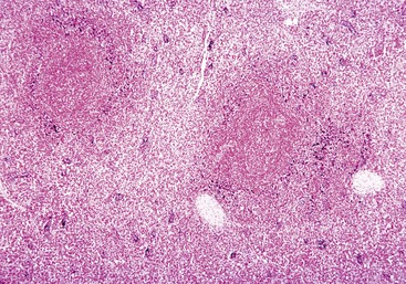
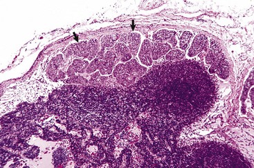
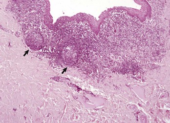
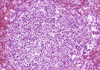
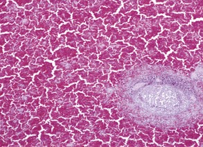
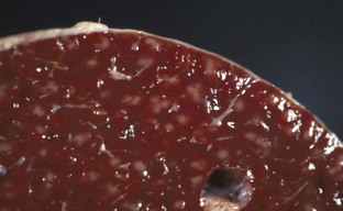
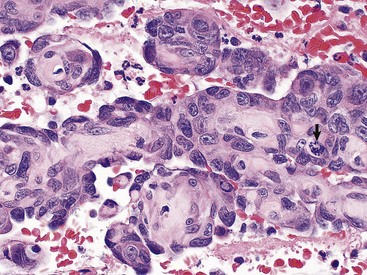
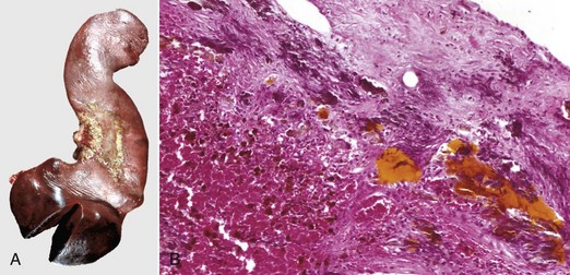
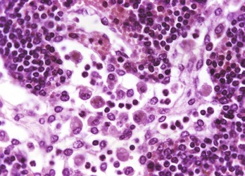
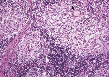
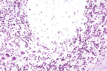
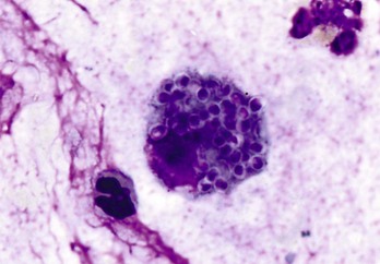
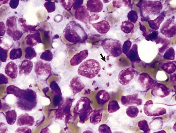
Fig. 13-40 Function of the marginal zone, splenic follicle, white pulp, spleen, calf injected intravenously with micronized carbon particles.
Carbon particles (black pigment) are present in macrophages of the marginal zone. Macrophages of the monocyte-macrophage system phagocytose blood-borne foreign material, bacteria, viruses, and senescent and/or damaged erythrocytes as in immune-mediated anemias and infections with hemotropic parasites. Eosin counterstain. (Courtesy Dr. M.D. McGavin, College of Veterinary Medicine, University of Tennessee.)
The sinusoidal, marginal sinus, and splenic cord macrophages are of bone marrow origin. They originate in the bone marrow, circulate in the blood as monocytes and migrate into the spleen. Fixed macrophages elsewhere in the body, in connective tissue, lymph nodes (sinus histiocytes), liver (Kupffer cells), lung (pulmonary intravascular macrophages and pulmonary alveolar macrophages), and brain (resident and perivascular microglial cells) are derived in a similar manner (see Chapters 5, 8, 9, and 14).
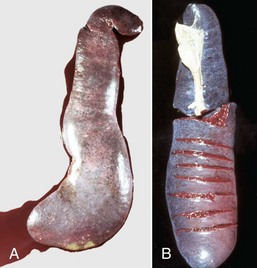
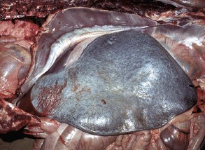
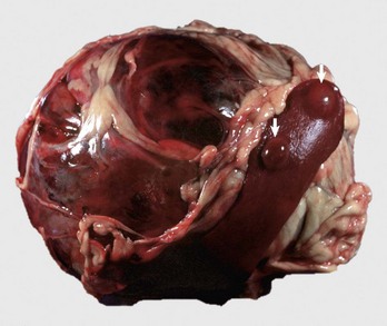
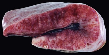
Storage or Defense Spleens: Spleens are classified as either storage or defense spleens, based on whether or not they can store significant volumes of blood. The spleens of domestic animals have both storage and defense functions, but are classified as storage spleens. Their trabeculae and capsules contain smooth muscle, which allows them to expand and contract. Equine, canine, and feline spleens all have considerable capacity. It has been claimed that the canine spleen can store one-third of the dog’s erythrocytes while the animal sleeps and the equine spleen holds one half of the animal’s circulating red cell mass, which is considered an advantage as it reduces the viscosity of the circulating blood. The spleens of ruminants and the pig are intermediate in the amount of smooth muscle and have limited storage capacity. Spleens, whose capsules and trabeculae have a low percentage of smooth muscle and elastic fibers and thus cannot expand and contract (rabbit and human), are designated as defense spleens. Storage spleens expand and contract quickly under the influence of the autonomic nervous system (sympathetic and vagal fibers, which reach the reticular walls of the red pulp vascular spaces; catecholamines; “flight or fight”) and other circulatory perturbations, such as in hypovolemic and/or cardiogenic shock (see the section on Small Spleens). Thus storage spleens may be either grossly enlarged and congested, or small with a wrinkled surface and a dry parenchyma (see the sections on Uniform Splenomegaly and Small Spleens). Also, the spleen responds to septicemia and acute hemotropic parasitic diseases by active hyperemia or acute passive congestion respectively, in the latter case the result of storing infected erythrocytes until they can be phagocytosed. Grossly congested spleens may be so enlarged that foci of white pulp are widely separated, making the white pulp look sparse and thus easily underestimated on gross examination.
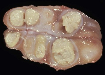
Hematopoietic Tissue: Although the liver is the primary site of hematopoiesis in the developing fetus, the spleen also makes a minor contribution. Shortly before or after birth, depending on the species, hematopoiesis ceases in the liver and spleen and the bone marrow becomes the primary hematopoietic organ. However, often under conditions of severe demand, for example in a severe and prolonged anemia, this increased demand can be met partly through reactivation of splenic hematopoiesis. This outcome is called extramedullary hematopoiesis (EMH). It is also an incidental finding in splenic nodular hyperplasia (see the section on Nodular Spleens with Firm Consistency).
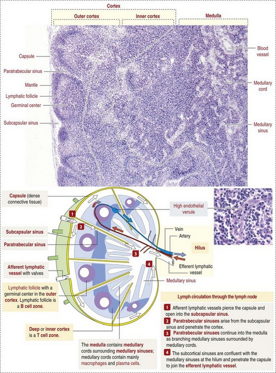
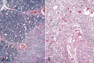
White Pulp: White pulp is grossly visible on the cut surface as distinct foci of grayish lymphoid tissue. Each focus consists of a periarteriolar lymphoid sheath and a splenic nodule surrounded by a marginal zone. There is considerable confusion regarding the terminology of splenic nodules or follicles. Histology books avoid the use of “lymphoid” as in “lymphoid nodule” or “lymphoid follicle”. Terms used include “lymphoid-like follicle,” “splenic follicle,” “splenic nodule” and even just “white pulp.” However, the pathology literature frequently uses the term “splenic lymphatic (or lymphoid) follicle”. This chapter uses the terms splenic nodule and splenic lymphoid follicle.
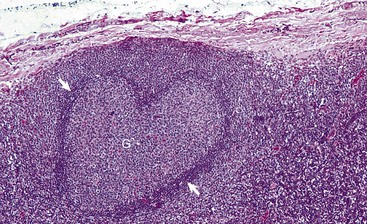
Normally, foci of white pulp are small and may not be visible on gross examination of a cross section of spleen. However, if nodules are enlarged either by lymphoid hyperplasia or by a neoplastic process (e.g., lymphoma), these foci initially become visible on the cut surface, as 0.5- to 1.0-mm white dots scattered through the red pulp. Also, in animals with storage spleens, the distention of the red pulp from stored blood may separate white pulp, making it look sparse.
Splenic white pulp is organized around branches of the splenic artery, the central arteriole, in the form of PALS, which is populated primarily by T lymphocytes (see Fig. 13-38). Primary splenic follicles are located eccentrically in PALS and are primarily comprised of B lymphocytes. When exposed to antigen, the latter develop germinal centers characteristic of secondary lymphoid follicles (see the section on Lymph Nodes).
The marginal zone is the third component of the splenic white pulp. It is located at the interface of the white and red pulp and surrounds the marginal sinus, which is immediately adjacent to the follicle. The marginal sinus is supplied with blood by radial branches of the central artery, which drain into the marginal sinus (see Fig. 13-38) and is the portal of entry into the spleen for recirculating B and T lymphocytes. The marginal zone consists of macrophages, dendritic cells, and T and B lymphocytes and is an important transit area for B and T lymphocytes, which after they leave the circulation, enter the marginal zone. From here, T lymphocytes migrate to the PALS. Those B lymphocytes that recognize antigens for which they have been programmed are activated and enter the follicle and proliferate to form plasmablasts. Dendritic cells in the marginal zone capture blood-borne antigens, process them, and preset them to the follicular lymphocytes.
Macrophages in the white pulp or marginal zone are phenotypically distinct from those in the red pulp. The latter function primarily to actively phagocytose particles, senescent erythrocytes, erythrocytes containing hemotropic parasites (e.g., Babesia spp.), and pathogenic bacteria and fungi and thus are responsible for “filtering” the blood. Many of the marginal zone macrophages are involved in phagocytosis and processing of antigen. However, some marginal zone macrophages actively phagocytose particulate matter in the blood (see Fig. 13-40) and bacteria in septicemias.
The responses of the spleen to injury (Box 13-7) include acute inflammation, hyperplasia of the monocyte-macrophage system, hyperplasia of lymphoid tissues, atrophy of lymphoid tissues, storage of blood or contraction to expel reserve blood, and neoplasia. These responses are reflections of the spleen’s three chief anatomic components: (1) monocyte-macrophage system, (2) lymphoid (immune) system, and (3) vasculature, chiefly the red pulp vascular spaces and their supporting reticular stroma (reticulum). Because the spleen is examined on the basis of red and white pulp, this approach is used in the following description.
Monocyte-Macrophage System: The distribution and function of macrophages in the spleen is described above under Structure and Function. These interactions are complex and their relationships to both innate and adaptive immunity, as well as cell turnover, are areas of intense study (see Chapter 5). The extent of phagocytosis by macrophages in the red pulp vascular spaces, which are attached to the reticulum forming the walls lining these spaces, splenic cords, and marginal sinuses, depends on the order in which they receive blood. In most species, the marginal sinus is the first, and consequently, particles and bacteria tend to be more concentrated here initially (see Fig. 13-40). However, again there are species differences, for example, in the cat, the marginal sinus is small and PAMS play a larger role in phagocytosis.
The spleen is able to mount a very strong response to blood-borne pathogens. In immunized rabbits injected intravenously with pneumococci, the blood was cleared of 98% of those bacteria within 15 minutes and 100% of an inoculum of 1 billion bacteria was removed from the blood within an hour. Also, the rabbit spleen removed 10 times the number of bacteria per gram as the liver. When 1 billion pneumococci per pound of body weight were injected into the splenic artery of a dog over a 5-minute period, all bacteria were removed from the blood in 65 minutes. After splenectomy, blood-borne organisms multiply rapidly and are disseminated widely in the body and may cause “overwhelming postsplenectomy infection.” It has also been shown that the phagocytic function of the spleen is critical in the control of plasmodium in humans with malaria and in cattle with babesiosis. To facilitate filtering, all of the blood in the body passes through the spleen at least once a day. In the dog, the blood flow and transit time depend on whether the spleen is contracted or distended. Blood flow is slower in the distended spleen, apparently because blood flows through the red pulp vascular spaces, rather than through the sinusoids as it does in the contracted canine spleen.
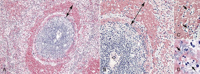
Sometimes in septicemias, the number of pathogenic bacteria arriving via the circulation can be so large that it exceeds the capacity of the splenic defenses to overcome them immediately. The result is acute splenic congestion and active hyperemia, followed by focal necrosis and or inflammation and a grossly enlarged and congested spleen. In the case of pyogenic bacteria, there may be neutrophils, other inflammatory cells, and bacteria throughout the red pulp. The marginal zone at the interface of the red and white pulp responds similarly to the red pulp vascular spaces and can be the initial site of response to blood-borne antigens and bacteria delivered by the radial branches of the central artery. As a result, it can be congested and in later stages (after only a few hours with highly pathogenic organisms) contain a diffuse spread or focal aggregates of neutrophils and macrophages. Histologically, the congestion forms a concentric ring, which may be incomplete, around the circumference of the splenic nodule (see the section on Anthrax).
Red pulp macrophages also proliferate in chronic hemolytic diseases in which there is a prolonged need to phagocytose erythrocytes and in chronic splenic congestion, which is usually a result of portal or splenic vein hypertension. The result is widened splenic cords. Unlike in humans, hepatic cirrhosis in animals does not cause chronic splenic congestion.
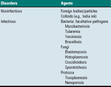
Macrophages in the red pulp (splenic cords and walls and lumens of red pulp vascular spaces) and marginal sinus also proliferate in response to fungi and facultative intracellular pathogens (e.g., Mycobacterium bovis) arriving hematogenously in the spleen. Their number may be augmented by monocytes recruited from the blood to form granulomatous inflammation. This inflammatory response can be diffuse (e.g. blastomycosis) or focal (tuberculosis).
White Pulp: The major system of the white pulp is the lymphoid system (PALS, splenic follicle), and the marginal zone, which lies at the junction of the red and white pulp but is not visible grossly.
The responses of the white pulp to injury are as follows:
• Activation or lymphocytolysis, necrosis, and reduction in size or disappearance (atrophy) of the follicle’s germinal center. These are followed by atrophy of the splenic follicles and ultimately atrophy of the lymph node.
• Phagocytosis of antibody, particulate material and microorganisms, macrophage hyperplasia (see the section on Monocyte-Macrophage System).
Splenic follicular hyperplasia is a response to antigenic stimuli and results in the formation of secondary follicles and consequently an increase in the size of the white pulp, which may be visibly enlarged. Splenic lymphoid follicles, lymphoid follicles in lymph nodes, and diffuse and nodular lymphatic tissue have a well-established sequence of morphologic changes, chiefly in the germinal center, which are helpful in tracking the stage of the B lymphocyte response. These sequential changes are similar to those described later in the Lymph Nodes section on Responses to Injury.
Splenic follicular atrophy occurs in response to lack of antigenic stimulation or from regression after antigenic stimulation has ceased, and from the effects of toxins, antineoplastic chemotherapeutic agents, microorganisms such as viruses and bacteria, radiation, malnutrition, wasting/cachectic diseases, and aging (see Box 13-5). The follicles are depleted of lymphocytes and with time, germinal centers and follicles disappear. The amount of the total lymphoid tissue is reduced and the spleen may be smaller.
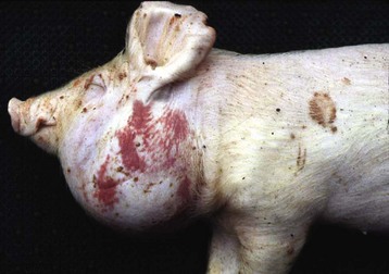
Monocyte-Macrophage System: The response of the monocyte-macrophage system in the marginal sinus and marginal zone to injury is phagocytosis and proliferation. Proliferation also occurs in the viral disease porcine PMWS where there is proliferation of both follicular macrophages and those in the deep cortex (see the section on Disorders of Pigs).
The three dimensional structure of a lymph node is complex and difficult to fully appreciate in a two-dimensional histological slide or diagram. It is convenient from the point of view of understanding the pathological response of the lymph node to consider them based on its anatomic components (Fig. 13-41), which are as follows:
Fig. 13-41 Schematic illustration of the histology of the lymph node and lymph circulation. (Adapted from Kierszenbaum AL: Histology and cell biology: an introduction to pathology, ed 2, St Louis, 2007, Mosby.)
• Stroma: fibrous capsule and reticulum
• Cortex: outer cortex (B lymphocytes), deep cortex (paracortex; T lymphocytes)
• Vasculature: arteries, arterioles, high endothelium venules, efferent veins, lymphatic sinuses and vessels
Stroma: The lymph node is almost completely enclosed by a fibrous capsule that is pierced by multiple afferent lymphatic vessels, which empty into the subcapsular sinus (see Fig. 13-41). Efferent lymphatic vessels and veins exit, and arteries enter at the hilus. The lymph node is supported by fibrous trabeculae and a reticulum of fibroblastic reticular cells. The reticulum also has an immunologic function of guiding lymphocytes and antigen-presenting cells to facilitate their interaction with B and T lymphocytes. Lymphocytes are such a predominant feature in lymph nodes that it is easy to overlook the fact that lymphocytes are transient cells, staying only a short time in the lymph node and that the lymph node itself is basically a fibrovascular structure within a dilated lymphatic.
Cortex: On gross examination of a cross-section of lymph nodes, two main areas are visible: cortex (outer) and medulla (inner) (see Fig. 13-41).
The outer cortex contains the lymphoid follicles. The morphologic and functional organization of the lymphoid follicle is similar to that of the splenic follicle, except there is no marginal zone. There are two types of lymphoid follicles, simply referred to as primary and secondary. Primary lymphoid follicles consist predominantly of densely packed resting B lymphocytes and follicular dendritic cells (see Chapter 5 for more complete discussion of dendritic cells). Mature naïve B lymphocytes expressing receptors for specific antigens exit primary lymphoid organs and circulate through the bloodstream, lymphatic vessels, and secondary lymphoid tissues. On their arrival at lymph nodes, B lymphocytes exit through high endothelial venules (HEVs) and home to the primary follicle. Those B lymphocytes that recognize the antigen for which they are expressing receptors are activated and proliferate to form secondary lymphoid follicles characterized by prominent germinal centers.
Germinal centers are areas with a specialized microenvironment that support the proliferation and further development of B lymphocytes to increase their antigen specificity (e.g., affinity maturation) and functional capacity (e.g., isotype switching). The germinal center has a “dark” zone, comprised of proliferating B lymphocytes, and an outer “light” zone comprised of nonproliferating B lymphocytes. Peripheral to that is the mantle zone that contains the inactive mature naïve B lymphocytes that have been displaced centrifugally as a result of the development and expansion of the germinal center. Plasma cell precursors (plasmablasts) formed in the germinal centers migrate from the cortex to the medullary cords.
The deep cortex (inner cortex, paracortex) consists of diffuse lymphoid tissue containing T lymphocytes and HEVs, through which T lymphocytes migrate from the blood into the deep cortex and B lymphocytes migrate to lymphoid follicles. Other T lymphocytes arrive via the afferent lymphatic vessels. T lymphocytes that are antigenically stimulated proliferate, but unless there is a marked increase in the number of T lymphocytes in the deep cortex, lymphoid hyperplasia here can be difficult to detect in H&E stained sections. Trabeculae, which are part of the stroma, are also in the cortex. They are collagenous bands extending radially inward from the capsule. They are often surrounded by trabecular (also called paratrabecular) sinuses, which are continuous with the subcapsular sinus and empty into the medullary sinuses (see Fig. 13-41).
Medullary Cords: The medulla consists of two major components: (1) medullary cords containing macrophages and lymphocytes—and in an immunologically stimulated lymph node, plasma cells actively secreting antibodies, which are released into the lymph in the medullary sinuses, and (2) the medullary sinuses lined by fibroblastic reticular cells. Macrophages (sinus histiocytes) cling to reticular fibers crossing the lumen of the sinus and actively phagocytose foreign material, cellular debris, and bacteria in the incoming lymph.
The porcine lymph node has a different structure, but its immunologic functions are unchanged from those of other domestic animals. There is one afferent lymphatic vessel that enters at the hilus and discharges lymph into the center of the node, from where it flows to the subcapsular sinus and then drains into several efferent lymphatic vessels, which pierce the capsule. This reversal of flow is associated with an “inverted” structural organization, in which the lymphatic follicles present in the cortex of other domestic animals are located in the central region of the lymph node, and the medullary region is located peripherally. Consequently, any blood in the sinuses is also highly visible through the capsule.
Vasculature: The vasculature of the lymph node includes afferent lymphatic vessels, arteries, and veins and their branches including the HEVs and the sinuses—subcapsular, trabecular, and medullary through which lymph from the afferent lymphatic vessels flows to the efferent lymphatic vessels. Medullary sinuses are described with the medulla. Response of these vessels to injury is as for other vasculature (see Chapter 10). Afferent and efferent lymphatic vessels are part of the Cardiovascular System and are also discussed in Chapter 10.
All lymph nodes receive afferent lymphatic vessels from specific areas of the body. For example, peripheral lymph nodes such as the popliteal and superficial inguinal drain the hind leg. Central lymph nodes, such as the tracheobronchial and mesenteric lymph nodes (lymphocenters), drain the lungs and intestinal tract, respectively. The term lymphocenter is used in veterinary anatomy for a lymph node or a group of lymph nodes that occur constantly in the same region and receive lymphatic vessels from the same region in all species. Multiple afferent lymphatic vessels drain into the subcapsular sinus, but lymph from one afferent lymphatic drains into the adjacent portion of the lymph node. The result is that only this portion (lobule), particularly in lymph nodes from large domestic animals, may respond immunologically or with inflammation. Similarly, a neoplastic embolus initially lodges in that portion of the lymph node drained by the branch of the afferent lymphatic vessel arising in the neoplastic site (Fig. 13-42).
Fig. 13-42 Subcapsular sinuses with metastatic carcinoma, lymph node, dog.
Subcapsular sinuses are sites for embolization, lodgement, invasion, and growth of neoplastic emboli (arrows), most commonly carcinomas. Emboli initially lodge in that portion of the lymph node drained by the branch of the afferent lymphatic vessel draining the primary carcinoma. H&E stain. (Courtesy Dr. M.D. McGavin, College of Veterinary Medicine, University of Tennessee.)
The lymph nodes are one of the first lines of defense against lymph and blood borne infectious and non-infectious agents. The functions of the lymph node are to filter lymph of particulate matter and microorganisms, and to facilitate the surveillance of incoming antigens and their interaction with B and T lymphocytes and to produce B lymphocytes. It is helpful to consider the paths taken by particles, molecules, and cells arriving at a lymph node. This is accomplished via two possible routes: (1) via the arterial blood supply or (2) via lymphatic afferents. T lymphocytes arriving by the arterial route migrate out of the HEVs into the T lymphocyte areas adjacent to the follicle (interfollicular portion of the deep cortex) (see Chapter 3).
Material arriving in the lymph can be subdivided into the following three types:
Particles and molecules in the lymph arriving via the afferent lymphatic vessels enter the subcapsular sinus, which has been likened to a swamp. Hydrostatic pressure is low, particles tend to settle, and reticular fibers crossing the sinus impede the flow. All these factors facilitate phagocytosis by the sinus macrophages. Larger molecules and particles flow from the subcapsular into the trabecular sinuses and then into the medullary sinuses, where particles and bacteria are phagocytosed by the sinus histiocytes. This is well illustrated by the concentration of carbon in the medulla of the tracheobronchial lymph nodes of animals with anthracosis (see Fig. 13-40).
Smaller molecules, such as free antigens and chemokines, cytokines and other soluble products of the immune and inflammatory responses, penetrate the floor of the subcapsular sinus and are carried along reticular fibers to the outer cortex adjacent to the HEVs, the site of migration of B and T lymphocytes into the cortex. Thus these molecules are brought into close association with B and T lymphocytes and dendritic cells, and this process facilitates the activation of antigen specific B lymphocytes, which develops into antibody secreting plasma cells.
Antigens can also reach the regional lymph node in antigen-presenting cells such as dendritic cells. These cells have been activated by uptake and processing of antigen and migrate via lymphatic vessels to the subcapsular sinus where they exit and migrate along the reticulum to the outer cortex where they present the antigen to the follicular B lymphocytes (see Chapter 5). The classic example is the intraepidermal dendritic cell (Langerhans’ cell) draining from the skin.
Responses to injury are listed in Box 13-8, and the responses are discussed on the basis of the following systems: monocyte-macrophage system, outer cortex, inner cortex, medullary sinuses, and medullary cords.
Monocyte-Macrophage System: Like the spleen, lymph nodes contain cells of the monocyte-macrophage system and the lymphoid system (see Box 13-8), and these cells are the first line of defense against infectious and non-infectious agents in the incoming lymph. As most of these agents arrive in the afferent lymph, the sinus macrophages (histiocytes) are the first to respond. This is evidenced by an increase in their number (hyperplasia, so-called sinus histiocytosis) chiefly in the medullary sinuses (Fig. 13-43). Additional inflammatory cells, neutrophils in acute lymphadenitis and monocytes in granulomatous lymphadenitis arrive in the blood and migrate into the parenchyma or the sinuses. Some of these leukocytes may contain viable intracellular pathogens (e.g., Mycobacterium spp. and fungi [Blastomyces dermatitidis]) or have cell-associated viruses (e.g., parvovirus) and infect the lymph node and then are disseminated throughout the lymphoid tissues of the body as a result of the normal trafficking of lymphocytes. Circovirus 2, the agent of porcine PMWS is unusual in that it causes an increase in macrophages in the lymphoid follicles and the deep cortex.
Fig. 13-43 Sinus histiocytosis, lymph node, medulla, dog.
The sinusoids (1) are filled with mostly macrophages and a few scattered neutrophils (small dark nuclei). Most of the macrophages are derived from the perisinusoidal macrophages, but some may arrive via the afferent lymphatics. The medullary cords (2) are filled with lymphocytes and plasma cells. Plasma cell precursors are formed in the germinal centers, mature into plasma cells, and migrate to the medullary cords. Thus the presence of large numbers of plasma cells in the medullary cords is indicative of ongoing production of antibody from an antigenic stimulus. (Courtesy Dr. M.D. McGavin, College of Veterinary Medicine, University of Tennessee.)
Outer Cortex: B Lymphocyte Areas: If the lymph tissue has not been antigenically stimulated, there may be no follicles or germinal centers visible, but within days of stimulation, a pale area, which is the site of a germinal center, appears (Fig. 13-44). It undergoes a predictable sequence of development, becomes larger, and contains mitotic figures, and lymphocytes proliferate. Because over 90% of these lymphocytes die by apoptosis in the germinal centers, nuclear debris is plentiful, and when this is phagocytosed by macrophages in the germinal center, these are named tingible body macrophages. Tingible is an old term meaning stainable, in this case referring to the phagocytosed nuclear debris. The immunoblasts formed here move to and mature in the medullary cords, which as a result are distended with plasma cells (medullary plasmacytosis). The concentration of medullary plasma cells correlates with the activity of the germinal centers. Thus an antigenically stimulated lymph node at the height of its response has numerous follicles (follicular hyperplasia) (Fig. 13-45), is enlarged, and has a tense capsule, and the cut surface may bulge. Histologically, there is follicular lymphoid hyperplasia and active germinal centers and medullary cord plasmacytosis. A subset of these B lymphocytes exit the lymph node and migrate to the bone marrow where they develop into fully differentiated plasma cells and secrete antibody. The majority of the serum antibody is secreted by plasma cells located in the bone marrow.
Fig. 13-44 Lymphoid hyperplasia, outer cortex: B lymphocyte areas, lymphoid follicles, lymph node, dog.
If the lymphoid follicle is antigenically stimulated by a bacterium for example, B lymphocyte areas undergo a predictable sequence of development and become larger and contain mitotic figures and B lymphocytes that proliferate. The central areas (germinal centers [G]) appear hypocellular as a result of rapid hypertrophy and hyperplasia of antigenically stimulated B lymphocytes followed by the death by apoptosis of more than 90% of these cells. The small numbers of immunoblasts formed in this proliferative response move to the mantle zone (arrows) of the follicle and then to the medullary cords, where they mature into plasma cells and secrete immunoglobulins. H&E stain. (Courtesy Dr. M.D. McGavin, College of Veterinary Medicine, University of Tennessee.)
Fig. 13-45 Follicular lymphoid hyperplasia, chronic demodicosis, caudal cervical (prescapular) lymph node, dog.
The number of lymphoid follicles (F) has increased (hyperplasia), and all of these have germinal centers (secondary follicles) indicating active proliferation of B lymphocytes to form plasma cells in response to an antigenic stimulus. H&E stain. (Courtesy Dr. M.D. McGavin, College of Veterinary Medicine, University of Tennessee.)
As the immune response declines, mitoses in the germinal centers become less frequent and the concentration of cells in the germinal centers is reduced with the result that the follicular stroma (dendritic cells and macrophages) becomes visible. This stage is designated follicular lymphoid depletion (sometimes inappropriately referred to as lymphoid exhaustion). B lymphocyte areas (lymphatic follicles) can atrophy, and histologically, there are reduced numbers of lymphocytes in the outer cortex, smaller and reduced numbers of follicles. Causes of this are a failure of adequate numbers of B lymphocytes to arrive from the bone marrow as in myelofibrosis and destruction of lymphocytes in the bone marrow by viruses, radiation, and toxins. Disappearance of germinal centers is normal once antigenic stimulation has ceased.
Deep Cortex: T Lymphocyte Areas: T lymphocytes can also respond by a limited degree by proliferation. An increase in T lymphocytes has been reported in the deep and interfollicular cortices and in the PALS in malignant catarrhal fever (MCF) in cattle and in the deep cortex in pigs with porcine reproductive and respiratory syndrome. A diffuse proliferation of macrophages occurs in the deep cortex in porcine circovirus 2 infection (PMWS).
Atrophy may indicate that there is a deficit in lymphocyte production in the bone marrow or in the differential selection in the thymus, or destruction by viruses, radiation, and toxins directly on the lymphocytes in the lymph node. Examination of H&E stained sections allows evaluation of follicular and germinal center activity and the concentration of plasma cells in the medullary cords and thus a reasonable estimate of B lymphocyte activity. However, because T lymphocytes cannot be differentiated from B lymphocytes in H&E stained sections, it is not possible to accurately evaluate their numbers in the deep cortex. For this, immunohistochemical staining of B and T lymphocytes is required. However, marked atrophy of the T lymphocyte areas can be detected histologically.
Medullary Sinuses: Responses to injury by the medullary sinuses are dilation and proliferation of the macrophages (histiocytes) in their lumens. Dilation can be caused by edema fluid draining from an edematous area (as in chronic cardiac failure), inflammatory edema fluid draining from an acutely inflamed area or inflammatory exudate in response to blood-borne bacterial emboli lodging in the lymph node. As the inflammation progresses (see the section on Acute Lymphadenitis), the sinuses become filled with inflammatory cells (neutrophils, macrophages) and hyperplastic sinus macrophages (see Fig. 13-43). Depending on the intensity of the inflammation, the adjacent parenchyma may be hyperemic and infiltrated with inflammatory cells. Sinus macrophages proliferate in response to a wide variety of particulate matter (see the section on Pigmentation), bacteria, and erythrocytes (erythrophagocytosis) draining from a hemorrhagic area.
Medullary Cords: As pointed out in the section on Structure and Function, after activation and proliferation of B lymphocytes in the follicle, the plasma cells produced migrate to and accumulate in the medullary cords, which at the height of the immune response are distended by plasma cells and some lymphocytes. As the immune response subsides, the number of plasma cells decreases and the medullary cords return to their resting state, populated by some lymphocytes and scattered plasma cells.
Although hemal nodes are often considered to be unique to ruminants, they have also been found in horses and primates. They are small, dark red-to-brown nodules. Their architecture resembles that of a lymph node with lymph follicles and sinuses, except that the hemal node sinuses are filled with blood (Web Fig 13-4). It was once thought that they had no afferent or efferent lymphatic vessels, but these have been identified. As erythrophagocytosis can be present, it is presumed that hemal nodes can filter blood and remove senescent erythrocytes, but their functional importance is not clear.
Histology books use the following terms: diffuse lymphatic tissue and solitary and aggregated lymphatic nodules to describe these forms of lymphatic tissue, but the immunology and pathology literature uses mucosa-associated lymphoid tissue (MALT). The importance of these three types of MALT is frequently overlooked because most of these tissues in the normal animal, apart from aggregated lymphatic tissue of the tonsil and Peyer’s patches, are visible only microscopically. The actual mass is significant, and MALT is essential for mucosal immunity. It is a site where antigen is phagocytosed and from which immune cells and antigen flow via afferent lymphatic vessels to regional lymph nodes. MALT consists of three distinct morphologic types:
• Diffuse lymphatic tissue includes scattered lymphocytes surrounded by dendritic cells and connective tissue in the mucosa of many organs. In normal animals, it is most obvious in the mucosa of the alimentary tract. The lymphocytes secrete not only IgA, but also IgG and IgM.
• Solitary lymph nodules, also in the mucosa, are small unencapsulated clusters of lymphocytes, principally B lymphocytes that secrete various types of immunoglobulin. They are surrounded by small numbers of dendritic cells and lymphocytes. In the resting or antigenically unstimulated state, they are usually not visible grossly.
• Aggregated lymphatic nodules are unencapsulated and consist of groups of lymph nodules. There are considerable differences between the different domestic animals in their locations and whether they are present at birth. Because this tissue is antigen dependent and responds to antigenic stimulus by hyperplasia, some of it is visible months or years after birth or infection.
The solitary lymph nodules and aggregated lymphatic tissues are classified by their anatomic locations and form the MALT. These lymphatic tissues include (1) the bronchus-associated lymphatic tissue (BALT), which is often at the bifurcation of the bronchi and bronchioles), (2) a ring of lymphoid tissue (pharyngeal and palatine tonsils [see below]) and lymph nodules (NALT, LALT, and ATALT [see below]) that surround the oral and nasal pharynges and encounter infectious microorganisms during ingestion and inhalation, and (3) the gut-associated lymphatic tissue (GALT), of which the most important and most obvious components are Peyer’s patches and the pharyngeal and palatine tonsils. Lymph nodules can also be present in the mucosa of the conjunctiva-associated lymphatic tissue (CALT) (Fig. 13-46), mammary gland, urinary bladder mucosa, nose (NALT), larynx (LALT), auditory tube (ATALT), and genital tract.
Fig. 13-46 Follicular lymphoid hyperplasia, conjunctiva, lymphoid nodules, cow.
The diffuse lymphoid tissue has increased in size, and there are two follicles with germinal centers (arrows). This reaction is a frequent response to conjunctivitis from irritants and bacteria. H&E stain. (Courtesy Dr. M.D. McGavin, College of Veterinary Medicine, University of Tennessee.)
The primary function of MALT is to protect mucosal barriers and thus they are strategically placed to act as sentinels, but they can also become a portal of entry for bacteria and this is particularly true of the tonsils and Peyer’s patches. Peyer’s patches in the ileum are overlaid by a special epithelium known as the follicle-associated epithelium (FAE), in which there are numerous M cells. These M cells deliver macromolecules (including antigens), particles, pathogenic bacteria, and viruses across the epithelium to an area rich in dendritic cells, which are able to transport the material into the GALT. Table 13-4 lists the functions of the MALT with different microorganisms.
The responses of MALT (diffuse lymphatic tissue and solitary and aggregated lymph nodules) to injury (Box 13-9) are similar to those of other lymphatic tissue: hyperplasia, atrophy, and inflammation.
Hyperplasia: After antigenic stimulation, activation of germinal centers, follicular hyperplasia (see Fig. 13-46), and increased production of plasma cell and antibodies occur. Lymph nodule hyperplasia can be marked in diseases, usually chronic diseases, for example, BALT in chronic Dictyocaulus spp. bronchitis or bronchiolitis in cattle, sheep, and goats; in Metastrongylus spp. bronchitis or bronchiolitis in pigs; and in some pneumonias. It is particularly marked in Mycoplasma spp. pneumonias of sheep and pigs and can be so extensive as to encircle bronchioles and bronchi and hence the designation of “cuffing pneumonia.”
Hyperplastic lymph nodules can be so enlarged that they become grossly visible as white plaques or nodules. They can be seen in the conjunctiva of the eyelids and the third eyelid in chronic conjunctivitis (see Fig. 13-46), the pharyngeal mucosa in chronic pharyngitis, the gastric mucosa in chronic gastritis, and the urinary bladder in chronic cystitis (follicular cystitis). The normal fetus has no detectable BALT, but it may be present in fetuses aborted because of infections.
Atrophy: Atrophy of the diffuse lymphatic tissue and lymph nodules has the same causes as those affecting lymph nodes. These causes include lack of antigenic stimulation, cachexia, malnutrition, aging, and viral infections. The microscopic lesions of BVDV infection of Peyer’s patches in ruminants (see Chapters 4 and 7) are distinctive and consist of lysis of cells of lymphoid follicles; in canine parvovirus infection, there is lymphoid necrosis of Peyer’s patches and atrophy of these tissues can be a sequela.
Inflammation: Granulomas can form in Peyer’s patches in paratuberculosis (Johne’s disease) in goats (see the section on Disorders of Ruminants).
Portals of entry used by microorganisms and other agents and substances to access the lymphatic system are summarized in Box 13-10. These portals include the blood vasculature (hematogenous) spread (cell-free or within leukocytes), direct penetration (trauma, wounds), afferent lymphatic vessels, or M cells and dendritic cells overlying MALT and tonsils.
Infectious agents, such as bacteria, enter the spleen by two routes: via the blood vasculature or by direct penetration. The spleen has no afferent lymphatic vessels and efferent lymphatic vessels drain only the capsule and trabeculae. The splenic capsule is thick, and thus inflammation from an adjacent peritonitis is unlikely to penetrate it. Direct penetration is extremely rare, but foreign bodies occasionally do penetrate as a sequela to traumatic reticulitis in cattle. Contraction of the reticulum extrudes a foreign body, usually a nail or wire through the wall of the reticulum. Most of these objects travel cranially to penetrate the diaphragm and pericardium, causing traumatic reticulopericarditis. Less frequently, some enter the visceral surface of the ventral extremity of the spleen, often causing a splenic abscess. In horses, on rare occasions, splenic abscesses can develop secondary to perforation of the gastric wall. Also, small pieces of wire have been found in these splenic abscesses, presumably originating from the GI tract. Equine splenic abscesses can be secondary to gastric ulcers (Gasterophilus intestinalis) or by extension on the granulomatous inflammation around Habronema spp. in the wall of the stomach.
Bacteria, viruses, and antigens enter the lymph node by two routes: afferent lymphatic vessels and via the blood vasculature (hematogenously).
Afferent Lymphatic Vessels: Infectious microorganisms, either cell free or within lymphocytes or monocytes, are transported in lymph to regional lymph nodes or lymphocenters. Here, some of these agents may escape removal by phagocytosis and are transported via the efferent lymphatic vessels to the next lymph node in the chain, where they can cause an inflammatory or immunologic response. This process can continue serially down a lymph node chain and if the agent is not removed it may eventually be transported via the lymphatic vessels to the circulating blood via the thoracic duct or truncus trachealis and disseminated throughout the body.
Blood Vasculature: Bacteria can arrive at lymph nodes hematogenously in septicemias and bacteremias. However, most pathogens arrive via afferent lymphatic vessels. B and T lymphocytes as part of their normal trafficking, arrive in the blood, enter the lymph node via the HEV and conduct surveillance for the specific antigens that they have been programmed to recognize. Macrophages containing microorganisms also arrive hematogenously (see Chapter 4).
Capsule: The lymph node is protected by a thick fibrous capsule, and thus direct penetration by inflammation, trauma, or neoplasms is rare. However, in sheep, in enzootic areas, during shearing, peripheral lymph nodes enlarged from caseous lymphadenitis (CLA) may be punctured by the comb of the shears, resulting in the shears becoming contaminated by pus. The infection may then transmit to other sheep through nicks and cuts in their skin.
Lymph nodules do not have afferent lymphatic vessels and thus antigens arrive hematogenously, in migrating macrophages or across mucous membranes. M cells of Peyer’s patches can transport infections agents. Live-attenuated Salmonella spp. bacteria induce dendritic cells in intestinal submucosal tissues to migrate to lymph follicles (B lymphocytes) and parafollicular (T lymphocytes) areas of Peyer’s patches. Other pathogenic bacteria known or believed to enter via the M cells from the intestine include Listeria monocytogenes, Mycobacterium avium ssp. paratuberculosis, and Yersinia pseudotuberculosis. The scrapie agent also accumulates in Peyer’s patches.
Defense mechanisms used by the lymphatic system to protect itself against microorganisms and other agents and substances are the innate and adaptive immune responses, which are discussed in Chapters 3 and 5. Viruses, bacteria, and particles arriving in the lymph and blood interact with cells of the monocyte-macrophage system through phagocytosis and antigen processing and presentation. Hyperplasia of the macrophages often occurs concurrently. Most of these macrophages are located in the medullary sinuses. If the material is antigenic, antigen processing and presentation are followed by an immune response resulting in proliferation of B lymphocytes and production of antibody and/or the proliferation of T lymphocytes. Other defense mechanisms are structural in nature and include the thick fibrous capsule of lymph nodes and the spleen, which protects them from external trauma.
Because the thymus involutes after sexual maturity, evaluating whether it is smaller than normal is difficult unless the change is extreme or age-matched control animals are available. Before sexual maturity, the thymus is easily identified as a lobular organ, white to gray with a thin capsule. After sexual maturity, the gland is often grossly indistinguishable from adipose connective tissue within the cranial mediastinum, although microscopic remnants may remain. An extremely small thymus in a neonatal animal should be considered abnormal and an indicator of a possible underlying primary or acquired immunodeficiency. Enlargement of the thymus is almost always caused by tumors. The cut surface should be examined for tumors and hematomas.
There is wide variation in location (ruminants versus the others), size, shape, and appearance of the cut surfaces between the spleens of domestic animals. At necropsy, the spleen is dissected free, and in the case of nonruminants, the gastrosplenic ligament is checked for torsion. The spleen is evaluated for size (splenomegaly, normal size, or atrophy) and the surface of its serosa and capsule for smoothness, nodularity, and ruptures.
Spleens vary widely in size within the same species and among the different species of domestic animals. Some pathologists wryly state “spleens come in two sizes: too large and too small.” The spleen can be enlarged (splenomegaly), normal in size, or small, and its surface can be smooth or nodular. The color of the capsular surface of the spleen also varies among species of domestic animals and depends on the opacity or translucence of the splenic capsule, which determines the visibility of the red pulp. The degree of opacity of the capsule is a function of its thickness and the amount of collagen. In the pig, dog, and cat, the splenic capsule is thin, and the surface of their spleens are red. The splenic capsules of horses and ruminants are thicker and usually appear gray because the color of the red pulp is not visible through the capsule. However, if the capsule has thick and thin areas, it is mottled gray and red. The tenseness of the capsule of normal and diseased spleens depends on how much the splenic parenchyma is distended, for example, by stored blood or infiltrating inflammatory cells or neoplasms. Storage spleens devoid of blood usually have a wrinkled surface. The appearance of the cut surface of the spleen in normal animals depends on the amount of stroma (e.g., trabeculae are prominent in ruminants); the size and visibility of the white pulp, which reflects the amount of lymphoid tissue; and whether the red pulp is congested from blood. The cut surfaces are examined for bulging (usually an indication of distention), visibility of the white pulp, and focal or diffuse lesions.
Enlarged spleens are classified into congested and noncongested. The cut surface of severely congested spleens may be red to bluish-black and exude blood and are often termed bloody spleens. Enlarged spleens that are not congested but firm are often called meaty spleens because of their firmness and texture. Little blood oozes from their cut surfaces, and the color of this surface depends on how much of the normal red pulp has been replaced by inflammatory cells, stored materials, or neoplastic cells (see the section on Splenomegaly and Table 13-5). The spleen may be measured and weighed but because of the wide variation in the dimensions and weight of normal spleens and the amount of blood stored, these data can be difficult to interpret.
In standard necropsy technique, the spleen is sliced transversely at 4 to 6 mm intervals, depending on whether it is small or large. The cut surfaces are checked for lesions and at this time specimens are taken for those tests that require fresh tissue (e.g., bacteriologic and virologic examinations). Cross-sections are placed in fixative (10% buffered neutral formalin). However, if the cut surface has bulged during fixation, the bulged tissue that is the best fixed is discarded at trimming. One solution is to trim off and discard the bulged area after 1 to 2 hours fixation in 10% BNF, trim the tissue to no more than 15 × 15 × 4 mm, and then replace it with the fixative.
Lymph nodes should be dissected free of fat and connective tissue and any firm attachment to adjacent tissues noted because this attachment can be an indication of neoplastic infiltration through the capsule. Gross examination includes determining the size (measurement or weight), the shape, and whether the capsule is intact. The cut surface is examined for bulging, edema, hyperemia or congestion, exudate, and color (see the section on Pigmentation); the ratio of cortex to medulla; changes in or obscuring of the normal architecture; and masses such as abscesses, granulomas, and neoplasms.
In live animals, microscopic examination is easily accomplished by cytologic evaluation of superficial lymph nodes. Aspirates provide excellent cellular detail, but for the study of architectural derangements, removal of a node for histologic evaluation is necessary.
The surgeon must handle lymph nodes carefully to minimize artifacts. Any compression (e.g., squeezing with forceps) causes artefacts in nuclei, usually pyknosis or “streaming” of nucleic acids. One end of the node should be removed and used to prepare impression smears. Depending on size, the remainder of the node or selected transverse slices are placed in fixative (10% BNF) for 1 hour before slicing. This period of fixation hardens the node and prevents artifactual bulging of the tissue from the cut surface. The lymph node, if large, should then be sliced transversely into parallel 3 to 5 mm thick sections to allow examination and ensure penetration of the fixative. To avoid compression artifacts and facilitate slicing, some pathologists prefer not to incise small lymph nodes before fixation. Lymph nodes that are either pathologically enlarged or normally large as those in large domestic animals need to be incised to allow fixative to penetrate rapidly.
Cross-sections of lymph node are preferred for histologic examination because they are usually sufficiently small to allow a full cross–section, including both the cortex and medulla to fit onto a microscope slide. The exception is the porcine lymph node with its different anatomic arrangement. Because the location and amount of cortex and medulla vary at different sites of the transverse cut, a longitudinal plane section is recommended as being the most representative.
Enlarged lymph nodes can be distributed in several different patterns in the body. First, all lymph nodes throughout the body (systemic or generalized) may be enlarged (lymphadenomegaly). This pattern is generally attributed to systemic infectious, inflammatory, or neoplastic processes. If a regional lymph node (i.e., a lymph node draining a specific area such as one of the limbs or one side of the oral cavity) is enlarged, then the area drained by that node must be checked for infectious, inflammatory, or neoplastic processes. Finally, enlargement of the mesenteric lymph nodes or GALT suggests that the infectious, inflammatory, or neoplastic process originates within the gut, although they can be infected hematogenously. Normal mesenteric lymph nodes are continuously receiving and responding immunologically to barrages of antigens and bacteria received via their afferent lymphatic vessels from the intestinal tract. This role is reflected in their appearance. They are larger and have numerous well-developed lymphoid follicles (follicular lymphoid hyperplasia) with active germinal centers. Often the sinuses contain histiocytes (sinus histiocytosis), presumably in response to the need to phagocytose incoming material.
Multiple lymph nodes of a chain may be enlarged because infection or metastases spread via the lymphatic vessels, from one lymph node to the next in the chain. Thus it is important to know the area drained by specific lymph nodes; for example, all lymph from the head ultimately drains to the medial retropharyngeal lymph nodes, and lymph from the foreleg drains to either the axillary or superficial cervical (prescapular) lymph node. However, there are some surprises. Besides the thoracic cavity, the caudal sternal and caudal mediastinal lymph nodes drain the cranial abdomen and may be enlarged in the absence of thoracic lesions. Similarly, the celiac lymph nodes of dogs, pigs, and horses receive some afferent lymphatic vessels from thoracic organs and lesions in them do not necessarily mean that disease originated in the abdomen.
Congenital disorders of the thymus are discussed in detail in Chapter 5. Summaries of the gross and microscopic morphologic changes are described in the sections on Disorders of Horses and Disorders of Dogs.
Thymic cysts can be found within the developing and mature thymus and in thymic remnants in the cranial mediastinum. These cysts are often lined by ciliated epithelium and represent developmental remnants of branchial arch epithelium and are usually of no significance.
Thymitis, an infrequent lesion, is seen in porcine circovirus 2 (PMWS) infection (see Disorders of Pigs and also Chapter 4), enzootic bovine abortion (EBA; see Chapter 18), and salmon poisoning disease of dogs (see Chapter 7). Variable degrees of acquired immunodeficiency may be caused by infectious agents (viruses), toxins, chemotherapeutic agents and radiation, malnutrition, aging, and neoplasia.
Infections that most commonly injure lymphoid tissues do so because they infect lymphocytes and destroy the lymphoid component of the thymus. Viruses, which can damage the lymphoid component of the thymus and are often found in other lymphoid tissues, include canine distemper virus, EHV-1 in aborted foals (Fig. 13-47), canine and feline parvovirus, FIV, BVDV, and hog cholera virus. In kittens infected with FIV, thymic lymphoid depletion is an early lesion, and in kittens with progressive FIV infection, ultimately there is complete loss of thymic architecture with only minimal thymic tissue remaining in the connective and adipose tissue of the mediastinum.
Fig. 13-47 Equine herpesvirus 1, spleen, aborted foal.
Most of the splenic follicle is occupied by nuclear debris, the result of lymphocytolysis. The splenic follicle is surrounded by red pulp. H&E stain. (Courtesy College of Veterinary Medicine, University of Illinois.)
Environmental toxins, such as halogenated aromatic hydrocarbons (e.g., polychlorinated biphenyls and dibenzodioxins, lead, and mercury) have a suppressive effect on the immune system. Fumonisins B1 and B2, which are secondary fungal metabolites produced by members of the genus Fusarium, cause lymphocytolysis in the thymic cortex. In weaned and grower pigs, aflatoxin causes thymic atrophy because of lymphocyte depletion. In the case of the halogenated aromatic hydrocarbons, there is a genetic basis for susceptibility that is mediated through a complex composed of the aromatic hydrocarbon receptor and the aromatic receptor nuclear transporter. In general, the end result is that these chemicals cause severe atrophy of the primary and secondary lymphoid organs. Other environmental toxins, such as the metals lead and mercury, cause toxic effects through the interaction of the metal ion with enzyme systems, cell membranes, or organelles. Similarly, chemotherapeutic agents in general specifically target enzyme systems and cellular components essential for cell replication. Thus they can cause inhibition of a cellular function rather than cause a morphologic change.
Anticancer drugs, radiomimetic anticancer drugs, or ionization radiation used in cancer therapy target dividing cells and destruction of lymphocytes can result in immunosuppression (Fig. 13-48). Lymphocytes are very sensitive to these agents. Most cytotoxic drugs used to treat cancer inhibit cell division by mechanisms that are centered on the function and activity of nucleic acids. Purine analogs (e.g., azathioprine) compete with purines in the synthesis of nucleic acids, whereas alkylating agents like cyclophosphamide cross-link DNA and inhibit the replication and activation of lymphocytes. Cyclosporin A specifically inhibits the T lymphocyte signaling pathway by interfering with the transcription of the IL-2 gene. Methotrexate, a folic acid antagonist, blocks the synthesis of thymidine and purine nucleotides. As chemotherapeutic drugs, they have the beneficial effect of targeting cancer cells and the detrimental effect of also targeting noncancerous immune cells, resulting in immunosuppression, the proverbial double-edged sword. Because of the desirable effect of these and other drugs, they are also used to prevent allograft rejection after transplantation. Corticosteroids are considered to be immunosuppressive, although the degree of suppression is highly variable among species, and in general, humans and rodents are corticosteroid sensitive, whereas domestic species are considered corticosteroid resistant.
Fig. 13-48 Effect of ionizing radiation, atrophy of the thymus, cortex and medulla.
A, Normal thymus. The cortex is heavily populated with numerous thymocytes. The medulla (bottom right) contains fewer of these cells. H&E stain. B, Thymus exposed to ionizing radiation. Note the depletion of lymphocytes in both cortex and medulla and the preservation of Hassall’s corpuscles (pink, concentric layers). H&E stain. (From Kumar V, Abbas AK, Fausto N: Robbins & Cotran pathologic basis of disease, ed 7, Philadelphia, 2005, Saunders.)
Ionizing radiation targets components of the cell’s DNA and through nonlethal mechanisms such as the generation of highly reactive free radicals can damage and be lethal to lymphoid tissues and cells (see Fig. 13-48).
Malnutrition and cachexia impairs thymic function because they induce lymphoid deletion and resultant thymic atrophy, with resultant reduction in thymic hormone output. The result is a decrease in the number of circulating T lymphocytes in the blood, depletion of T lymphocytes from the secondary lymphoid organs, and impairment of T lymphocyte functions. Consequently, blood concentration of lymphocytes is decreased in starvation and immunosuppression. This effect of starvation is mediated by the hormone leptin, whose concentration in the blood is proportional to the mass of fat in the body.
As part of the general effects of aging in cells (see Chapter 1), all lymphoid organs decrease in size (atrophy) with advancing age and thus have reduced lymphocyte populations. In the case of the thymus, this reduction in size occurs normally after sexual maturity and is more appropriately termed thymic involution. The term involution should be reserved for normal physiologic processes in which an organ either returns to normal size after a period of enlargement (e.g., postpartum uterus) or regresses to a more primitive state (e.g., thymic involution) (see the section on Methods of Gross and Microscopic Examination of the thymus). Atrophy, although most commonly used to denote a pathologic decrease in the size of a cell, tissue, or organ, can also be used to indicate a decrease in size of an organ that depends on hormonal stimulation. Stated simply, involution should be reserved for nonpathologic physiologic processes, whereas atrophy may or may not imply a pathologic process.
Because the thymus has both lymphoid and epithelial components, it is possible for either or both components to serve as cells of origin of neoplasm. Thymic lymphoma is a T lymphocyte neoplasm of young animals, particularly in cats and cattle and is less frequently seen in dogs (Fig. 13-49) (see the section on Hematopoietic Neoplasia). Clinical findings reflect the presence of a large mass in the cranial mediastinum. This mass can often be forced into the thoracic inlet with gentle compression of the cranial ribs. Thymomas are usually benign neoplasms that occupy the cranial mediastinum, usually of older animals. They are significantly less common than thymic lymphoma and are only distinguishable microscopically by the presence of neoplastic epithelial cells. Variable numbers of lymphocytes are present within thymomas but are not neoplastic. Nonneoplastic lymphocytes often outnumber the neoplastic epithelial cells. Thymomas have been associated with myasthenia gravis (which may be accompanied by megaesophagus) and polymyositis (immune-mediated) in dogs. A rare condition, thymic hyperplasia, which results from the formation of B lymphocyte follicles within the thymus, has also been reported in association with myasthenia gravis in dogs and cats.
Asplenia or the failure of a spleen to develop in utero occurs occasionally in animals. This condition is usually incidental, because congenital asplenia is so rare, it has not been possible to be certain of the effect on the animal’s immune status. Splenic aplasia is well known in certain strains of mice, but as these are usually maintained under either germ-free or specific pathogen–free (SPF) conditions the effect of asplenia cannot be evaluated. However, congenitally asplenic mice have a high mortality rate to experimental plasmodium infection. Splenectomy in the adult results in an increased susceptibility to pathogenic bacteria, particularly those with polysaccharide-rich capsular antigens such as pneumococci.
Congenital immunodeficiency diseases have been described in the discussions on the thymus in the sections on Disorders of Horses and Disorders of Dogs.
The procedure for gross examination of the spleen is described in the discussion in the section on Methods of Gross and Microscopic Examinations; this examination involves deciding whether the spleen was enlarged (splenomegaly), normal, or small. Enlarged spleens are classified as congested and noncongested. The cut surface of severely congested spleens may be red to bluish-black and exude blood; these spleens are often termed bloody spleens. Enlarged spleens that are not congested but firm are often called meaty spleens because of their firmness and texture. Little blood oozes from their cut surfaces, and the color of this surface depends on how much of the normal red pulp has been replaced by inflammatory cells, stored materials, or neoplastic cells (see the section on Splenomegaly and Table 13-5). The spleen may be measured and weighed but because of the wide variation in the dimensions and weight of normal spleens and the amount of blood stored, these data can be difficult to interpret. The diseases and disorders associated with splenomegaly are discussed using the following categories:
TABLE 13-5
Common Causes of Uniform Splenomegaly in Domestic Animals
EIA, Equine infectious anemia; IMHA, immune-mediated hemolytic anemia.
• Uniform splenomegaly with a bloody consistency: bloody spleen (Fig. 13-50, A)
Fig. 13-50 Uniform splenomegaly.
A, Congested bloody spleen. This condition occurs secondary to compromises in vascular flow into and out of the spleen (e.g., vovulus), from intravenous barbiturates (e.g., euthanasia or anesthesia), and from acute hyperemia from septicemia. B, Meaty spleen. This condition most commonly results from proliferation of cells, most frequently macrophages in the red pulp and increased phagocytosis in, for example, septicemias, bacteremias, and hemolytic diseases. (Courtesy College of Veterinary Medicine, University of Illinois.)
• Uniform splenomegaly with a firm consistency: meaty spleen (Fig. 13-50, B)
Table 13-5 lists the common causes of uniform splenomegaly.
Uniform Splenomegaly with a Bloody Consistency—“Bloody” Spleen: The common causes of a bloody spleen are (1) congestion (gastric volvulus with splenic entrapment, splenic volvulus, barbiturate euthanasia, anesthesia, and sedation), (2) acute hyperemia (inflammation, anthrax), and (3) acute hemolytic anemia (autoimmune disorder or hemotropic parasite).
Splenic Volvulus and Gastric Volvulus with Splenic Entrapment: Volvulus of the spleen occurs mainly in pigs and dogs, and volvulus of the spleen and stomach together occurs in dogs (gastric volvulus), usually deep-chested ones (see Chapter 7).
In contrast to ruminants, in which the spleen is firmly attached to the rumen, the spleen of dogs and pigs is attached loosely to the stomach by the gastrosplenic ligament. It is the twisting of the spleen around this ligament that results initially in occlusion of the veins, causing splenic congestion, and later in occlusion of the artery, causing splenic infarction.
In dogs, the spleen is uniformly and markedly enlarged and may be blue-black from cyanosis (see Fig. 13-50, A). It is often folded back on itself (visceral surface to visceral surface) in the shape of the letter “C.” Treatment for this condition is splenectomy, but removal of the spleen renders the animal susceptible to certain organisms (as discussed previously), such as hemotropic mycoplasma infections (previously known as haemobartonellosis and eperythrozoonosis) in dogs and cattle, and babesiosis and theileriosis in cattle in endemic areas.
Barbiturate Euthanasia, Anesthesia, or Sedation: Acute passive congestion is seen most dramatically at necropsy in horses and dogs that have been euthanized or anesthetized by intravenous injection of barbiturate. The swollen spleen is thought to be the result of splenic relaxation of smooth muscle in the capsule and trabeculae, induced by the anesthetic When a dog under barbiturate anesthesia and with a markedly congested spleen from the anesthetic was injected with 1 mL of 1 : 1000 adrenaline into the splenic artery, the splenic volume was reduced by 75%.
Grossly, the spleen is extremely enlarged (Fig. 13-51), and the cut surface is notably congested and bulges and oozes blood. The splenic capsule can be fragile and easily ruptured. Histologically, as a result of the distention of the red pulp by blood, the normal architecture of the splenic parenchyma is almost obliterated by masses of blood cells, chiefly erythrocytes (Fig. 13-52). Because of the splenic distention, lymphoid tissues (periarteriolar lymphoid sheaths and splenic follicles) are widely separated and trabeculae and the capsule are thin.
Fig. 13-51 Splenic congestion from barbiturate euthanasia, horse.
The spleen is enlarged and congested from storage of blood. (Courtesy Dr. M.D. McGavin, College of Veterinary Medicine, University of Tennessee.)
Fig. 13-52 Splenic congestion from barbiturate euthanasia, dog.
The red pulp vascular spaces are markedly distended by blood. One focus of white pulp—a splenic follicle with a pale germinal center—is present in the lower right. H&E stain. (Courtesy Dr. M.D. McGavin, College of Veterinary Medicine, University of Tennessee.)
Electric stunning of pigs at slaughter occasionally results in a large congested spleen, but the mechanism is unknown. It is important because it could be confused with a pathologically congested spleen. Splenic congestion in acute cardiac failure is rarely seen in animals.
Acute Hyperemia: Acute septicemias may cause acute hyperemia and concurrent acute congestion of marginal zones first and then the splenic cords. Microbes are transported hematogenously to these sites where they are rapidly phagocytosed by macrophages. Enormous numbers of intravenous bacteria can be cleared by the spleen from the blood in 20 to 30 minutes, but when this defensive mechanism is overwhelmed, the outcome is usually fatal. The response of the spleen depends on the duration of the disease. In acutely fatal cases, such as anthrax and fulminating salmonellosis, distention by blood may be the only finding. If the animal survives longer, as in swine erysipelas and the less virulent forms of salmonellosis, there may be time for neutrophils and macrophages to accumulate in the marginal zones and splenic cords (Fig. 13-53) and for macrophages to undergo hyperplasia.
Fig. 13-53 Active hyperemia, anthrax, marginal zones, spleen, monkey.
Also see Fig. 13-40 for schematic illustration of the marginal zone. A, Acute septicemias may cause active hyperemia (inflammation) and concurrent acute congestion of the marginal zone (double-headed line) and then of the splenic cords (not shown). H&E stain. B, Higher magnification of A. Marginal zone (double-headed line); central arteriole (C) of the follicle. H&E stain. C, Higher magnification of B. Note the small aggregates of neutrophils (active hyperemia) within the marginal zone (arrows). H&E stain. D, Higher magnification of B. Note the accumulation of the vegetative form (arrows) of anthrax (light blue and bacillus-shaped) within the marginal zone. This form produces anthrax toxins, which cause severe tissue injury resulting in inflammation and cell death. H&E stain. (Courtesy Dr. J.F. Zachary, College of Veterinary Medicine, University of Illinois. Photographed from slides provided by Toxicology Battelle Columbus to the Wednesday Slide Conference [2003-2004, Conference number 13, Case number 1], Armed Forces Institute of Pathology, Department of Veterinary Pathology.)
In human medicine, it has been generally accepted that acute sepsis (septicemia) can cause acute splenitis, loosely defined as splenomegaly, which is the result of acute splenic congestion and neutrophilic infiltrates. A retrospective study in humans did not find splenitis, but rather there was little significant change in the spleen. However, in mice injected intraperitoneally with Burkholderia mallei (glanders bacillus), there was notable splenomegaly and histologically the spleen was infiltrated initially (within hours) with neutrophils and later (>24 hours) by pyogranulomatous inflammation. Although the Kupffer cells of the liver make up 80% to 90% of the body’s macrophages in some animals, in these mice the greatest number of bacilli was present in the spleen.
Grossly in acute septicemias in domestic animals, the spleen is moderately enlarged and red from congestion, and the cut surface oozes blood. Microscopically, there is congestion of the marginal sinuses and red pulp vascular spaces, followed hours later infiltrations of neutrophils and a day or so later by granulomas in the marginal sinuses.
Anthrax: Anthrax is caused by Bacillus anthracis and is primarily a disease of ruminants, especially cattle and sheep (see Chapters 4, 7, 9, and 10). Bacillus anthracis is a Gram-positive, large rod-shaped, endospore-forming bacterium, which grows in aerobic to facultative anaerobic environments. If spores are ingested, they replicate locally in the intestinal tract, spread to regional lymph nodes, and then disseminate systemically, through the bloodstream, resulting in septicemia. Bacillus anthracis produces exotoxins, which degrade endothelial cell membranes and enzyme systems.
Grossly, the spleen is uniformly enlarged and dark red to bluish-black, and contains abundant unclotted blood. In peracute cases, the only histologic lesion may be marked congestion of the marginal sinuses and the splenic red pulp vascular spaces. At low magnification, congestion of the marginal sinus may appear as a circumferential red ring around the splenic follicle and there is marked lymphocytolysis of follicles and PALS. If the animal lives a little longer, neutrophils are scattered or in small foci in the marginal sinuses and red pulp vascular spaces (see Fig. 13-53). Bacilli are present intravascularly, presumably because death is so rapid from anthrax toxin that there is insufficient time for phagocytosis to take place. Impression smears of peripheral blood usually sampled at the ear vein or from blood exuding from their nostrils, oral cavity, or anus (see Chapters 4 and 7) can contain Gram-positive rod-shaped bacteria. Anthrax cases are not normally necropsied because exposure to air causes the bacteria to sporulate and anthrax spores are extremely resistant and contaminate the environment.
Acute Hemolytic Anemias: In hemolytic diseases, such as acute babesiosis, and during the hemolytic crises in equine infectious anemia, and some forms of immune mediated hemolytic anemia because of the need to remove large numbers of sequestered parasitized and altered erythrocytes from the circulation, the spleen is grossly enlarged and congested and the cut surface oozes blood. Histologically, there is dilation of the red pulp vascular spaces and erythrophagocytosis in the splenic cords. If the condition becomes chronic, hyperplasia of the red pulp macrophages takes place, and because of the decreased numbers of parasitized erythrocytes sequestered waiting to be phagocytosed, congestion is reduced.
Uniform Splenomegaly with a Firm Consistency—“Meaty” Spleen: The three basic mechanisms leading to uniform splenomegaly with a firm “meaty” consistency are as follows:
a. Hyperplasia of cells: such as lymphocytes producing biologic molecules like immunoglobulin, chemokines, and amyloid
b. Inflammatory cells: macrophages
c. Hemopoietic cells: in extramedullary hematopoiesis
d. Diffuse primary neoplasms: primary splenic lymphoma, visceral mast cell tumor (cat)
e. Diffuse metastatic neoplasms: lymphosarcoma, mast cell tumors, myeloid or erythroid tumors, plasma cell tumors
3. Storage of material: as in storage diseases and amyloidosis
The appearance of the cut surface of a “meaty” spleen varies with the extent of the change in the red and white pulp. In the early stages, red pulp has its normal color, but it becomes paler as lesions caused by any of these mechanisms become more extensive. The white pulp is not visible in normal spleens, but in cases of marked lymphoid hyperplasia, it may be visible on the cut surface as whitish foci, up to 1 to 2 mm in diameter, scattered through the red pulp. Moderately enlarged, firm spleens with visible white pulp are indicative of “reactive spleens,” or lymphoma (lymphosarcoma).
Phagocytosis: Splenomegaly from an increased splenic phagocytosis could be considered a response to workload, a sort of “workload hyperplasia” of the macrophages in the splenic cords and red pulp vascular spaces to the need to phagocytose organisms in prolonged bacteremias or parasitemias from hemotropic organisms. While acute hemolytic anemias cause splenomegaly with congestion, the more chronic types of chronic hemolytic disease have less hemolysis and erythrophagocytosis and the spleen is firm (meaty) and red and not as congested. Because of the chronicity of the antigenic stimulus, there may be a concurrent hyperplasia of T and B lymphocyte areas. Equine infectious anemia has cyclical periods of viremia, immunologically mediated damage to erythrocytes and platelets, and phagocytosis to remove altered erythrocytes and platelets. These cycles result in proliferation of red pulp macrophages, hyperplasia of hematopoietic cells (EMH) to replace those lost, and hyperplasia of lymphocytes in T and B lymphocyte areas.
Cells Producing Biologic Molecules: Follicular Lymphoid Hyperplasia: Lymphoid hyperplasia has been described in detail in the section on Responses to Injury. The change in the splenic follicle is similar to that which occurs in the lymphoid follicle. Follicles become large and visible on gross examination (Fig. 13-54). Many of the chronic diseases previously listed above induce an immune response resulting in follicular lymphoid hyperplasia. In contrast to follicular lymphoid hyperplasia, in MCF in cattle there is T lymphocyte hyperplasia of the PALS.
Fig. 13-54 Lymphoid hyperplasia, cross section of spleen, dog.
Each of the 1- to 3-mm pale beige foci consists of hyperplastic periarteriolar lymphoid sheaths and splenic follicles. These structures are not visible in the normal spleen but become enlarged and visible from marked lymphoid hyperplasia or from lymphoma. (Courtesy Dr. S. Wolpert, USDA/FSIS; and Noah’s Arkive, College of Veterinary Medicine, The University of Georgia.)
Inflammatory Cells: Uniformly firm spleens resulting from infectious diseases are usually the result of chronic diseases. Splenitis caused by intracellular bacteria and fungi are commonly associated with uniformly enlarged firm spleens the result of macrophage hyperplasia and diffuse granulomatous disease and there may be some lymphoid hyperplasia.
Diffuse granulomatous diseases: These diseases occur (1) in response to bacteria that are intracellular facultative pathogens of macrophages, which include but are not limited to mycobacteria (Mycobacterium bovis, tuberculosis), Brucella spp. (brucellosis), and Francisella tularensis (tularemia) (see the section on Large Lymph Nodes); and (2) in systemic mycoses (see the section on Large Lymph Nodes), such as blastomycosis (Blastomyces dermatitidis). Some of these organisms may also produce nodular enlarged spleens with the formation of granulomas (see the section on Nodular Spleens with Firm Consistency). Histoplasma capsulatum causes a marked proliferation of cells of the monocyte-macrophage system, and the resultant accumulation of macrophages in the spleen may enlarge this organ to several times its normal size (Fig. 13-55, A and B). Leishmania spp. also cause proliferation of macrophages, and large areas of the spleen may be replaced by macrophages. These cells also infiltrate the bone marrow and portal areas of the liver.
Fig. 13-55 Histoplasmosis, spleen, dog.
A, There is uniform splenomegaly (meaty spleen) and the surface of the spleen is mottled from the diffuse granulomatous infiltrate. B, Cross-section of spleen. The red pulp has been almost completely replaced by diffuse noncaseous granulomatous inflammation. (Courtesy Department of Veterinary Biosciences, The Ohio State University; and Noah’s Arkive, College of Veterinary Medicine, The University of Georgia.)
Extramedullary Hematopoiesis: Hormonal or physiologic signaling mechanisms within the spleen initiate the synthesis of progenitor cells from stem cells in an attempt to fulfill the cellular demands of the systemic circulation. However, splenic EMH is often found incidentally, and many consider its presence in the spleen to be within normal limits, at least in dogs. It usually involves cells of the erythroid, myeloid, and megakaryocytes lines, but one type usually predominates. Splenic enlargement from extramedullary hematopoiesis may be minimal, and EMH is often not detectable on gross examination. Extramedullary hematopoiesis may also occur in cases of chronic anemia and in conditions such as chronic respiratory disease or chronic cardiovascular disease, in which the circulation is not able to adequately maintain systemic partial pressure of oxygen (pO2) concentrations. Extramedullary myelopoiesis may occur in suppurative bacterial diseases, such as canine pyometra, in which there is an excessive demand for neutrophils that exceeds the supply from the bone marrow. EMH is also present in splenic nodular hyperplasia.
Primary Neoplasms: Primary neoplastic diseases of the spleen arise from cell populations that normally exist in the spleen and include hematopoietic components, such as lymphocytes, mast cells and macrophages, and stromal cells, such as fibroblasts, smooth muscle, and endothelium. Primary splenic lymphoma, although rare, can produce a uniform splenomegaly (Fig. 13-56, A). Microscopically, the normal white pulp follicles are displaced, and the red pulp is occupied to varying degrees by neoplastic lymphocytes (Fig. 13-56, B). The different types of lymphoma in domestic animals are discussed in the section on Hematopoietic Neoplasia. In cats, visceral mast cell tumor typically presents as a uniform splenomegaly. In the advanced stages of disease, acute and chronic leukemias cause uniform splenomegaly. The splenic red pulp appears hypercellular from the extensive infiltration of tumor cells. It should be appreciated that in some cases many of these different types of primary splenic neoplasms produce nodular lesions, which are discussed later.
Fig. 13-56 Lymphoma (lymphosarcoma).
A, Spleen and liver, dog. The spleen is grossly enlarged with pale subcapsular nodules. The mottled appearance of the liver is caused by infiltration of malignant lymphocytes into the portal areas. B, Spleen, cow. The pale horizontal band on the upper right is a trabecula. The remainder of the spleen is diffusely infiltrated by malignant lymphocytes, which have completely obliterated all normal architecture. Note the absence of any normal red or white pulp. H&E stain. (A courtesy College of Veterinary Medicine, University of Illinois. B courtesy Dr. M.D. McGavin, College of Veterinary Medicine, University of Tennessee.)
Metastatic (Secondary) Neoplasms: The spleen is not a common site of metastatic neoplasia, but when it occurs, it generally causes nodules in the spleen—not a uniform splenomegaly. Metastatic neoplasms of the spleen, which cause uniform splenomegaly, can originate from the same hematopoietic cell populations as those that cause primary neoplastic disease, but the neoplastic cells metastasize to the spleen hematogenously. Lymphoma is the most common metastatic neoplasm of the spleen.
Amyloid: The accumulation of amyloid in the spleen may occur with primary (AL) or secondary (AA) amyloidosis (see Chapters 1 and 5). Rarely can this accumulation be so severe as to cause uniform splenomegaly (Fig. 13-57), in which the spleen is firm, rubbery to waxy, and beige to orange. Microscopically, amyloid is usually in the splenic follicles. If these deposits are large enough, they are visible as approximately 2 mm gray nodules resembling sago pearls and the lesion has been called sago spleen. Sago is a starch extracted from the pith of sago palm stems and is processed into food in the form of gray or white opaque 2 mm diameter spherules. Splenic red pulp is rarely affected.
Lysosomal Storage Diseases: Storage diseases constitute a large heterogeneous group of genetically determined and acquired disorders, which result from the lack of an enzyme required in the metabolism of a specific substrate. Storage diseases typically occur in animals less than 1 year of age. In general, these substrates are lipids and/or carbohydrates that accumulate in the cells, as the result of the lack of normal processing within lysosomes. Major categories of stored materials include mucopolysaccharides, sphingolipids, lipids, glycoproteins, glycogen, and mucolipids. Macrophages are the “cell of last resort” to phagocytose and degrade substances or microorganisms, but if the macrophages within the spleen are unable to degrade these substrates, they essentially serve to store them in an unprocessed form. Ultimately, the mass of this undigested substrate results in a uniformly enlarged firm spleen, which may be pale red, depending on the amount of unprocessed lipid or carbohydrate that has accumulated.
Splenic Nodules with a Bloody Consistency: The most common disorders of the spleen with bloody nodules are (1) hematomas, (2a) hematomas induced by hyperplastic lymphoid nodules, (2b) hematomas induced by splenic vascular neoplasms, (3) incompletely contracted areas of the spleen, (4) acute splenic infarcts, and (5) vascular neoplasms (hemangiosarcomas);. The term nodule has been applied rather loosely here. In some of these conditions, such as incompletely contracted areas of the spleen, the elevated area of the spleen is not as well defined as the term nodule would imply.
Hematomas: Bleeding into the red pulp, which is confined by the splenic capsule, produces a red to dark red soft bulging, usually solitary mass of varying size (2 to 15 cm in diameter) (Fig. 13-58). Resolution of a splenic hematoma progresses over days to weeks, through the stages of coagulation and breakdown of the blood into a dark red-brown soft mass (Fig. 13-59, A), infiltration by macrophages that phagocytose erythrocytes and break down hemoglobin to form bilirubin and hemosiderin (Fig. 13-59, B), and repair leading to tissue replacement by a scar. On occasions, the capsule (splenic capsule and visceral peritoneum) over the hematoma can rupture resulting in hemoperitoneum, hypovolemic shock, and death.
Fig. 13-58 Hematoma, spleen, dog.
The ventral extremity of the spleen has a large hematoma on its visceral surface. Note the two nodules (arrows) of splenic hyperplasia (dorsal extremity), a common site for hematomas to occur (see Fig. 13-63). (Courtesy College of Veterinary Medicine, University of Illinois.)
Fig. 13-59 Subcapsular hematoma, spleen, dog.
A, Note the separation of the splenic capsule from the underlying parenchyma by a mass of blood. B, The yellow material is bilirubin, resulting from the breakdown of erythrocytes in the subcapsular hemorrhage. H&E stain. (Courtesy Dr. M.D. McGavin, College of Veterinary Medicine, University of Tennessee.)
The origin or cause of many hematomas is unknown. Some are due to trauma, but two types are induced by (1) splenic hyperplastic lymphoid nodules and (2) splenic vascular neoplasms.
Hematomas Associated with Splenic Hyperplastic Nodules: Hematomas occur associated with these nodules, and it has been postulated that as the splenic follicles become hyperplastic they distort the adjacent marginal zone and marginal sinus, interfering with its drainage. The result is accumulation of pooled blood within and surrounding the hyperplastic nodule. Blood is unable to find its way into sinusoids or red pulp vascular spaces, and this leads to hypoxia and necrosis and hematoma formation.
Hematomas Induced by Splenic Vascular Neoplasms: Hematomas can also occur in the spleen secondary to the rupture of splenic neoplasms of primary or secondary vascular origin such as hemangiomas and hemangiosarcomas. Such tumors may arise from neoplastic transformation of endothelial cells in the spleen (primary tumors) or from metastases to the spleen (secondary tumors) from any location in the body, the more common sites include blood vessels of the right auricle, skin, and liver.
Incompletely Contracted Areas of the Spleen: Incompletely contracted areas of the spleen are areas that fail to empty during contraction induced by circulatory shock (hypovolemic, cardiogenic, or septic) or by a parasympathetic response, as in flight or fright situations. Also in the dog, microthrombi of disseminated intravascular coagulation (DIC) may block vascular flow in sinusoids and vascular spaces, resulting in incompletely contracted areas of the spleen. Acute splenic infarcts can be indistinguishable from areas of incompletely contracted splenic parenchyma. Incompletely contracted areas appear as numerous dark red to black, raised, soft, blood-filled areas of various sizes. These areas are usually at the margins of the spleen, and the intervening tissues are depressed and red. The latter areas are normal, contracted splenic red pulp devoid of blood. Incompletely contracted areas were previously confused with splenic infarcts and sometimes hematomas, but they are now recognized to be caused by failure of smooth muscle in some areas to contract, resulting in incomplete splenic evacuation of stored blood.
Acute Splenic Infarcts: Splenic infarcts occur principally in the subcapsular areas of the spleen, which are regarded as having poor perfusion and reduced venous return. In the dog, these areas are occupied by the splenic sinusoids. Infarcts are the result of splenic vein thrombosis in cattle with portal thrombosis, arterial thrombosis in bovine theileriosis, and hypercoagulable states such as those in immune hemolytic anemias. In classic swine fever, endothelial damage to the follicular artery is thought to be responsible for the infarction. Infarction from emboli is rare, and the cause for infarction may not be evident.
Acute splenic infarcts, which are initially hemorrhagic, are not always clearly visible in the early stages but appear as discrete, deeply congested areas with distention of the overlying capsule. As the lesion develops, it becomes somewhat wedge-shaped and gray-white with the base at the splenic capsule (Fig. 13-60). Later, a scar forms. Spleens distended with blood are prone to thrombosis and infarction.
Vascular Neoplasms: The most common causes of nodular spleens with a bloody consistency are benign and malignant neoplasms of vascular endothelial cell origin. Hemangiosarcomas (malignant) commonly occur, but hemangiomas (benign) are relatively uncommon. It may be differentiate between the two on gross examination.
Grossly, hemangiomas are usually solitary masses, dark red to bluish-purple, friable, and usually covered by a thin shiny serosa. Unlike hemangiosarcomas, there are no metastases to the liver or peritoneal mesothelium. Hemangiomas are composed of well-differentiated endothelial cells, which differentiate into relatively well-formed vascular spaces. Grossly, hemangiosarcomas can be difficult to differentiate from hemangiomas and hematomas. They are dark red to bluish-purple, friable, and usually covered by a thin serosa (Fig. 13-61), and they commonly occur as numerous large, discrete, and coalescing masses, scattered randomly throughout the spleen and effacing normal splenic architecture. The volume of the actual tumor may be small when compared with that of the associated hemorrhage. They also may give rise to hepatic, pulmonary, and/or peritoneal transcoelomic metastases, the latter usually occurring by “seeding” of the peritoneal cavity. Hemangiosarcomas are composed of anaplastic endothelial cells, which form haphazardly arranged and poorly defined vascular spaces (Fig. 13-62) that most often contain liquid (unclotted) blood.
Fig. 13-61 Hemangiosarcoma, spleen, dog.
A, There are multiple nodules on the dorsal extremity and a large nodule on the ventral extremity of the spleen. B, The ventral mass has been incised to reveal the stroma of the hemangiosarcoma. (Courtesy Dr. M.D. McGavin, College of Veterinary Medicine, University of Tennessee.)
Fig. 13-62 Hemangiosarcoma, spleen, dog.
Note the haphazardly arranged vascular channels lined by anaplastic endothelial cells. Mitotic figure (arrow). H&E stain. (Courtesy Dr. J.F. Zachary, College of Veterinary Medicine, The University of Illinois.)
Splenic hemangiosarcomas can occur as primary masses that arise within the spleen or as metastases from distant sites, including skin or right atrium. Primary splenic hemangiosarcomas metastasize to the liver early and frequently; therefore evaluation of the abdomen must include a detailed examination of the entire abdominal cavity for hepatic and peritoneal metastases. Splenic hemangiosarcomas have a poor prognosis.
Splenic Nodules with a Firm Consistency: The most common disorders of the spleen with firm nodules are (1) splenic nodular hyperplasia, (2) fibrohistiocytic nodules, (3) primary neoplasms, (4) secondary (metastatic) neoplasms, (5) granulomas, and (6) abscesses.
Splenic Nodular Hyperplasia: Splenic nodular hyperplasia is most commonly seen in the spleen of older dogs and is often an incidental finding. Hyperplastic nodules are usually hemispherical and up to 2 cm or larger in diameter protruding from the surface (Fig. 13-63) and on cross-section have intermixed areas of red and white (Web Fig. 13-5). Nodules have no deleterious effect unless they result in a large hematoma, which can rupture and cause hemoperitoneum (see the section on Disorders of Dogs).
Fig. 13-63 Nodular hyperplasia, spleen, dog.
Several hemispherical 2- to 4-cm diameter nodules are protruding from the capsular surface. They are likely an aging change, formed by a mixture of hyperplastic extramedullary hemopoietic cells, and are prone to rupture from trauma, resulting in hemoabdomen and exsanguination. (Courtesy Dr. M.D. McGavin, College of Veterinary Medicine, University of Tennessee.)
Web Fig. 13-5 Nodular hyperplasia, spleen, dog.
A, A hemispheric 4-cm diameter nodule is protruding from the capsular surface. B, Cross-section of the nodular mass showing intermixed red and white areas composed of red blood cells and proliferating lymphocytes. (A courtesy College of Veterinary Medicine, University of Illinois. B courtesy Dr. M.D. McGavin, College of Veterinary Medicine, University of Tennessee.)
Fibrohistiocytic Nodules: See the section on Disorders of Dogs.
Primary Neoplasms: The primary neoplastic diseases of the spleen that result in nodular enlarged firm spleens commonly include lymphoma, histiocytic sarcoma, leiomyoma, leiomyosarcoma, and myelolipomas. These neoplasms may be solitary or multiple and are locally extensive. They are firm, are raised above the capsular surface, but usually confined by the capsular surface, and bulge from the cut surface. Because of the cell of origin (mesenchymal-spindle cells [fibroblasts, myocytes]), the cut surface of these spindle cell neoplasms may have a fibrillar appearance. Myxosarcomas have a distinctively mucinous or slimy character to the cut surface.
Malignant fibrous histiocytomas in canine spleens are considered a continuum of proliferations of fibrous and histiocytic cells (fibrohistiocytic nodule) normally found in the splenic reticular meshwork of the red pulp. They are most often seen as fibrous and histiocytic cellular proliferations in association with hyperplastic lymphoid cells. As the ratio of fibrohistiocytic cells to lymphoid cells increases, the malignant potential of the nodules also increases. Grossly, these tumors are often homogeneous and white and bulge from the cut surface of the spleen. To our knowledge, there is no evidence that malignant fibrous histiocytoma of the canine spleen is derived from the same cell of origin as the soft-tissue sarcoma of the same name (occasionally called giant cell tumor of soft parts) that occurs in dogs and cats.
Myelolipomas (neoplasms composed of approximately equal quantities of hematopoietic cells and adipose tissue) also may form nodules in the spleen, and these are softer than other mesenchymal-spindle cell neoplasms. Similarly, benign tumors of adipocytes (lipomas) can occur as a single neoplasm and cause splenomegaly.
In cats, the most common neoplasms (primary and secondary) forming nodules in the spleen are, in descending order of frequency, mast cell tumor, lymphoma (lymphosarcoma), myeloid neoplasms, and hemangiosarcomas.
Secondary (Metastatic) Neoplasms: Metastatic neoplastic diseases of the spleen that result in enlarged nodular firm spleens (Fig. 13-64) can arise from hematopoietic stem cells, from mesenchymal cells (sarcomas), or epithelial cells (carcinomas). These neoplasms may be solitary or multiple and highly invasive and can involve large areas of the spleen. They are firm nodular masses, usually confined by the capsule, and may bulge from the cut surface (see Fig. 13-64). Metastatic neoplasms of hematopoietic origin may be lymphoid or myeloid (see the section on Hematopoietic Neoplasia).
Fig. 13-64 Metastatic carcinoma, spleen, cow.
The white mass is an undifferentiated carcinoma, which has metastasized to the spleen. Note the lobular texture of the mass and how it bulges from the cut surface. (Courtesy Dr. M.D. McGavin, College of Veterinary Medicine, University of Tennessee.)
Metastatic neoplasms of mesenchymal origin (sarcomas) include fibrosarcomas, leiomyosarcomas, and osteosarcomas. The cut surface of mesenchymal neoplasms may have a fibrillar appearance, and they can be difficult to cut if osteoid and/or mineralized bone is present. Metastatic neoplasms of epithelial origin (carcinomas) include most of the common carcinomas (mammary, prostatic, lung, and endocrine). The cut surface of epithelial neoplasms may have a lobulated bulging appearance (see Fig. 13-64).
Granulomas (Chronic Infectious Diseases): Some of the microorganisms listed earlier as causing diffuse granulomatous splenitis and uniform splenomegaly can also cause focal lesions. Organisms, such as Brucella abortus and Mycobacterium bovis, cause focal granulomas and nodules in the spleens of pigs. Porcine circovirus 2 causes multiple foci of giant cells in the red pulp without splenomegaly (Table 13-6).
Abscesses (Acute Infectious Diseases): Abscesses in the spleen (multifocal chronic suppurative splenitis) are relatively rare but can develop after septicemia and/or bacteremia, usually from pyogenic bacteria such as Streptococcus spp., Rhodococcus equi (Fig. 13-65), Arcanobacterium pyogenes (Fig. 13-66), and Corynebacterium pseudotuberculosis. The bacteria are filtered by the monocyte-macrophage system in the spleen but are not killed and replicate within the splenic red pulp to form abscesses of various sizes, composition, and consistency. Abscesses bulge from the capsule of the spleen or from cut surfaces. The exudate can vary in texture and color depending on the inciting organism. In most cases, the content is white to yellow-white and moderately thick and with time becomes encapsulated.
Fig. 13-65 Multiple subcapsular splenic abscesses, Rhodococcus equi, spleen, horse. (Courtesy Dr. P. Carbonell, School of Veterinary Science, University of Melbourne.)
Fig. 13-66 Chronic multifocal suppurative splenitis, splenic abscesses, Arcanobacterium pyogenes, spleen, cow.
Multiple encapsulated abscesses are present throughout the parenchyma of the spleen, the result of a previous bacteremia. (Courtesy Department of Veterinary Biosciences, The Ohio State University; and Noah’s Arkive, College of Veterinary Medicine, The University of Georgia.)
Although there are a large number of diseases and conditions commonly associated with bacteremia, including navel ill, joint ill, chronic respiratory infections, bacterial endocarditis, chronic skin diseases, castration, tail docking, and ear trimming and/or notching, these rarely result in visible splenic abscesses. Streptococcus equi ssp. equi, the cause of equine strangles, is the prototypical bacterium of acute bacterial infections. The classic lesion of strangles is a nasopharyngitis with lymphadenitis of the regional lymph nodes, usually the mandibular and retropharyngeal. If the organism becomes bacteremic, it commonly causes abscesses in liver, kidney, synovial structures, mesenteric and mediastinal lymph nodes, and occasionally in the spleen. Bastard strangles is the term given to the form of the disease characterized by Streptococcus equi ssp. equi abscesses anywhere in the body other than the pharyngeal area.
In cattle, splenic abscesses can be the result of direct penetration by a foreign body from the reticulum (see the sections on Spleen and Portals of Entry). In horses, Gasterophilus and Habronema spp. have been reported causing perforating gastric ulcers and abscesses in the adjacent spleen. Splenic abscesses in horses have been found to contain short lengths of wire, which apparently have been ingested and migrated from the stomach.
The most common diseases or conditions associated with small spleens are (1) developmental anomalies, (2) aging changes, (3) wasting and/or cachectic diseases, (4) splenic contraction, (5) radiation of the spleen, and (6) splenic trauma.
Splenic Hypoplasia: Immunodeficiency diseases can result in small spleens (splenic hypoplasia), as well as small thymuses and lymph nodes. Small spleens occur most commonly with primary immunodeficiency diseases of young animals and involve defects in T or B lymphocytes or a combination. Severe combined immunodeficiency (SCID) in Arabian foals is a hereditary disease in which affected foals lack T and B lymphocytes and therefore is characterized by notable lymphoid hypoplasia of primary and secondary lymphoid tissues (see Fig. 13-81). Grossly, these spleens are exceptionally small, firm, and pale red. Spleens from affected animals lack lymphoid follicles and PALS, and there are few to no plasma cells. These diseases and their pathologic findings are discussed in Chapter 5.
Congenital Accessory Spleens: Accessory spleens can be either congenital or acquired (see the section on Splenic Trauma). Congenital accessory spleens are usually small and located in the gastrosplenic ligament.
Splenic Fissures: Fissures in the splenic capsule are elongated grooves whose axes run parallel to the borders of the spleen. This developmental defect is seen most commonly in horses but also occurs in other domestic animals. The surface of the fissure is smooth and covered by the normal splenic capsule.
Aging Changes: As part of the general aging change of cells as the body ages, there is reduction in the number of B and T lymphocytes in secondary lymphoid organs. This results in lymphoid atrophy and a sequela is a small spleen. Grossly, the organ is small and the capsule may be wrinkled. Microscopically, there is reduction in the white pulp; both PALS and splenic follicles may be undetectable, and if present, follicles lack germinal centers. Sinuses also lack blood, possibly because of anemia, and are collapsed, resulting in a condensation of their walls, which makes the red pulp appear fibrous.
Wasting/Cachectic Diseases: Any chronic disease, such as starvation, systemic neoplasia, and malabsorption syndrome, may produce cachexia. Starvation, although having a marked effect on the thymus with resultant atrophy of the T lymphocyte areas in the spleen and lymph nodes, has little or no effect on the B lymphocyte areas.
Splenic Contraction: Contraction of the spleen is a result of contraction of the smooth muscle in the capsule and trabeculae of storage and intermediate type spleens. It can be induced by the activation of the autonomic system and catecholamine release, which can occur in “fight or flight” situations, and in heart failure and cardiogenic, hypovolemic, and septic shock. It is also present in acute splenic rupture that has resulted in hemorrhage (hemoperitoneum). The contracted spleen is small, its surface is wrinkled, and the cut surface is dry.
Ionizing Radiation: There is atrophy of lymphoid tissue and with chronic radiation some red pulp fibrosis.
Splenic Trauma: Also see Miscellaneous Disorders of the Spleen-Splenic Rupture
Accessory Spleens Secondary to Splenic Trauma: Accessory spleens are the result of splenic rupture and seeding of the adjacent peritoneum with small implants of spleen (see the section on Disorders of Dogs).
Hemosiderosis: Hemosiderin is a form of storage iron derived chiefly from the breakdown of erythrocytes, which normally occurs in the spleen. Thus some splenic hemosiderin is to be expected, and the amount varies with the species. It is most extensive in the horse. Excessive amounts of splenic hemosiderin are seen either from a reduced rate of erythropoiesis (less demand for iron) or from rapid destruction of erythrocytes, as in hemolytic anemias (increased stores of iron) such as those caused by immune hemolytic anemias or by hemotropic parasites. Excess hemosiderin may also occur in chronic heart failure and from the injection of iron dextran. Focal accumulations of hemosiderin in the capsule or parenchyma can be a sequela to hemorrhage, for example from trauma. Intraparenchymal deposits can also be sequelae to hematomas and infarcts. Hemosiderin is also present in siderotic plaques.
Siderotic Plaques: Siderotic plaques are also known as siderocalcific plaques and Gamna-Gandy bodies. Grossly, they are gray-white to yellowish, firm, dry encrustations on the splenic capsule. Usually, they are most extensive along the margins of the spleen but can be elsewhere on the capsule (Fig. 13-67) and sometimes in the parenchyma. Microscopically in an H&E-stained section, they are frequently multicolored: yellow (bilirubin in early cases), golden brown (hemosiderin) (Fig. 13-68), and blue (calcium stained by hematoxylin) (Web Fig. 13-6). As they are often present in older dogs, they have been classified as a senile change, but they are likely sequelae to previous hemorrhages induced by trauma of the spleen.
Fig. 13-67 Siderotic plaques, spleen, macroscopic view, dog.
Note the yellow-white plaques on the capsular surface and along the border of the spleen. These plaques are likely the result of healing of sites of previous trauma and hemorrhage. The yellow color is attributable to hemosiderin and bilirubin, the white color to cacium and fibrosis. (Courtesy Dr. M.D. McGavin, College of Veterinary Medicine, University of Tennessee.)
Fig. 13-68 Siderotic plaque, spleen, microscopic view, dog.
The mass present on the capsular surface of the spleen on the right half of the illustration is a siderotic plaque formed by fibrous connective tissue of healing and hemosiderin pigment (blue color from stain reaction) and bilirubin pigment (orange color) from the breakdown of the heme molecule of hemoglobin from red blood hemorrhaged into the capsule from trauma. Prussian blue reaction. (Courtesy Dr. M.D. McGavin, College of Veterinary Medicine, University of Tennessee.)
Web Fig. 13-6 Siderotic plaques and nodules, spleen, dog.
A, Multiple, sometimes confluent raised yellow-white plaquelike foci are present on the capsular surface of the body of the spleen. Note the nodular hyperplasia (incised). B, The plaque lies in the fibrous connective tissue of the capsule and consists chiefly of the pigments hemosiderin (blue) and bilirubin (orange) in fibrous connective tissue from healing. The pigments result from the breakdown of erythrocytes and the heme molecule in a capsular hemorrhage. Prussian blue reaction. (A courtesy College of Veterinary Medicine, University of Illinois. B courtesy Dr. M.D. McGavin, College of Veterinary Medicine, University of Tennessee.)
Splenic Rupture: Splenic rupture is not infrequent in animals, especially in dogs, and is most commonly caused by trauma, such as from an automobile accident or being kicked by other animals. Thinning of the capsule from splenomegaly can render the spleen more susceptible to rupture, and this occurs at sites of infarcts, hematomas, hemangiomas, hemangiosarcomas (dogs), and malignant lymphoma (cattle).
On gross examination, in acute cases when the capsule is ruptured, the spleen is markedly contracted in response to blood loss (Fig. 13-69). The rupture can be incomplete and involve just the red pulp, but leave the splenic capsule or the visceral peritoneum intact and covering the hematoma (see Fig. 13-59). In more severe cases, there may be a tear in the capsule, or the spleen may be broken into two or more pieces (see the section on Accessory Spleens). Small pieces of splenic parenchyma may be scattered on the peritoneum (sometimes called splenosis or splattered spleen syndrome) (Fig. 13-70). Clotted blood may adhere to the surface at the rupture site. If the rupture is not fatal, the spleen heals by scarring. There may be a capsular scar or two or more separate pieces of spleen adjacent to each other in the gastrosplenic ligament may be joined by a fibrous band. Exactly how functional the small accessory “spleens” are is questionable, although in human medicine, it is thought they may have some immune function and have a protective effect against postoperative infection. Because they have areas of erythrophagocytosis, it is presumed they are capable of removing senescent erythrocytes, but as most of them are supplied by small arteries and thus have a limited blood supply it is unlikely they are effective in filtering the body’s blood. Also, it is generally considered that at least half of the original splenic mass is required for protection against blood-borne bacterial infections. Consequently, if the ruptured spleen is removed, it is unlikely that these splenic implants are capable of the spleen’s full function.
Fig. 13-69 Acute splenic rupture, spleen, dog.
The spleen has been almost transected by recent trauma. Because of the loss of blood, the spleen has contracted, the surface is crinkled, and the exposed surface of the parenchyma is dry. (Courtesy Dr. M.D. McGavin, College of Veterinary Medicine, University of Tennessee.)
Fig. 13-70 Multiple “spleens,” dog.
The spleen had been broken into several parts, and the rupture sites have healed by fibrosis. These small pieces of spleen on the gastrosplenic ligament, sometimes referred to as daughter or accessory spleens, are functional but not very effective in filtration because of their relatively paltry blood supply. (Courtesy Dr. H.B. Gelberg, College of Veterinary Medicine, Oregon State University.)
The bovine spleen has been reported to spontaneously rupture in acute septicemic anthrax.
Chronic Splenic Infarcts: In the early stage, splenic infarcts are hemorrhagic and may elevate the capsule (see the section on Nodular Spleens with a Bloody Consistency). However, as the lesions age, they diminish in size, become fibrotic, and may be depressed below the level of the surface of the adjacent capsule.
At necropsy the pathologist has to evaluate the size of lymph nodes, thus disorders are discussed on that basis.
Small Lymph Nodes: The most common diseases or conditions associated with small lymph nodes are as follows:
Lymph Node Hypoplasia/Immunodeficiency Syndromes: Neonatal animals with primary immunodeficiency diseases of either B lymphocytes or T lymphocytes, or both, often have extremely small to undetectable lymph nodes. In dogs and horses with SCID, lymphoid tissues, including lymph nodes from affected animals, are characterized by an absence of lymphoid follicles and corticomedullary differentiation, and there are few to no lymphocytes and plasma cells (see Chapter 5).
Congenital Hereditary Lymphedema: Congenital hereditary lymphedema has been reported in certain breeds of cattle and dogs. Grossly, the most severely affected animals have generalized subcutaneous edema (see Fig. 2-11) and fluid in the serous cavities and are often stillborn. Histologically, in severe cases, lymphatic vessels are aplastic, and peripheral and central lymph nodes are hypoplastic.
Lack of Antigenic Stimulation: Size of the lymph node depend on whether it is “resting” or whether it is actively phagocytosing foreign material and/or responding to antigenic stimulation. In SPF animals, the lymph nodes are small because they have not been antigenically stimulated. Histologically, there is a small number of primary lymphoid follicles and few or no secondary follicles. This appearance can also be true of peripheral lymph nodes, such as the popliteal, of normal neonatal animals. However, those lymph nodes constantly receiving antigenic material and perhaps bacteria, such as lymph nodes draining the gut, are large, with active lymphoid follicles containing germinal centers. The number of follicles increases or decreases with changes in the intensity of the antigenic stimuli, and the germinal centers go through a cycle of activation, depletion, and rest as described previously. As the antigenic response wanes, germinal centers become depleted of lymphocytes and lymphoid and lymphoid follicles and lymph nodes become smaller.
Cachexia and Malnutrition: Any chronic disease, such as starvation, systemic neoplasia, or malabsorption syndrome, ultimately produces cachexia. As described in the sections on Inflammatory and Degenerative Disorders and Wasting/Cachectic Diseases, starvation mainly reduces the production of T lymphocytes, resulting in atrophy of T lymphocyte areas but with little or no direct effect on B lymphocyte areas. This lymphoid atrophy can be the result of either lack of essential nutrients or physiological and emotional stress, which can be associated with the release of catecholamines and glucocorticoids. Thus cachexia and malnutrition have the potential to result in an immunosuppressed state.
Aging: Aging in the lymphoid system, as in aging of other organs results in reduced function and loss of cells followed by atrophy of all lymphatic tissues (see Chapter 1). Consequently, lymph nodes are small. The cortex is reduced, there is a loss of B lymphocytes and T lymphocytes, and lymphoid follicles may be absent.
Viral Infections: Many viral infections of animals target lymphocytes and cause the destruction of lymphoid tissue. In BVDV in the mesenteric lymph nodes, there is lymphocytolysis and necrosis of the germinal centers.
Canine distemper virus preferentially infects lymphoid, epithelial, and nervous cells. The distemper virus spreads from the tonsil and tracheobronchial lymph nodes to the spleen, bone marrow, and distant lymph nodes, where it causes lymphoid necrosis. The cortices of lymph nodes of dogs infected with canine distemper are depleted of lymphocytes 6 to 9 days after exposure. This loss of lymphocytes is also reflected hematologically by a profound lymphopenia. Although some viruses destroy lymphoid tissue, others can stimulate lymphoid tissue (e.g., Aleutian mink disease virus, maedi-visna virus, and MCF virus) or cause neoplasia (e.g., FeLV, BLV, and Marek’s disease). Inclusion bodies, typical of porcine inclusion body rhinitis, are found in other epithelia and in lymph nodes.
Radiation: The response of lymphoid tissue to irradiation has been described in the section on Responses to Injury. After large doses of radiation and because of the susceptibility of lymphocytes and their rapid destruction, lymph nodes can quickly become smaller, but they can be restored to normal weeks later if the bone marrow can supply lymphocytes. With chronic radiation, besides the atrophy, there is fibrosis.
Large Lymph Nodes: Causes of large lymph nodes are as follows:
1. Lymphoid hyperplasia-follicular (B lymphocyte) and diffuse (T lymphocyte) lymphoid hyperplasia
3. Lymphadenitis—chronic, including encapsulated abscesses and granulomatous inflammation, either diffuse or focal
Lymphoid Hyperplasia: Lymphoid hyperplasia may involve the follicles, deep cortex, or both. It is a common response after splenectomy of cattle with hemotropic parasite infections (babesiasis, theileriasis) and presumably is a compensatory response and results in marked generalized lymph node enlargement. Lymph nodes can be very large and have tense, distended capsules, and both cortex and medulla are hyperplastic.
Follicular lymphoid hyperplasia can involve large numbers of lymph nodes, as in a systemic disease, or can be localized to a regional lymph node draining an inflamed area. It is a common response, provided the animal survives for several days or longer after the initial antigenic stimulation. Follicular lymphoid hyperplasia can be present in the initial stages of a disease, but this may be followed by loss of lymphocytes from the follicles from lymphocytolysis as in many viral diseases (e.g., EHV-1). With time, the lymph follicles become progressively depleted and active proliferation in the germinal centers ceases.
Follicular lymphoid hyperplasia is evident in any regional lymph node draining an area in which there are inflammatory products or antigens (e.g., tuberculin from a tuberculin test or an injected vaccine). It is also particularly notable in lymph nodes draining areas of chronic inflammation (e.g., mammary lymph nodes in chronic bovine mastitis). Follicular lymphoid hyperplasia is characterized by proliferation of lymphoid follicles (see Fig. 13-45) and have active germinal centers to produce plasma cells to secrete antibody and an increase in T lymphocytes in the paracortical areas. In acute inflammation, these changes commence after a few days.
Grossly, when there is notable lymphoid hyperplasia, lymph nodes are enlarged, the capsule may be tense, and on incision the parenchyma bulges. The cortex may be increased in width.
Microscopically, lymph nodes are enlarged chiefly because of the expansion of the cortex by increased numbers of lymphoid follicles (follicular lymphoid hyperplasia), most of which have active germinal centers with numerous mitotic figures. Plasma cells precursors are generated here and then migrate to the medullary cords where they develop into antibody secreting plasma cells. After about 10 days or more, secondary lymphoid follicles can become depleted of mitotically active cells and lymphocytes. The result is pale germinal centers consisting primarily of stromal and precursor cells. Medullary cords originally densely packed with plasma cells also become depleted, approximately a couple of weeks after the antigenic stimulus ceases.
Diffuse (T lymphocyte) lymphoid hyperplasia. This is rarely detected in routine histologic examination but is seen in MCF of cattle, trypanosomiasis and a compensatory response in cattle after splenectomy, and in circovirus type 2 infections in pigs (see the section on Disorders of Pigs).
Acute Lymphadenitis: Acute lymphadenitis is usually the result of a regional lymph node draining an inflammatory site and becoming infected (e.g., the medial retropharyngeal lymph nodes in acute rhinitis, tracheobronchial lymph nodes in pneumonia [Fig. 13-71], and the supramammary [mammary] lymph node in acute mastitis). In cross-section, the affected lymph node is hyperemic and swollen with a tensed capsule (Fig. 13-72). In some instances, the afferent lymphatic vessels may also be inflamed (lymphangitis). The material draining to the regional lymph node may be bacteria, inflammatory products including mediators, a sterile irritant, or even parasites (such as Demodex canis). In septicemic diseases, such as bovine anthrax, the lymph nodes are grossly markedly congested, and histologically all lymphatic sinuses from the subcapsular to the medullary are filled with blood. Examination of these lymph nodes should include culturing for bacteria, examination of smears, and histologic sections for bacteria and fungi.
Fig. 13-71 Acute lymphadenitis, tracheobronchial lymph nodes, pig.
The nodes are enlarged and reddened from draining the pneumonic cranial lung lobes. Because of the “reversed” anatomical arrangement in the porcine lymph node, the “medullary” sinuses filled with blood are obvious at the surface. Note the red consolidation of the dorsal portion of the cranial lung lobes. (Courtesy Dr. M.D. McGavin, College of Veterinary Medicine, University of Tennessee.)
Fig. 13-72 Acute lymphadenitis, lymph node, dog.
Acute lymphadenitis usually occurs when a regional lymph node drains an inflammatory site via afferent lymphatic vessels and becomes infected, most commonly with microorganisms. Note that this lymph node is firm, hyperemic (acute), and swollen with a tensed capsule. The cut surface bulges as the result of edema and the surface is wet with blood, lymph, and pus from acute inflammation. (Courtesy Dr. M.D. McGavin, College of Veterinary Medicine, University of Tennessee.)
Grossly in acute lymphadenitis, lymph nodes are enlarged and may be soft or firm, depending on the amount of edema and exudate. The cut surface may be red, from hyperemia of local blood vessels, blood that has drained from an inflammatory site into the sinuses, or hemorrhage. Acute lymphadenitis should be differentiated from passive congestion of a lymph node diffusely reddened by congestion, from either post mortem (hydrostatic) congestion or from interference with the venous return, as occurs in the cervical tissues secondary to increased intrathoracic pressure in acute bovine bloat. When incised, the parenchyma may bulge, and the surface may be wet with blood, lymph, or pus (see Fig. 13-72). Suppuration is usually the result of pyogenic bacteria (e.g., Streptococcus equi ssp. equi in horses; Streptococcus porcinus in pig; and Arcanobacterium pyogenes in cattle and sheep). Microscopically, the lymph node is hyperemic. In the initial stages, neutrophils and usually erythrocytes are present in the sinuses, which are distended with lymph or exudate. After the passage of a day or so, numerous macrophages enter the sinuses (sinus histiocytosis), particularly the medullary sinuses (Fig. 13-73).
Fig. 13-73 Acute lymphadenitis (early), lymph node, medulla, dog.
Medullary sinus with adjacent medullary cords. The lumen of the medullary sinus contains numerous macrophages (large cells, sinus histiocytosis) and a few neutrophils. This is the type of early response seen when a lymph node drains an inflamed area. The medullary cords are packed with lymphocytes and some plasma cells. H&E stain. (Courtesy Dr. M.D. McGavin, College of Veterinary Medicine, University of Tennessee.)
In equine strangles caused by Streptococcus equi ssp. equi, the mandibular lymph nodes are the most often affected, but the retropharyngeal and parotid lymph nodes may be involved (Fig. 13-74). Multiple abscesses 1 to 10 cm in diameter may be present in the mandibular lymph nodes and may coalesce and rupture and discharge pus to the surface of the skin through a fistulous tract.
Fig. 13-74 Acute suppurative lymphadenitis, equine strangles (Streptococcus equi ssp. equi), dorsal view of larynx, left and right retropharyngeal lymph nodes, horse.
The lymph nodes are grossly distended with pus. (Courtesy College of Veterinary Medicine, University of Illinois.)
Focal areas of necrosis in lymph nodes are a common feature in many diseases, including toxoplasmosis, salmonellosis, tularemia, yersiniosis, Tyzzer’s disease, and feline infectious peritonitis.
If inflammation in the lymph node continues for several days or longer, the lymph node is also enlarged by follicular hyperplasia and plasmacytosis of the medullary cords from the response of the immune system.
Chronic Lymphadenitis: Chronic lymphadenitis may be a chronic suppurative lymphadenitis with encapsulated abscesses as in ovine caseous lymphadenitis, granulomatous (diffuse or focal) or mixed (a mixture of microabscesses and fibrosis), follicular lymphoid hyperplasia, plasmacytosis and sinus histiocytosis. In chronic suppurative inflammation, abscesses range in size from small—causing no increase in the size of the lymph node—to large, even large enough to occupy the whole lymph node. In chronic recurrent lymphadenitis, as in the mammary (supramammary) lymph node draining a bovine udder with chronic mastitis, the lymph node is firm, and on cross-section, abscesses, chronic fibrosis, and follicular lymphoid hyperplasia are visible (see Web Fig. 13-5). Histologically, there are microabscesses or abscesses, fibrosis, follicular lymphoid hyperplasia, medullary cord plasmacytosis, and sinus histiocytosis.
Chronic Suppurative Lymphadenitis (Encapsulated Abscesses): If an infection does not resolve then the result may be abscess formation. The classic example of chronic suppurative lymphadenitis is caseous lymphadenitis, a disease of sheep and goats caused by Corynebacterium pseudotuberculosis (Figs. 13-75 and 13-76) (see Disorders of Ruminants). It is also the cause of ulcerative lymphangitis in cattle and horses and pectoral abscesses in horses.
Fig. 13-75 Caseous lymphadenitis, Corynebacterium pseudotuberculosis, lymph node, sheep.
The whole lymph node has been replaced by an abscess containing mostly semifluid yellowish pus. This is an early stage of caseous lymphadenitis, before the pus has become inspissated and caseous. (Courtesy Dr. K. Read, College of Veterinary Medicine, Texas A&M University; and Noah’s Arkive, College of Veterinary Medicine, The University of Georgia.)
Fig. 13-76 Chronic caseous lymphadenitis, Corynebacterium pseudotuberculosis, lymph node, sheep.
The lymph node has been sliced longitudinally, exposing three chronic abscesses enclosed by thick fibrous capsules and containing yellowish caseous pus. (Courtesy Dr. W. Crowell, College of Veterinary Medicine, The University of Georgia; and Noah’s Arkive, College of Veterinary Medicine, The University of Georgia.)
Granulomatous Lymphadenitis: Granulomatous lymphadenitis may be focal as in tuberculosis, focal coalescing as in blastomycosis and cryptococcosis, or diffuse as in histoplasmosis. Porcine circovirus 2 induces a diffuse granulomatous lymphadenitis with diffuse proliferation of macrophages in the lymphoid follicles and deep cortex.
Focal granulomatous lymphadenitis: The classic example of focal granulomatous lymphadenitis is tuberculosis caused by Mycobacterium bovis, but the more pathogenic members of the Mycobacterium avium complex can cause similar lesions in cattle and pigs. Initially, lesions in the lymphatic system are in the regional lymph nodes (e.g., the tracheobronchial lymph nodes in the case of pulmonary tuberculosis), but once tuberculosis is disseminated, for example, by trafficking macrophages, lymph nodes throughout the body will have lesions. Mycobacterium bovis lesions in lymph nodes are characterized by the formation of caseating granulomas. These are often multiple (Fig. 13-77) but can become coalescing and occupy the whole lymph node. Grossly, the lesions are pale, caseous, and often mineralized in cattle. Microscopically, the granulomas have central necrotic debris surrounded by a layer of epithelioid macrophages interspersed with scattered Langhans’ giant cells and lymphocytes. Peripheral to this is a layer of lymphocytes, and in old lesions the granuloma may be surrounded by a fibrous capsule. Pigs ingesting one of the mycobacteria of the Mycobacterium avium complex may have caseous lesions confined to the retropharyngeal lymph nodes, and these lesions are self-limiting. In bovine Johne’s disease, the mesenteric lymph nodes draining the infected intestine can have noncaseous granulomas (Fig. 13-78). Lesions may be seen in the spleen and Peyer’s patches.
Fig. 13-77 Tuberculosis (Mycobacterium bovis), lymph node, ox.
The normal architecture of the lymph node has been completely obliterated by multiple caseating granulomas, typical of Mycobacterium bovis lesions. (Courtesy Dr. M.D. McGavin, College of Veterinary Medicine, University of Tennessee.)
Fig. 13-78 Johne’s disease (Mycobacterium avium ssp. paratuberculosis), lymph node, ox.
Several noncaseating granulomas (pale areas) have replaced the normal lymphoid tissue (blue). Note the Langhans’ giant cell (arrow). H&E stain. (Courtesy College of Veterinary Medicine, University of Illinois.)
Chronic demodicosis is an unusual example of a focal granuloma. A foreign body granuloma develops around Demodex that have drained to a regional lymph node from an area of skin affected with chronic demodicosis. There is also marked follicular hyperplasia (see Fig. 13-45).
Diffuse granulomatous lymphadenitis: Blastomycosis and cryptococcosis are examples of focal coalescing granulomatous lymphadenitis. Both of these diseases frequently involve a regional lymph node draining an affected area (e.g., the tracheobronchial lymph nodes in the case of pulmonary infections). In advanced cases, the lymph node may be enlarged, the cut surface pale, and its normal architecture totally or almost completely obliterated (Web Fig. 13-7). In cryptococcosis in cats, there may be little or no inflammatory response (Web Fig. 13-8), and the enlargement of the lymph node is due mainly to a large mass of organisms (Cryptococcus neoformans). For discussions of histoplasmosis and leishmaniasis, see the section on Disorders of Dogs. A diffuse granulomatous lymphadenitis characterized by diffuse proliferation of macrophages in the lymphoid follicles and deep cortex is seen in porcine PMWS (porcine circovirus 2; see the section on Disorders of Pigs).
Web Fig. 13-7 Cryptococcosis (Cryptococcus neoformans), left mandibular lymph node, cat.
The lymph node is grossly enlarged, the incised surface is bulging and pale, and the normal architecture has been completely effaced by masses of cryptococcus. (Courtesy Dr. M.D. McGavin, College of Veterinary Medicine, University of Tennessee.)
Web Fig. 13-8 Cryptococcosis, left mandibular lymph node, cat.
The pale area in the upper center is occupied by Cryptococcus neoformans. In H&E stained sections, the capsule of the organism, which is thick, does not stain. The mass of cryptococci is bordered by lymphocytes of the cortex. Note the complete absence of any inflammation (usually granulomatous), which frequently occurs in feline cryptococcosis. H&E stain. (Courtesy Dr. M.D. McGavin, College of Veterinary Medicine, University of Tennessee.)
Lymphosarcoma: Lymphoma can be primary or metastatic and both appear grossly similar. The different types of lymphoma are discussed in the section on hematopoietic neoplasia. The most common primary neoplasm is lymphoma (lymphosarcoma). Lymphoma can involve a wide range of organs and tissues, including alimentary (tonsils, stomach, and intestine), liver, respiratory (larynx and trachea), heart, kidneys, nerves, uterus, thymus, spleen, lymph nodes, and retrobulbar tissue. The actual incidence in any organ depends on the species of domestic animal and the type of lymphoma in that species. See appropriate chapters for each organ for the classification and distribution of lesions.
There is notable enlargement of lymph nodes (Figs. 13-79), and on cross-section the normal architecture (cortex and the medulla) may be obliterated by the malignant cells (Fig. 13-80). The cut surface is pale, is often homogeneous, and bulges (see the section on Hematopoietic Neoplasms).
Secondary (Metastatic) Neoplasms: Carcinomas typically metastasize to regional lymph nodes, at least initially. The hematogenous route is typical of metastasis of sarcomas but is often seen with carcinomas with systemic metastases. Thus metastatic carcinomas in regional lymph nodes are a common finding, and these lymph nodes may at least temporarily prevent further dissemination of the neoplasm. An incomplete list of metastatic tumors includes squamous cell carcinoma, mammary carcinoma, gastric carcinoma, pulmonary carcinoma, osteogenic sarcoma, malignant melanoma, and malignant mast cell tumor.
If the tumor has induced an inflammatory response at its primary site (e.g., from ulceration such as that caused by an ulcerating squamous cell carcinoma or a perforating gastric carcinoma), an acute lymphadenitis occurs in the regional lymph node.
Histologically, single cells or clusters of neoplastic cells travel via the afferent lymphatic vessels and are deposited in a sinus, usually the subcapsular sinus. Here, the cells proliferate and can ultimately occupy the whole lymph node. They can also send more malignant cells to the next lymph node in the chain via lymph drainage.
Red: This color may be caused by erythrocytes draining blood from a hemorrhagic or acutely inflamed area, or from hyperemia of the lymph node in acute lymphadenitis. Because of the peculiar anatomy of the porcine lymph node, with large sinuses under the capsule, blood in these nodes is very obvious. It is frequently seen in acute septicemias, in which endotoxin-induced vasculitis or DIC have caused hemorrhages. Histologically, initially there are erythrocytes in the sinuses, and these rapidly undergo erythrophagocytosis by sinus histiocytes, which rapidly increase in number (sinus histiocytosis). If the animal lives, hemosiderin deposits are present within 7 to 10 days in the perisinusoidal macrophages, which will give the lymph node a grossly brown color. Uniformly reddened lymph nodes can also been seen in dependent areas in postmortem hypostatic congestion.
Black: Carbon pigment from pulmonary anthracosis (see Chapter 9) drains to the tracheobronchial lymph nodes and from skin tattoos drains to the regional lymph node. The tattoo ink used in black animals is often green. These pigments are usually visible in the medulla, the location of most of the sinus macrophages.
• Melanin: In chronic dermatitis, melanocytes may be destroyed and their melanin released into the dermis (so-called pigmentary incontinence), and the pigment is then transported in macrophages to the regional lymph node. In animals with heavily pigmented mouths, the mandibular lymph nodes often contain numerous melanophages, presumably caused by the chronic low level inflammation often present in the oral cavity. Also, lymph nodes draining areas of congenital melanosis, such as in porcine lungs, may have melanin deposits. Malignant melanomas metastasizing to the regional lymph node may be brown to black.
• Parasitic hematin: This pigment produced by Fascioloides magna in the livers of cattle and Fasciola hepatica in the livers of sheep is transported to the hepatic (portal) lymph nodes. Parasitic hematin imparts a brown coloration to lymph nodes.
• Hemosiderin: Hemosiderin imparts a brown coloration to lymph nodes. This coloration is caused by the arrival of breakdown products (e.g., hemoglobin) of erythrocytes in the afferent lymph from a congested (e.g., chronic passive congestion of the lungs), hemorrhagic, or inflamed area or from the phagocytosis of altered or parasitized erythrocytes in hemolytic anemias. Another cause is drainage of iron dextran to the lymph node from an intramuscular injection site.
• The medullas of mesenteric lymph nodes of cattle and sheep are often discolored by a brown to blackish pigment, whose exact nature is unclear. It is considered an incidental finding.
Green: Rarely, lymph nodes have a greenish tinge. Causes are green tattoo pigment, usually used in black animals, ingestion of green-blue algae, massive accumulation of eosinophils, for example, in metastatic bovine mast cell tumor and in mutant Corriedale sheep with a genetic defect for the excretion of bilirubin by hepatocytes. As chlorophyll breakdown products, such as phylloerythrin, share the same excretory pathway as bilirubin, if this is defective, they are not excreted into the biliary canaliculi and are circulated in the blood. It is thought that the green color, which can be intense in these sheep, is caused by one of these products.
Miscellaneous colors: Animals injected intravenously with a dye (e.g., methylene blue or trypan blue) may have generalized discoloration of lymph nodes, or if the injection is subcutaneous (e.g., drug), then the discoloration may be in the regional lymph node. Icterus lymph nodes are stained similarly to other tissues. Mesenteric lymph nodes of sheep draining intestine affected with Johne’s disease caused by the pigmented strain of Mycobacterium avium spp. paratuberculosis may be orange.
Inclusion Bodies: Many viruses produce inclusion bodies, and some of these occur in lymph nodes. These viruses include EHV-1 in horses, bovine adenovirus, cytomegalic virus in inclusion body rhinitis and porcine circovirus 2 of pigs, herpesvirus of pseudorabies in pigs, parvovirus in dogs, and feline panleukopenia virus.
Emphysema: Emphysema in lymph nodes is a consequence of emphysema in their drainage fields and is seen most frequently in tracheobronchial lymph nodes in bovine interstitial emphysema and in porcine mesenteric lymph nodes in intestinal emphysema (see Chapter 7). The appearance of the lymph node varies with the extent of the emphysema. In severe cases, the lymph node is light, puffy, and filled with discrete gas bubbles, and the cut surface may be spongy. Histologically, the sinuses are distended with gas and their walls are lined by macrophages and giant cells. This change has been considered a foreign body reaction to the gas bubbles. Similar lesions are seen in afferent lymphatic vessels.
MALT is involved in a variety of ways with bacteria and viruses, and these are summarized in Table 13-4. These interactions include being a portal of entry for pathogens, such as Salmonella spp., Rhodococcus equi, and BVDV; a site of replication for viruses (e.g., BVDV); occasionally a site infected hematogenously (panleukopenia virus and parvovirus); and a site of gross or microscopic lesions in some diseases caused by viruses. Bovine coronavirus, BVDV, Rinderpest virus, malignant catarrhal fever (MCF) virus, feline panleukopenia virus, and canine parvovirus all cause lymphocyte depletion. Bacteria such as Salmonella spp., Rhodococcus equi, Yersinia pestis, Mycobacterium bovis, M. avium ssp. paratuberculosis (goats), and Listeria monocytogenes infect Peyer’s patches and cause disease.
Severe combined immunodeficiency (SCID)
SCID in Arabian foals is a hereditary disease in which affected foals lack T and B lymphocytes and therefore is characterized by notable lymphoid aplasia or hypoplasia of primary and secondary lymphoid tissues (Fig. 13-81). Reflecting the immunodeficiency, necropsy findings are often severe bronchopneumonia and a small thymus, spleen, and lymph nodes. The thymus may be difficult to identify or may consist of a few isolated lobules within the mediastinal fat and microscopically, it usually consists of a few islands of lymphocyte-like cells and Hassall’s corpuscles. Grossly, spleens are exceptionally small, firm, and pale red. The spleen is smaller than normal because of a marked reduction in the white pulp owing to absence of germinal centers (see Fig. 13-81) and periarteriolar lymphoid sheaths. Microscopically, they lack lymphoid follicles and PALS, and there are few to no plasma cells. Grossly, peripheral lymph nodes and internal lymph nodes may be small and difficult to identify because of the absence of lymphocytes. Congenital immunodeficiency diseases are also discussed in detail in Chapter 5.
Streptococcus equi ssp. equi, the etiologic agent of equine strangles, causes lesions typical of an acute bacterial infection. The classic lesion of strangles is a nasopharyngitis with lymphadenitis of the regional lymph nodes, usually the mandibular and retropharyngeal (see Fig. 13-74). This lymphadenitis can progress to the formation of abscesses and ultimately the abscesses may rupture and discharge pus through a sinus to the skin surface. If the organism becomes bacteremic, it commonly causes abscesses in liver, kidney, synovia, mesenteric and mediastinal lymph nodes, and occasionally in the spleen. Bastard strangles is the term given to the form of the disease characterized by Streptococcus equi ssp. equi abscesses anywhere in the body other than the pharyngeal area (see the discussion on acute infectious diseases in the section on Nodular Spleens with Firm Consistency).
The intestinal and pulmonary lesions have been described in Chapter 7 and 9, respectively. Rhodococcus equi enters the intestinal wall at Peyer’s patches, causing a granulomatous inflammation and abscess formation and necrosis, which results in ulceration. The organisms drain to the regional lymph node (mesenteric, colonic, and cecal), causing a granulomatous lymphadenitis with granulomas and abscesses. Intestinal mucosal lesions are multiple and over sites of GALT. The diffuse lymphatic tissue in the lamina propria has increased numbers of macrophages and giant cells, many of which contain phagocytosed Gram-positive bacteria.
Equine lymphoma is divided into three anatomic types on the basis of the location of the majority of the lesions. These types are alimentary, which affects the rostral (upper) small intestine and its regional lymph nodes: abdominal with lesions in widespread lymph node involvement, large intestine and its regional lymph nodes, spleen, liver, peritoneum, and segmentally in the small intestine; multicentric, which can be a mediastinal mass that may extend into the heart and lungs and peripheral lymph nodes and abdominal cavity; and cutaneously (see Chapter 17).
As the lesions of Johne’s disease have been described in detail in Chapter 7, only those affecting the lymphatic system are mentioned here. Bovine Johne’s disease is characterized by a chronic diffuse noncaseating (in contrast to sheep and goats) granulomatous enteritis affecting the submucosa and mucosa of the ileum, cecum, and colon. Secondary to this, there is a similar type of granulomatous lymphadenitis in the regional lymph node (see Web Fig. 13-7). Unlike bovine Johne’s disease, which has a diffuse noncaseating (lepromatous) granulomatous inflammation in the intestine and lymph node, sheep and goats have caseous granulomas that may mineralize. Also, granulomas are scattered in other lymph nodes; in organs, such as the liver, lung, and spleen; and throughout the body.
Anthrax is caused by Bacillus anthracis and is primarily a disease of ruminants, especially cattle and sheep. It is discussed in the section on Uniform Splenomegaly of Bloody Consistency in this chapter and also in Chapter 4.
Lymphocytolysis and necrosis of the germinal centers of the mesenteric lymph nodes are found in the follicles of the GALT. Lesions are grossly striking in Peyer’s patches where a fibrinonecrotic pseudomembrane covers the damaged GALT and outlines Peyer’s patches (see Chapters 4 and 7).
The two forms of bovine lymphoma are a BLV-associated type and a sporadic type that is not associated with BLV (see the section on Bone Marrow), and the distribution of the lesions varies with the two types. In the BLV-associated type, lymphomas are located in the superficial, pelvic, and abdominal lymph nodes (see Fig. 13-80) and in the abdominal wall, extradurally in the vertebral canal, kidney, heart (right atrium), retrobulbar space, uterus, liver, and spleen (splenomegaly). The non-BLV lymphoma, designated sporadic form, is subdivided into thymic, cutaneous, multicentric, and calf form. The calf form affects calves at birth and up to 6 months of age and is rapidly progressive, with grossly visible infiltrations of neoplastic lymphocytes into all lymph nodes, liver, spleen, kidneys, and bone marrow. Bovine thymic lymphoma most often occurs in beef cattle 6 to 24 months of age and is characterized by massive thymic enlargement. The cause is unknown, and the occurrence of a concurrent leukemia is unusual. Microscopically, lymphomas are dominated by lymphocytes, which are homogeneous in size, shape, nuclear morphology, and nuclear-cytoplasmic ratio. The multicentric type has widespread symmetric lymph node enlargement and lesions in the liver spleen, kidneys (diffuse), and sometimes in skeletal muscle (see the section on Bone Marrow).
Splenic abscesses can be the result of direct penetration by a foreign body from the reticulum (see the sections on Spleen and Portals of Entry).
Caseous lymphadenitis, a chronic suppurative lymphadenitis, is a disease of sheep and goats caused by Corynebacterium pseudotuberculosis. In sheep, the bacterium enters the skin through wounds, such as shearing cuts, and then drains to the regional lymph node. This node is usually either the superficial cervical (prescapular) or the subiliac (prefemoral) lymph node because the cuts are frequently on the legs, and these are a portal of entry for C. pseudotuberculosis. A suppurative lymphadenitis develops. Initially, there are multiple microabscesses with numerous eosinophils in the sinuses. These microabscesses coalesce and caseate and become encapsulated by fibrous tissue. However, they continue to enlarge, a process that results in the characteristic concentric laminations, which can often be seen on a cross-section of an old abscess. On gross examination, the pus in the abscess is initially greenish (because of the eosinophils) and semifluid (see Fig. 13-75), but it becomes caseous with age (see Fig. 13-76), loses its green color, and becomes inspissated. Old abscesses can reach a diameter of 4 to 5 cm. Similar abscesses may be found in the lungs, especially in older sheep. The abscesses in goats are usually more numerous and frequently involve lymph nodes of the head and neck.
Postweaning multisystem wasting syndrome
PMWS, or porcine circovirus 2, is also discussed in Chapters 4 and 9. The major findings at necropsy are poor body condition, generalized lymphadenopathy, and interstitial pneumonia. In the lymphatic system, the disease is characterized by B and T lymphocyte depletion and a granulomatous response. The lymphocyte depletion affects all lymphatic tissues—spleen, lymph node, MALT (tonsil and Peyer’s patches), and thymus. Microscopically, in the lymph node the characteristic lesions are granulomatous lymphadenitis and lymphocyte depletion. The increase in macrophages is chiefly in the follicles but also to a lesser degree diffusely throughout the deep cortex. The microscopic lesions in the follicles include depletion of lymphocytes, germinal center necrosis, and proliferation of macrophages with the formation of syncytial cells. Many of the macrophages contain inclusion bodies—intracytoplasmic, basophilic, round, and often multiple. Similar lesions are seen in the Peyer’s patches. In the spleen, the T lymphocyte depletion affects the PALS and in the thymus there is a diffuse infiltration of macrophages.
Porcine reproductive respiratory syndrome (PRRS) is caused by a virus transmitted by contact with body fluids—saliva, mucus, serum, urine, and mammary secretions and from contact with semen during coitus. The virus infects macrophages at the site of contact. Infected macrophages migrate to lymphoid tissues (nasal or tonsillar in oral infections), infect and replicate in similar cells in these tissues, and then disseminate in macrophages throughout the body. The result is a reduction in the phagocytic and functional capacity of macrophages of the monocyte-macrophage system and as a consequence, reduction in resistance to common porcine bacterial and viral pathogens. The major lesions are interstitial pneumonia, generalized lymphadenopathy, and lymphocytic infiltrates into a wide variety of organs. Gross lesions are most likely in the tracheobronchial and mediastinal lymph nodes, which may be enlarged, pale, and firm. Microscopically, the lesions in the lymph nodes, tonsils, and spleens consist of follicular and deep cortical (T lymphocyte) hyperplasia, apoptosis of cells in the follicular germinal centers, and sometimes multinucleated macrophages.
In jowl abscess, Streptococcus porcinus colonizes the oral cavity, which results in infection of the tonsils and the regional lymph nodes. The mandibular lymph nodes are the most often affected, but the retropharyngeal and parotid lymph nodes may be involved (Fig. 13-82). Multiple abscesses 1 to 10 cm in diameter may be present in the lymph nodes.
X-linked severe combined immunodeficiency
X-linked SCID (XSCID) has been reported in basset hounds, Jack Russell terrier, and Welsh corgi breeds of dogs. The thymus of these dogs is small and often obscured by mediastinal fat. Tonsils, lymph nodes, and Peyer’s patches usually cannot be identified at necropsy. Microscopically, the thymic tissue consists of small dysplastic lobules with a variable number of Hassall’s corpuscles. Congenital immunodeficiency diseases are also discussed in detail in Chapter 5.
Thymic hemorrhage and hematomas have been reported in dogs, especially young dogs. Many of these dogs die unexpectedly from hypovolemic shock as a result of massive thymic and mediastinal hemorrhage. A variety of causes have been implicated. These include rupture of dissecting aortic aneurysms, trauma from automobile accidents or excessive pulling on a collar, and ingestion of anticoagulant rodenticides (warfarin, dicumarol, diphacinone, and brodifacoum). In the last type, hemorrhage causes expansion of thymic lobules and interlobular septa and appears to originate in the medulla.
Splenic myeloid metaplasia, with histiocytosis and hypersplenism, in the dog is characterized by severe, diffuse, and persistent splenomegaly, and the splenic capsule may be smooth or have multiple and confluent nodules. There is often random vascular thrombosis, producing grossly visible splenic infarcts. Microscopically, extramedullary hematopoiesis is present with interspersed foci of prominent macrophages (PAMS). The process effaces the splenic red pulp and is responsible for the splenic enlargement. This disease may be rather benign, not involving other organs, in which case splenectomy is generally curative. However, EMH may be systemic, involving bone marrow and liver, and this is indicative of a poor prognosis.
For more discussion on splenic hematomas induced by lymphoid hyperplastic nodules, splenic hematomas induced by splenic vascular neoplasms, splenic hematomas induced by lymphoid hyperplastic nodules, hemangiomas and hemangiosarcomas, and acute splenic infarcts, see the section on Nodular Spleens with Bloody Consistency.
Splenic nodular hyperplasia is most commonly seen in the spleen of older dogs and is often an incidental finding. This lesion has also been called canine nodular splenic hyperplasia and splenoma. Nodules are formed by hyperplastic lymphoid cells or mixed accumulations of hyperplastic erythroid, myeloid, and megakaryocytic cells (EMH) with lymphoid cells. The nodules can be a single discrete nodule or multiple coalescing firm nodules protruding from the surface but covered by the splenic capsule. The capsular surfaces and cut surfaces often have a mottled red-white pattern because of the intermingling of erythrocytes and leukocytes. Hyperplastic nodules are usually hemispheric and up to 2 cm or larger in diameter (see Fig. 13-63) and on cross-section have intermixed areas of red and white (see Web Fig. 13-5). Nodules have no deleterious effect unless they interfere with the drainage of blood from the adjacent marginal sinus and cause a large hematoma, which can rupture and cause hemoperitoneum. Rupture is usually caused by trauma or even a misjudged jump from a couch. These masses must be distinguished from other types of nodules in the spleen, including those of hematoma, and fibrohistiocytic nodules, hemangioma, hemangiosarcoma, and primary or metastatic neoplasms.
Fibrohistiocytic nodules are splenic masses in dogs that are typically solitary and spheric. They are composed of a mixed population of histiocytoid spindle cells, hematopoietic cells, plasma cells, and lymphocytes. These nodules likely represent a continuum between lymphoid nodular hyperplasia and malignant fibrous histiocytoma. A higher lymphoid-to-fibrohistiocytic cell ratio is associated with better long-term survival. Increased mitotic figures and fibrohistiocytic proportion are associated with metastasis and mortality, likely representing transformation into malignant fibrous histiocytoma.
Malignant fibrous histiocytomas in canine spleens are considered a continuum of proliferations of fibrous and histiocytic cells (fibrohistiocytic nodule) normally found in the splenic reticular meshwork of the red pulp (see Nodular Spleens with Firm Consistency.
Accessory spleens can be acquired after rupture of the spleen from traumatic injury in dogs (see Fig. 13-70). Fragments of spleen may be implanted onto peritoneal surfaces or embedded in the pancreas. Here, they become vascularized and functional. Implanted fragments grossly and histologically resemble normal splenic tissue. They have red and white pulp areas and a thick fibromuscular capsule. These features are important in differentiating accessory spleens from peritoneal implanted fragments (metastases) of hemangiosarcoma, which have a thin shiny serosal covering and a poor prognosis. It is thought that hyperplastic splenic nodules are more friable than normal spleen and thus are more likely to rupture and be a source of accessory spleens.
See the discussion on miscellaneous disorders in the section on the Spleen.
Histoplasmosis caused by Histoplasma capsulatum is a diffuse disease of the monocyte-macrophage system and causes a marked proliferation of macrophages in a wide variety of tissues including spleen, lymph nodes, liver, lungs, and intestine. The dimorphic fungus Histoplasma capsulatum grows as a mold in soil and as a yeast in animal tissue. The fungus is distributed throughout the world, in major river valleys, and in temperate and tropical climates; Histoplasma capsulatum grows especially well in soil enriched by bird feces. The greatest incidence of disease is in dogs; the incidence is lower in cats.
In most animals, the organism is inhaled and results in mild self-limiting infections in the lungs, but causes enlargement of tracheobronchial lymph nodes. Dogs and cats are usually asymptomatic. Because the fungus is confined to monocytes and macrophages, its spread beyond the respiratory tract is assumed to occur by hematogenous and lymphogenous dissemination of infected cells. Disseminated histoplasmosis in dogs and cats results in GI or hepatic disease of long duration.
Disseminated histoplasmosis is characterized by neutrophilia and monocytosis in some animals. Nonregenerative anemia is common because of the chronic inflammation. Nonspecific changes as a result of the damage to the liver are elevated serum alkaline phosphatase activity and hyperbilirubinemia. The total serum protein may be low, normal, or increased, depending on factors such as extent and duration of the diarrhea and emaciation.
Cytology is useful for the diagnosis of histoplasmosis. The least invasive procedures include examination of cells of body fluids, tracheal wash preparations, and aspirates of bone marrow and lymph nodes. The organisms are often visible in macrophages (Web Fig. 13-9).
Web Fig. 13-9 Histoplasmosis, feline transtracheal wash.
A macrophage is laden with small, oval, encapsulated yeast forms. Wright’s stain. (Courtesy Dr. M.M. Fry, College of Veterinary Medicine, University of Tennessee.)
Dogs dying of this disease are emaciated. The large bowel is thickened with mucosal corrugations caused by infiltration of the submucosa and lamina propria by macrophages, lymphocytes, and plasma cells. Lymph nodes are uniformly enlarged (Fig. 13-83), and normal architecture may be obscured (Fig. 13-84). In contrast to lymphoma, the nodes are firm when incised.
Fig. 13-83 Mesenteric lymph node, diffuse granulomatous lymphadenitis, histoplasmosis, dog.
Note that this lymph node is firm and swollen with a tensed capsule. The cut surface is grayish white and bulges from granulomatous inflammation (see Fig. 13-84). (Courtesy Dr. M.D. McGavin, College of Veterinary Medicine, University of Tennessee.)
Fig. 13-84 Histoplasmosis, lymph node, dog.
Diffuse granulomatous lymphadenitis. Most of the field is occupied by macrophages, many of which have phagocytosed Histoplasma capsulatum. H&E stain. (Courtesy Dr. M.D. McGavin, College of Veterinary Medicine, University of Tennessee.)
Histologically, in the lymph nodes, coalescing granulomas replace the normal cortical lymphoid tissue. Typical yeast organisms, which are 2- to 4-mm-wide hematoxylinophilic dots surrounded by a clear halo, are present in variable numbers in epithelioid macrophages (see Fig. 13-84). The spleen and liver are enlarged and firm, and the liver is diffusely gray. Affected organs can be imprinted on glass slides for cytologic evaluation.
Leishmaniasis is a disease of the monocyte-macrophage system caused by protozoa of the genus Leishmania. It occurs in dogs and other animals and is endemic, in parts of the US, Europe, Mediterranean, Middle East, Africa, and Central and South America. It may be seen in any part of the world in dogs that have resided in endemic areas. The protozoa proliferate by binary fission in the gut of the sand fly and become flagellated organisms, which are introduced into mammals by insect bites; they then assume a nonflagellated form in macrophages.
The two types of lesions are cutaneous and/or visceral. The cutaneous lesions in dogs are ulcers at the site of insect bites. These ulcers are directly attributable to proliferation of the organism within macrophages; inflammation with the accumulation of neutrophils, lymphocytes, and plasma cells; and focal disruption of the dermis and epidermis. In the visceral form of the disease, dogs are emaciated and have general enlargement of abdominal lymph nodes. Dogs have a nonregenerative anemia and a polyclonal hypergammaglobulinemia with total protein concentrations that may exceed 100 g/L (10 g/dL).
At necropsy, dogs with visceral leishmaniasis are emaciated and have an enlarged liver, spleen, and lymph nodes (see Web Fig. 3-15). Histologically, the lymph node sinuses are filled with macrophages (sinuses histiocytosis), which are filled with intracytoplasmic organisms (see Web Fig. 3-15). Lymph node aspirates contain macrophages, in which there are organisms. Imprints made of these enlarged organs have macrophages containing numerous round organisms approximately 2 µm in diameter. They have a vesicular nucleus and a small kinetoplast, which aids in distinguishing them from Histoplasma capsulatum (Web Fig. 13-10). The bone marrow is usually hyperplastic. Late in the disease, there is usually atrophy of the lymph nodes and spleen.
Canine distemper virus preferentially infects lymphoid, epithelial, and nervous cells (see Chapter 14). It spreads by aerosols of secretions, and the virus is phagocytosed and transported by macrophages to the adjacent lymphoid tissue—tonsil or tracheobronchial lymph nodes and disseminated to the spleen, bone marrow, and distant lymph nodes, where it causes lymphoid necrosis. The cortices of lymph nodes of dogs infected with canine distemper are depleted of lymphocytes 6 to 9 days after exposure. Accompanying this is a profound lymphopenia a result of the depression of lymphopoiesis in the bone marrow.
Parvovirus is cytolytic to dividing cells and attacks these in the bone marrow, liver, kidney, heart (myocytes) vessel, intestinal epithelium, and lung in neonatal dogs. The most striking lesions are in the dividing cells in the crypts of the small intestine. Hemorrhage from these drains to the regional lymph nodes. Because of a lack of supply of lymphocytes from the bone marrow, there is lymphoid depletion, followed by atrophy in the thymus and depletion of lymphocytes in the B and T lymphocyte areas in lymph nodes and MALT. Also in lymph nodes and MALT, follicular lymphocytes are lost from lymphocytolysis.
Lymphomas are common tumors in dogs, and there are five anatomic types: multicentric with generalized lymph node enlargement and often with splenic and hepatic involvement; alimentary (Fig. 13-85); thymic (see Fig. 13-49); cutaneous; and epidural (Fig. 13-86). The histologic appearance of the cell types is discussed in the section on Bone Marrow.
Fig. 13-85 Alimentary lymphoma (lymphosarcoma), stomach, cat.
Note the notable thickening of the stomach, which occurred because of infiltration with neoplastic cells. Although uncommon, the mucosal epithelium is focally ulcerated. (Courtesy Dr. M.D. McGavin, College of Veterinary Medicine, University of Tennessee.)
Fig. 13-86 Lymphoma (lymphosarcoma), vertebral canal, epidural space, cow.
Bilateral ventrally located soft pink masses compress the spinal cord. In addition to lymph nodes, lymphoma in cattle often involves other locations such as abomasum, vertebral canal, kidney, heart, retroorbital space, and uterus. (Courtesy Dr. J.M. King, College of Veterinary Medicine, Cornell University.)
Thymomas are usually benign neoplasms that occupy the cranial mediastinum, usually of older animals. They are significantly less common than thymic lymphoma and are only distinguishable microscopically by the presence of neoplastic epithelial cells. Variable numbers of lymphocytes are present within thymomas but are not neoplastic, and these nonneoplastic lymphocytes often outnumber the neoplastic epithelial cells. Thymomas have been associated with myasthenia gravis (which may be accompanied by megaesophagus) and polymyositis (immune-mediated) in dogs. A rare condition, thymic hyperplasia, which results from the formation of B lymphocyte follicles within the thymus, has also been reported in association with myasthenia gravis in dogs and cats.
Feline panleukopenia (parvovirus)
Parvovirus causes lymphocytolysis of proliferating cells, including those in the bone marrow. As the thymus solely depends on bone marrow for the supply of lymphocytes, the result is thymic atrophy and collapse of the cortex. Similarly, in all secondary lymphatic organs such as spleen, lymph node and MALT (Peyer’s patches), B lymphocyte (follicles) and T lymphocyte (inner cortex) areas become hypocellular. Also, follicular lymphocytes are destroyed by lymphocytolysis and microscopically only the pale staining stromal and stem cells remain.
Lymphosarcoma: In cats, lymphoma is one of the manifestations of FeLV infections. Distribution of lesions varies widely but they are similar to those in canine lymphoma—mediastinal (thymic) (see Fig. 13-49) and multicentric and alimentary (see Figs. 7-79, 7-179, and 13-85). The thymus is an important site of lymphoma in cats. The tumors are large white or gray mediastinal masses that result in displacement of adjacent structures and in pleural fluid accumulation. In cats, the fluid is frequently chylous (see the discussion on lymph nodes in the section on Disorders of Domestic Animals).
In cats, the most common neoplasms (primary and secondary) forming nodules in the spleen are, in descending order of frequency, mast cell tumor, lymphoma (lymphosarcoma), myeloid neoplasms, and hemangiosarcomas. Splenic hyperplastic nodules and hematomas are less frequent than in the dog.
Alexandre-Pires, G. Intermediary spleen microvasculature in Canis familiaris—morphological evidence of a closed and open type. Anat Histol Embryol. 2003;32(5):263–270.
Avery, PR, Avery, AC. Molecular methods to distinguish reactive and neoplastic lymphocyte expansions and their importance in transitional neoplastic states. Vet Clin Pathol. 2004;33(4):196–207.
Beutler E, Lichtman MA, Coller BS, et al, eds. Williams hematology, ed 6, New York: McGraw-Hill, 2001.
Boudreaux, MK. Characteristics, diagnosis, and treatment of inherited platelet disorders in mammals. J Am Vet Med Assoc. 2008;233(8):1251–1259.
Carrade, DD, et al. Canine granulocytic anaplasmosis: a review. J Vet Intern Med. 2009;23(6):1129–1141.
Chaplin, DD. Lymphoid tissues and organs. In Paul WE, ed.: Fundamental immunology, ed 5, Philadelphia: Lippincott Williams & Wilkins, 2003.
Charles, JA. Lymph nodes and thymus. In: Sims LD, Glastonbury JRW, eds. Pathology of the pig: a diagnostic guide. Victoria, Australia: Barton, A.C.T.: Pig Research and Development Corp, 1996.
Cowell, RL, et al. Diagnostic cytology and hematology of the dog and cat, ed 3. St. Louis: Mosby; 2008.
Ganz, T. Hepcidin: a key regulator of iron metabolism and mediator of anemia of inflammation. Blood. 2003;102(3):783–788.
George, JW, et al. Changes to bovine hematology reference intervals from 1957 to 2006. Vet Clin Pathol. 2010;39(2):138–148.
Harvey, JW. Atlas of veterinary hematology: blood and bone marrow of domestic animals. Philadelphia: Saunders; 2001.
Harvey, JW. Pathogenesis, laboratory diagnosis, and clinical implications of erythrocyte enzyme deficiencies in dogs, cats, and horses. Vet Clin Pathol. 2006;35(2):144–156.
Hoover, EA, Mullins, JI. Feline leukemia virus infection and diseases. J Am Vet Med Assoc. 1991;199(10):1287–1297.
Jacobs, RM, Messick, JB, Valli, VE, Tumors of the hemolymphatic system. Tumors in domestic animals ed 4. Ames: Iowa State Press; 2002.
Jain, NC, Blue, JT, Grindem, CB, et al. Proposed criteria for classification of acute myeloid leukemia in dogs and cats. Vet Clin Pathol. 1991;20(3):63–82.
Janeway, CA, Travers, P, Walport, M, et al. Immunobiology: the immune system in health and disease, ed 5. New York: Garland Publishing; 2001.
Kaushansky, K. Lineage-specific hematopoietic growth factors. N Engl J Med. 2006;354(19):2034–2045.
Kierszenbaum, AL. Histology and cell biology: an introduction to pathology, ed 2. St Louis: Mosby; 2007.
Kumar, V, Abbas, AK, Fausto, N, et al. Robbins & Cotran pathologic basis of disease, ed 8. Philadelphia: Saunders; 2009.
McOrist, S, Sims, LD. The spleen. In: Sims LD, Glastonbury JRW, eds. Pathology of the pig: a diagnostic guide. Victoria, Australia: Barton, A.C.T.: Pig Research and Development Corp, 1996.
McQuiston, JH, McCall, CL, Nicholson, WL. Ehrlichiosis and related infections. J Am Vet Med Assoc. 2003;223(12):1750–1756.
Messick, JB. Hemotropic mycoplasmas (hemoplasmas): a review and new insights into pathogenic potential. Vet Clin Pathol. 2004;33(1):2–13.
Meyer, J, et al. Clinical, laboratory, and histopathologic features of equine lymphoma. Vet Pathol. 2006;43(6):914–924.
Sellon, DC, Fuller, FJ, McGuire, TC. The immunopathogenesis of equine infectious anemia virus. Virus Res. 1994;32(2):111–138.
Shelton, GH, Linenberger, ML. Hematologic abnormalities associated with retroviral infections in the cat. Semin Vet Med Surg (Small Anim). 1995;10(4):220–233.
Sims LD, Glastonbury JRW, eds. Pathology of the pig: a diagnostic guide. Victoria, Australia: Barton, A.C.T.: Pig Research and Development Corp, 1996.
Spangler, WL, Kass, PH. Pathologic and prognostic characteristics of splenomegaly in dogs due to fibrohistiocytic nodules: 98 cases. Vet Path. 1998;35(6):488–498.
Spangler, WL, Kass, PH. Splenic myeloid metaplasia, histiocytosis, and hypersplenism in the dog (65 cases). Vet Pathol. 1999;36(6):583–593.
Stockham, SL, Scott, MA. Fundamentals of veterinary clinical pathology, ed 2. Ames: Blackwell Publishing; 2008.
Valli, VE, Jacobs, RM, Parodi, AL, et al, Histologic classification of hematopoietic tumors of domestic animals. second series. World Health Organization international histological classification of tumors in domestic animals. Armed Forces Institute of Pathology: Washington, DC, 2002.
Valli, VEO. Lymphoreticular tissues. In Jubb KVF, Kennedy PC, Palmer H, eds.: Pathology of domestic animals,, ed 4, San Diego: Academic Press, 1993.
Vardiman, JW, Thiele, J, Arber, DA, et al. The 2008 revision of the World Health Organization (WHO) classification of myeloid neoplasms and acute leukemia: rationale and important changes. Blood. 2009;114(5):937–951.
Wardrop, KJ. The Coombs’ test in veterinary medicine: past, present, future. Vet Clin Pathol. 2005;34(4):325–334.
Weiss DJ, Wardrop KJ, eds. Schalm’s veterinary hematology, ed 6, Ames: Wiley-Blackwell, 2010.
Weiss, DJ. A retrospective study of the incidence and the classification of bone marrow disorders in the dog at a veterinary teaching hospital (1996-2004). J Vet Intern Med. 2006;20(4):955–961.
Workman, HC, Vernau, W. Chronic lymphocytic leukemia in dogs and cats: the veterinary perspective. Vet Clin North Am Small Anim Pract. 2003;33(6):1379–1399.
Tests to Evaluate Platelet Function Or Immune-Mediated Thrombocytopenia
• Bleeding time (template bleeding time, buccal mucosal bleeding time). This assay assesses primary hemostasis (platelet plug formation) by measuring the time interval between inflicting of standardized wound and cessation of bleeding. Sedation may be required. In small animals, the test is usually performed on the buccal mucosa; in large animals, it may be performed on the distal limb. Prolonged bleeding time may be because of a platelet function defect, von Willebrand disease, or a vascular defect. The sensitivity of this test is low; reference intervals are species- and site-dependent (can perform test on a normal animal as a control). This test is contraindicated in cases of thrombocytopenia because significant thrombocytopenia can cause a prolonged bleeding time (invalidates interpretation of test results).
• Clot retraction test. This assay assesses retraction of a clot, in which platelets play an essential role. This is a crude test that is rarely performed. Different protocols are described. Significant thrombocytopenia invalidates interpretation of test results.
• Tests to characterize platelet function abnormalities more specifically are available through specialized laboratories.
• Aggregometry—to assess platelet aggregation in response to different physiologic agonists.
• Adhesion assays—to assess the ability of platelets to adhere to a substrate (e.g., collagen).
• Flow cytometry—to assay for expression of surface molecules.
• PFA-100—an instrument that simulates a damaged blood vessel, by measuring time for a platelet plug to occlude an aperture; to date, this instrument has mainly been used in research applications.
• Thromboelastography (TEG)—global assessment of hemostasis (platelets, coagulation, and fibrinolysis) based on viscoelastic analysis of whole blood.
• Tests for immune-mediated thrombocytopenia (IMT)
• Flow cytometry—to detect immunoglobulin bound to the platelet surface, using a fluorescent-labeled antibody.
• Bone marrow immunofluorescent antibody (IFA) test—to detect bound immunoglobulin. Sometimes referred to as the “antimegakaryocyte antibody test,” this assay actually detects the presence of immunoglobulin nonspecifically: a smear of a bone marrow aspirate is incubated with a fluorescent-labeled antibody to species-specific immunoglobulin.
• Activated partial thromboplastin time (aPTT or PTT)
• Required sample: citrated plasma.
• Measures time for fibrin clot formation after addition of a contact activator, calcium, and a substitute for platelet phospholipid.
• Deficiencies/dysfunction in intrinsic and/or common coagulation pathway (all factors except for VII and XIII) causes prolongation of PTT.
• Insensitive test—prolongation requires 70% deficiency.
• Other causes of prolongation include polycythemia (less plasma per unit volume, so excess amount of citrate is available to chelate calcium) and heparin therapy.
• Activated clotting time (ACT)
• Required sample: nonanticoagulated whole blood in special ACT tube (diatomaceous earth as contact activator).
• Used in practice setting—performed by warming sample to body temperature, monitoring for clot formation; normal clotting times are within 60 to 90 seconds in dogs, 165 seconds in cats.
• Less sensitive version of PTT—prolongation requires 95% deficiency.
• One-stage prothrombin time (OSPT or PT)
• Required sample: citrated plasma.
• Measures time for fibrin clot formation after addition of tissue factor (TF; thromboplastin), calcium, and a substitute for platelet phospholipid.
• Deficiencies/dysfunction in extrinsic (factor VII) and/or common coagulation pathway cause prolongation of PT.
• Proteins induced by vitamin K antagonism or absence (PIVKA) test
• Required sample: citrated plasma.
• Essentially, a version of the PT using an especially sensitive thromboplastin reagent.
• PIVKA are inactive (uncarboxylated) vitamin K–dependent factors; an increase in PIVKA is not specific for vitamin-K antagonism but may be an earlier and more sensitive detector than PT or PTT.
• Required sample: citrated plasma.
• Measures time for fibrin clot formation after thrombin (factor IIa) is added.
• Defects directly involving formation and/or polymerization of fibrin prolongs this test (i.e., if the lesion is upstream of the conversion of fibrinogen to fibrin, the TT will be normal). Hypofibrinogenemia or dysfibrinogenemia causes prolongation of the TT.
• Required sample: citrated plasma.
• Fibrinogen concentration measured based on time to clot formation after addition of thrombin; this is essentially the same as the TT mentioned earlier and is a more accurate method than the heat precipitation method.
• Decreased fibrinogen may be because of increased consumption (disseminated intravascular coagulation [DIC]) or decreased production (liver disease).
• Increased fibrinogen is associated with inflammation, renal disease, and dehydration.
• Fibrin degradation products (FDPs)
• Required sample: special FDP tube.
• Used in the practice setting.
• Performed by adding blood to a special tube containing thrombin and a trypsin inhibitor (sample clots almost instantly in normal dogs and cats) and incubating two dilutions of serum (1 : 5 and 1 : 20) with polystyrene latex particles coated with sheep anti-FDP antibodies (should be negative in normal dogs and cats, but positives have been reported in normal cats).
• Required sample: citrated plasma.
• To date, only validated in dogs and horses.
• Assay detects a specific type of FDP resulting from breakdown of cross-linked fibrin; concentration of plasma D-dimer indicates the degree of fibrinolysis; often used as part of a DIC panel; can be used as a negative predictor to rule out pathologic thrombosis (e.g., pulmonary thromboembolism); also increased when there is appropriate clotting.
*Dr. G.P. Searcy, Western College of Veterinary Medicine, University of Saskatchewan contributed to this chapter in the third edition.
†Contributor of the section on the bone marrow and blood cells.
‡Contributor of the section on the lymphatic system.