ALTERATIONS OF DIGESTIVE FUNCTION


The gastrointestinal tract is a continuous hollow organ that extends from the mouth to the anus. It includes the esophagus, stomach, small intestine (duodenum, jejunum, ileum), large intestine (ascending, transverse, descending, and sigmoid colon), and rectum. The accessory organs of digestion include the salivary glands, liver, gallbladder, and pancreas.
Disorders of the gastrointestinal tract disrupt one or more of its functions. Structural and neural abnormalities can slow, obstruct, or accelerate the movement of chyme at any level of the gastrointestinal tract. Inflammatory and ulcerative conditions of the gastrointestinal wall disrupt secretion, motility, and absorption. Inflammation or obstruction of the liver, pancreas, or gallbladder can alter metabolism and result in local or systemic symptoms, or both. Many clinical manifestations of gastrointestinal tract disorders are nonspecific and they can be caused by a variety of impairments. These manifestations are described in the next section.
DISORDERS OF THE GASTROINTESTINAL TRACT
Clinical Manifestations of Gastrointestinal Dysfunction
Anorexia
Anorexia is lack of a desire to eat despite physiologic stimuli that would normally produce hunger. Anorexia is a nonspecific symptom that is often associated with nausea, abdominal pain, diarrhea, and psychologic distress. Disorders of other organ systems, including cancer, heart disease, and renal disease, are often accompanied by anorexia (see p. 1480 for a discussion of anorexia nervosa).
Vomiting
Vomiting is the forceful emptying of stomach and intestinal contents (chyme) through the mouth. In the brain stimulation of receptors (e.g., dopamine (D2), serotonin, opioid, acetylcholine and substance P) in the chemoreceptor trigger zone of the area postrema in the fourth ventricle leads to vomiting. The vestibular system initiates vomiting (motion sickness) via the eighth cranial nerve. Several types of intestinal, vagal, or sympathetic stimuli also initiate the vomiting reflex, including the presence of ipecac or copper salts in the duodenum; severe pain; distention of the stomach or duodenum; torsion or trauma affecting the ovaries, testes, uterus, bladder, or kidney; and activation of the chemoreceptor trigger zone in the medulla. 5-Hydroxytryptamine (5-HT, i.e., serotonin) stimulates the vomiting center and appears to be released from enterochromaffin cells in the intestinal wall and possibly from neurons in the brainstem.1–3 5-HT type 3 receptor antagonists are effective antiemetics and have been used to treat nausea and vomiting associated with postoperative vomiting and cancer chemotherapy. Apomorphine, levodopa, and bromocriptine are dopamine D2 agonists that cause nausea and vomiting. Metoclopramide, domperidone, and haloperidol are D2 antagonists and are effective antiemetics.
Nausea and retching usually precede vomiting. Nausea is a subjective experience that is associated with many different conditions, including visceral pain, labyrinthine stimulation (i.e., motion) and use of opiate medications. Specific neural pathways have not been identified for nausea. Hypersalivation and tachycardia are common associated symptoms. Retching begins with deep inspiration. The glottis closes, intrathoracic pressure falls, and the esophagus becomes distended. Simultaneously the abdominal muscles contract, creating a pressure gradient from abdomen to thorax. The lower esophageal sphincter and body of the stomach relax, but the duodenum and antrum of the stomach go into spasm. The reverse peristalsis and pressure gradient force chyme from the stomach and duodenum up into the esophagus. Because the upper esophageal sphincter is closed, chyme does not enter the mouth. As the abdominal muscles relax, the contents of the esophagus drop back into the stomach. This process may be repeated several times before vomiting occurs. A diffuse sympathetic discharge causes the tachycardia, tachypnea, and sweating that accompany retching and vomiting. The parasympathetic system mediates copious salivation, increased gastric motility, and relaxation of the upper and lower esophageal sphincters.
Vomiting is usually associated with nausea and follows retching. The duodenum and antrum of the stomach produce retrograde peristalsis while the body of the stomach and esophagus relax. When the stomach is full of gastric contents, the diaphragm is forced high into the thoracic cavity by strong contractions of the abdominal muscles. The higher intrathoracic pressure forces the upper esophageal sphincter to open, and chyme is expelled from the mouth. Then the stomach relaxes and the upper part of the esophagus contracts, forcing the remaining chyme back into the stomach. The lower esophageal sphincter then closes. The cycle is repeated if there is a volume of chyme remaining in the stomach.
Spontaneous vomiting that is not preceded by nausea or retching is called projectile vomiting. Projectile vomiting is caused by direct stimulation of the vomiting center by neurologic lesions (e.g., increased intracranial pressure, tumors, or aneurysms involving the brain stem [see Chapter 16]). The metabolic consequences of vomiting are fluid, electrolyte, and acid-base disturbances (see Chapter 3).
Constipation
Constipation is difficult or infrequent defecation and is estimated to affect 2% to 28% of the population.4,5 Constipation must be individually defined because patterns of bowel evacuation differ greatly among individuals. Constipation usually means a decrease in the number of bowel movements per week, hard stools, and difficult evacuation. Normal bowel habits range from two or three evacuations per day to one per week. Constipation is not significant until it causes health risks or impairs quality of life.
PATHOPHYSIOLOGY Chronic constipation can be caused by neurogenic disorders of the large intestine in which neurotransmitters are altered or neural pathways are absent or degenerated and colon transit time is delayed.6 An example is Hirschsprung disease (congenital megacolon)—the absence of ganglion cells in the myenteric plexus of the large intestine causes loss of propulsive movements that move feces into the rectum (see Chapter 40). Other disorders associated with constipation include acquired megacolon (enlarged or dilated colon), hypothyroidism, pelvic hiatal hernia, multiple sclerosis, spinal cord trauma, cancer, cerebrovascular disease, and irritable bowel disease with constipation (Box 39-1).
Many functional or mechanical conditions can slow intestinal transit time. Muscle weakness or pain caused by abdominal surgery can impair or inhibit defecation. Normally the abdominal muscles are used to create the intra-abdominal pressure required to evacuate the rectum. Weakness or pain can interfere with the generation of adequate intra-abdominal pressure. Lesions of the anus, such as inflamed hemorrhoids, fissures, or fistulae, make defecation painful because of stretching. With the urge to defecate, the sphincter becomes hypertonic, and the stool is not eliminated.
A low-residue diet (the habitual consumption of highly refined foods) decreases the volume and number of stools and causes constipation. A sedentary lifestyle, lack of regular exercise, and insufficient hydration are common causes of constipation. Lack of access to toilet facilities and consistent suppression of the urge to empty the bowel are other causes. Depression often impairs bowel evacuation, partly because depressed individuals tend to be sedentary and lack the motivation to eat a healthy diet. The problem is made worse if antidepressant drugs (e.g., anticholinergics) are used to treat the depression. Anticholinergics block parasympathetic impulses in the gastrointestinal tract, thereby impairing motility. Aging may result in changes in neuromuscular function, causing constipation or use of medications that cause constipation.7
Excessive use of antacids containing calcium carbonate or aluminum hydroxide often results in constipation. Opiates, particularly codeine, tend to inhibit bowel motility.8
CLINICAL MANIFESTATIONS Changes in bowel evacuation patterns—such as less frequent defecation, smaller stool volume, difficulty in evacuating the rectum, or a feeling of bowel fullness and discomfort—require investigation.
EVALUATION AND TREATMENT The individual’s medical history, physical examination, and stool diaries provide precise clues regarding the nature of constipation. Functional constipation (i.e., constipation resulting from lifestyle or bowel habits) usually has a long history. Dysfunctional constipation is more likely to be sudden. Sudden-onset constipation can accompany the development of organic lesions and requires careful evaluation.
The Rome III criteria for constipation includes two of the following that occur for 12 weeks (consecutive not required) in the previous 12 months9:
1. Straining during 25% of defecations
2. Lumpy or hard stool in at least 25% of defecations
3. Sensation of incomplete evacuation in at least 25% of defecations
4. Manual maneuvers to facilitate at least 25% of defecations
5. Sensation of anorectal blockage/obstruction in at least 25% of defecations
The individual’s description of frequency, stool consistency, associated pain, and presence of blood is significant. Blood may be present as a result of bleeding hemorrhoids or a neoplastic lesion of the colon. Cramping abdominal pain may be symptomatic of partial bowel obstruction. In assessing frequency, it is important to discover whether evacuation was stimulated by enemas or cathartics (laxatives). Palpation discloses colonic distention, masses, and tenderness. Stool transit time is evaluated. Digital examination of the rectum is performed to assess sphincter tone and detect anal lesions. Proctosigmoidoscopy is used to visualize the lumen directly. A barium enema may be required if no lesions are directly visualized and symptoms continue after simple treatment. Colonic transit studies and anal manometry may be useful.
The treatment for dysfunctional constipation is to manage the underlying lesion or disease. Management of functional constipation likewise depends on its cause. Irritable bowel syndrome with constipation is presented on p. 1476. Treatment usually consists of bowel retraining, in which the individual establishes a satisfactory bowel evacuation routine without becoming preoccupied with bowel movements. Biofeedback training can be effective for dyssynergic defecation (failure to relax the pelvic floor).10 Moderate exercise, increased fluid and fiber intake, bulk supplements (e.g., Metamucil, Konsyl), stool softeners, and laxative agents are useful for some individuals. Enemas can be used to establish bowel routine, but they should not be used habitually. Lubiprostone is a drug used for the treatment of chronic constipation.9
Diarrhea
Diarrhea is an increase in the frequency of defecation and the fluidity, volume, and weight of feces and is often a protective response. Three or more stools per day are considered abnormal. Many factors determine stool volume and consistency, including water content of the colon and the presence of unabsorbed food, unabsorbable material, and intestinal secretions. Stool volume in the normal adult averages less than 200 g/day. Stool volume in children depends on age and size. An infant may pass up to 100 g/day. The adult intestine processes approximately 9 L of luminal content per day; 2 L is ingested, and the remaining 7 L consists of intestinal secretions. Of this volume, 99% of the fluid is absorbed—90% (7 to 8 L) in the small intestine and 9% (1 to 2 L) in the colon. Normally, approximately 150 ml of water is excreted daily in the stool.
PATHOPHYSIOLOGY Diarrhea in which the volume of feces is increased is called large-volume diarrhea. Large-volume diarrhea generally is caused by excessive amounts of water or secretions or both in the intestines. Small-volume diarrhea, in which the volume of feces is not increased, usually results from excessive intestinal motility. The three major mechanisms of diarrhea are osmotic, secretory, and motility.11 (Specific mechanisms of diarrhea in children are described in Chapter 40.)
In osmotic diarrhea a nonabsorbable substance in the intestine draws water into the lumen by osmosis. The excess water and the nonabsorbable substance cause large-volume diarrhea. Magnesium, sulfate, and phosphate are poorly absorbed ions and can increase intraluminal osmotic pressure. Lactase deficiency is the most common cause of osmotic diarrhea and loss of pancreatic enzymes can be a contributing factor. In this condition the nonabsorbable substance is milk sugar, or lactose. Lactose remains in the intestinal lumen because it is not digested or absorbed (see p. 1470). Excessive ingestion of synthetic, nonabsorbable sugars (e.g., sorbitol) has a similar effect. Osmotic diarrhea disappears when ingestion of the osmotic substance stops. Malabsorption related to bile salt deficiency, small intestine bacterial overgrowth, and celiac disease also cause diarrhea.12
Secretory diarrhea is a form of large-volume diarrhea caused by excessive mucosal secretion of chloride- or bicarbonate-rich fluid or inhibition of net sodium absorption. Primary causes are bacterial enterotoxins (particularly those released by cholera or strains of Escherichia coli) and neoplasms (such as gastrinoma or thyroid carcinoma). These tumors produce hormones that stimulate intestinal secretion.
Large-volume diarrhea also can result from excessive motility of the intestine. The cause is usually a lesion that impairs autonomic control of motility, such as diabetic neuropathy. Excessive motility decreases transit time, mucosal surface contact, and opportunities for fluid absorption. Therefore, a larger volume of stool reaches the rectum, producing urgency and frequency of elimination.
Small-volume diarrhea usually is caused by an inflammatory disorder of the intestine, such as ulcerative colitis or Crohn disease. Inflammation of the colon causes cramping pain, urgency, and frequency. Small-volume diarrhea also can be caused by fecal impaction, a severe form of constipation. This diarrhea consists of secretions (mucus and fluid) produced by the colon to lubricate the impacted feces and move it toward the anal canal. These secretions flow around the impaction and cause low-volume, secretory diarrhea.
Motility diarrhea is caused by resection of the small intestine (short bowel syndrome), surgical bypass of an area of the intestine, or fistula formation between loops of intestine. Food is not mixed properly, and there is impaired digestion and increased motility.
CLINICAL MANIFESTATIONS Diarrhea can be acute or chronic, depending on its cause. Systemic effects of prolonged diarrhea are dehydration, electrolyte imbalance, metabolic acidosis, and weight loss. Manifestations of acute bacterial or viral infection include fever, with or without cramping pain. Fever, cramping pain, and bloody stools accompany diarrhea caused by inflammatory bowel disease. Steatorrhea (fat in the stools) and diarrhea are common signs of malabsorption syndromes.
EVALUATION AND TREATMENT A thorough history is taken to document the onset and frequency of diarrhea. Exposure to contaminated food or water is indicated if the individual has traveled in foreign countries or areas where drinking water might be contaminated. Iatrogenic diarrhea is suggested if the individual has undergone abdominal radiation therapy, intestinal resection, or treatment with selected drugs (e.g., antibiotics, diuretics, antihypertensives, laxatives). Physical examination helps the clinician to identify underlying systemic disease. Stool culture, examination of stool specimens for blood, abdominal roentgenograms, and intestinal biopsies provide more specific data.13
Treatment for diarrhea includes restoration of fluid and electrolyte balance, management of distressing symptoms, and treatment of causal factors. In older adults and children, dehydration and electrolyte imbalance may be severe and require intravenous fluid therapy. Nutritional deficiencies need to be corrected in cases of chronic diarrhea or malabsorption. Substances that solidify stools decrease frequency and water content. Natural bran and commercial preparations of psyllium, such as Konsyl and Metamucil, are inexpensive and effective treatments for mild diarrhea. Loperamide (an opiate) or diphenoxylate and atropine (Lomotil) suppress motility, relieve cramping, and reduce stool volume and frequency.
Abdominal Pain
Abdominal pain is the presenting symptom of a number of gastrointestinal diseases and can be acute or chronic; it is usually associated with tissue injury. (The physiology of pain is described in Chapter 15.) Abdominal pain may be generalized to the abdomen or localized to a particular abdominal quadrant. The pain is often described as sharp, dull, or colicky. The causal mechanisms of abdominal pain are mechanical, chemical mediators of inflammation, or ischemic. Generally the abdominal organs are not sensitive to mechanical stimuli, such as cutting, tearing, or crushing. These organs are, however, sensitive to stretching and distention, which activate nerve endings in hollow as well as solid structures. The onset of pain is associated with rapid distention; gradual distention causes little pain. Traction on the peritoneum caused by adhesions, distention of the common bile duct, or forceful peristalsis resulting from intestinal obstruction causes pain because of increased tension. Capsules that surround solid organs, such as the liver and gallbladder, contain pain fibers that are stimulated by stretching if these organs swell.
Biochemical mediators of the inflammatory response, such as histamine, bradykinin, and serotonin, stimulate pain nerve endings and produce abdominal pain. The edema and vascular congestion that accompany chemical, bacterial, or viral inflammation also cause painful stretching. Obstruction of blood flow from the distention of bowel obstruction or mesenteric vessel thrombosis produces the pain of ischemia, and increased concentrations of tissue metabolites stimulate pain receptors.
Abdominal pain can be parietal (somatic), visceral, or referred. Parietal pain arises from the parietal peritoneum. This pain is more localized and intense than visceral pain, which arises from the organs themselves. Nerve fibers from the parietal peritoneum travel with peripheral nerves to the spinal cord, and the sensation of pain corresponds to skin dermatomes T6 and L1. Parietal pain lateralizes because, at any particular point, the parietal peritoneum is innervated from only one side of the nervous system.
Visceral pain arises from a stimulus (distention, inflammation, ischemia) acting on an abdominal organ.14 Chronic low-grade inflammation can cause pain hypersensitivity with involvement of neurokinins, serotonin, and voltage-gated ion channels.15 Pain is usually felt near the midline in the epigastrium (upper midabdomen), midabdomen, or lower abdomen. The pain is poorly localized, is dull rather than sharp, and is difficult to describe. Its location is generally related to the corresponding skin dermatomes of the affected organ and may be referred pain. Visceral pain is diffuse and vague because nerve endings in abdominal organs are sparse and multisegmented. Pain arising from the stomach, for example, is experienced as a sensation of fullness, cramping, or gnawing in the midepigastric area.
Referred pain is visceral pain felt at some distance from a diseased or an affected organ. Referred pain is usually well localized and is felt in skin or deeper tissues that share a central afferent pathway with the affected organ. Generally referred pain develops as the intensity of a visceral pain stimulus increases. Intense gallbladder pain is, for example, referred to the back between the scapulae (shoulder blades). The pain may begin as a vague discomfort in the right epigastric region and then, as inflammation worsens, progress to a sharp, localized, referred pain between the shoulder blades.
Gastrointestinal Bleeding
Numerous disorders cause bleeding in the gastrointestinal tract, and the bleeding can occur from more than one site. Upper gastrointestinal bleeding, which is defined as bleeding in the esophagus, stomach, or duodenum, is commonly caused by bleeding peptic ulcers. Other causes include esophageal or gastric varices, a Mallory-Weiss tear at the esophageal gastric junction from severe retching, cancer, or angiodysplasias.16 Lower gastrointestinal bleeding—bleeding below the ligament of Treitz or bleeding from the small bowel (jejunum or ileum), colon, or rectum—can be caused by polyps, inflammatory disease, diverticulosis, cancer, vascular ectasias, or hemorrhoids.17 Acute, severe gastrointestinal bleeding is life threatening. Mortality depends on the volume and rate of blood loss, associated disease, age, and effectiveness of treatment.
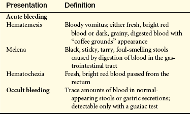
The presentation of gastrointestinal bleeding is summarized in Table 39-1. Acute blood loss is usually characterized by hematemesis (the presence of blood in the vomitus), hematochezia (bright red or burgundy blood from the rectum), or melena (dark, tarry stools). Occult bleeding is usually caused by slow, chronic blood loss that is not obvious and results in iron deficiency anemia as iron stores in the bone marrow are slowly depleted. Physiologic response to gastrointestinal bleeding depends on the amount and rate of the loss (Figure 39-1). Changes in blood pressure and heart rate are the best indicators of massive blood loss in the gastrointestinal tract. Blood losses of 1000 ml or more over a short time cause a decrease in cardiac output, a decrease in systolic and diastolic blood pressure, and an increase in pulse rate. With losses of 1000 ml or more, the heart rate is greater than 100 beats/minute and systolic blood pressure is less than 100 mmHg. During the early stages of blood volume depletion, the peripheral vascular compartment constricts to shunt blood to vital organs, including the brain (see Chapters 30 and 46). Signs that this is happening are postural hypotension (a drop in blood pressure that occurs with a change from the recumbent position to a sitting or upright position), lightheadedness, and loss of vision. If blood loss continues, hypovolemic shock progresses. Diminished blood flow to the kidneys causes decreased urine output and may lead to oliguria (low urine output), tubular necrosis, and renal failure. Ultimately, insufficient cerebral and coronary blood flow causes irreversible anoxia and death.
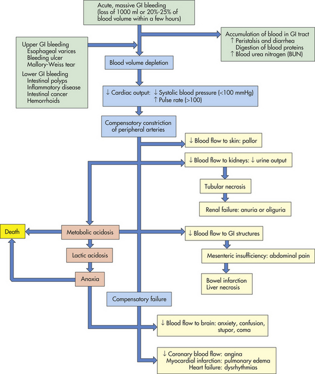
The accumulation of blood in the gastrointestinal tract is irritating and increases peristalsis, causing vomiting or diarrhea, or both. If bleeding is from the lower gastrointestinal tract, the diarrhea is frankly bloody. Bleeding from the upper gastrointestinal tract also can be rapid enough to produce bright red stools, but generally some digestion of the blood components will have occurred, producing melena. The digestion of blood proteins originating from massive upper gastrointestinal bleeding is reflected by an increase in blood urea nitrogen (BUN) levels (see Figure 39-1).
The hematocrit and hemoglobin values are not the best indicators of acute gastrointestinal bleeding because plasma and red cell volume are lost proportionately. As the plasma volume is replaced, the hematocrit and hemoglobin values begin to reflect the extent of blood loss. The interpretation of these values is modified to account for exogenous replacement of fluids and the hydration status of the tissues. Iron deficiency anemia is associated with obscure bleeding.18
Disorders of Motility
PATHOPHYSIOLOGY Dysphagia is difficulty swallowing. It can result from mechanical obstruction of the esophagus or a functional disorder that impairs esophageal motility. Mechanical obstructions can be intrinsic or extrinsic. Intrinsic obstructions originate in the wall of the esophageal lumen. Tumors, strictures, and diverticular herniations (outpouchings) are all causes of intrinsic mechanical obstruction. Extrinsic mechanical obstructions originate outside the esophageal lumen and narrow the esophagus by pressing inward on the esophageal wall. The most common cause of extrinsic mechanical obstruction is tumor.
Functional dysphagia is caused by neural or muscular disorders that interfere with voluntary swallowing or peristalsis. Disorders that affect the striated muscles of the upper esophagus interfere with the oropharyngeal (voluntary) phase of swallowing. Typical causes of functional dysphagia in the upper esophagus are dermatomyositis (a muscle disease) and neurologic impairments caused by cerebrovascular accidents, Parkinson disease, or achalasia.19
Achalasia is a rare disorder related to (1) denervation of smooth muscle in the middle and lower portions of the esophagus, and (2) failure of the lower esophageal sphincter (LES) to relax causing functional obstruction of the lower esophagus.20 Achalasia results from an unknown cause of an autoimmune destruction of myenteric ganglion cells and atrophy of smooth muscle cells. Food accumulates above the obstruction, distends the esophagus, and causes dysphagia. As hydrostatic pressure increases, food is slowly forced past the obstruction into the stomach.
CLINICAL MANIFESTATIONS Clinical manifestations of dysphagia vary according to the cause and location of the obstruction. Distention and spasm of the esophageal muscles during eating or drinking may cause a mild or severe stabbing pain at the level of obstruction. Discomfort occurring 2 to 4 seconds after swallowing is associated with upper esophageal obstruction. Discomfort occurring 10 to 15 seconds after swallowing is more common in obstructions of the lower esophagus. If the cause of obstruction is a growing tumor, dysphagia begins with difficulty swallowing solids and advances to difficulty swallowing semisolids and liquids.21 Dysphagia is experienced with both solids and liquids if the cause is loss of neuromotor function. Retrosternal pain, regurgitation of undigested food, unpleasant taste, vomiting, and weight loss are common manifestations of all types of dysphagia. Aspiration of esophageal contents can lead to pneumonia.
EVALUATION AND TREATMENT Knowledge of the individual’s history and clinical manifestations contributes significantly to a diagnosis of dysphagia. Videofluoroscopy and intraluminal ultrasound are used to visualize the contours of the esophagus and identify structural defects. Manometry and intraluminal impedance monitoring documents the duration and amplitude of abnormal pressure changes associated with obstruction or loss of neural regulation.22 Esophageal endoscopy is performed to examine the esophageal mucosa, obtain biopsy specimens, or perform corrective surgery.
The individual is taught to manage symptoms by eating slowly, eating small meals, taking fluid with meals, and sleeping with the head elevated to prevent regurgitation and aspiration. Anticholinergic drugs may alleviate symptoms. Definitive treatments include mechanical dilation of the esophageal sphincter and surgical separation of the lower esophageal muscles with a longitudinal incision (myotomy). Myotomy widens the passage into the stomach.23
Gastroesophageal Reflux Disease
Gastroesophageal reflux disease (GERD) is the reflux of chyme from the stomach to the esophagus. The LES may relax spontaneously and transiently 1 to 2 hours after eating, permitting gastric contents to regurgitate into the esophagus. The acid is usually neutralized and cleared from the esophagus by peristaltic action within 1 to 3 minutes, and sphincter tone is restored. Gastroesophageal reflux that does not cause symptoms is known as physiologic reflux. In nonerosive reflux disease (NERD), individuals have symptoms of reflux disease but no visible esophageal mucosal injury.24 Esophageal hypersensitivity is common. In some individuals, however, a combination of factors causes injury and an inflammatory response to reflux called reflux esophagitis. Risk factors for GERD include obesity and Helicobacter pylori.25 GERD may be a trigger for asthma or chronic cough.26
PATHOPHYSIOLOGY Normally the resting tone of the LES maintains a zone of high pressure that prevents gastroesophageal reflux. In individuals who develop reflux esophagitis, this pressure tends to be lower than normal from either transient relaxation or weakness of the sphincter. Vomiting, coughing, lifting, bending, or obesity increases abdominal pressure contributing to the development of reflux esophagitis. Delayed gastric emptying contributes to reflux esophagitis by (1) lengthening the period during which reflux is possible and (2) increasing the acid content of chyme. Disorders that delay emptying include gastric or duodenal ulcers, which can cause pyloric edema; strictures that narrow the pylorus; and hiatal hernia, which can weaken the LES.27
The severity of the esophagitis depends on the composition of the gastric contents, the length of time they are in contact with the esophageal mucosa, and epithelial resistance to acid.28 If the chyme is highly acidic, or contains pepsin, bile salts, and pancreatic enzymes, reflux esophagitis can be severe. In individuals with weak esophageal peristalsis, refluxed chyme remains in the esophagus longer than usual. This increases the amount of time the esophageal mucosa is exposed to acids, pepsin, bile, and enzymes.
The presence of H. pylori in lowering the severity of reflux disease is controversial; some studies report that cytotoxin associated gene-A (CagA) strains lower risk of GERD.29 However, there is uncertainty about the possible negative effect of eradicating H. pylori infection on GERD and esophageal adenocarcinoma and in relation to treatment with proton pump inhibitors.30–32
Reflux esophagitis causes inflammatory responses in the esophageal wall, such as hyperemia, increased capillary permeability, edema, tissue fragility, erosion, and ulcerations (Figure 39-2). Fibrosis, basal cell hyperplasia, and elongation of papillae are common.33 Precancerous lesions (Barrett esophagus, see p. 1497) can be a long-term consequence.34
CLINICAL MANIFESTATIONS The clinical manifestations of reflux esophagitis are heartburn, regurgitation of acidic chyme, and upper abdominal pain within 1 hour of eating. The symptoms worsen if the individual lies down or if intra-abdominal pressure increases (e.g., as a result of coughing, vomiting, or straining at stool). Symptoms may be present when no acid is in the esophagus.33 Heartburn also may be experienced as chest pain, which requires ruling out cardiac ischemia. Edema, fibrosis (strictures), esophageal spasm, or decreased esophageal motility may result in dysphagia. Alcohol or acid-containing foods, such as citrus fruits, can cause discomfort during swallowing. There also is an association between acid reflux and laryngitis, asthma, and chronic cough.35,36
EVALUATION AND TREATMENT Diagnosis of reflux esophagitis is based on the history and clinical manifestations, which are usually chronic and relapsing. Ambulatory pH monitoring evaluates acidity near the LES. Esophageal endoscopy shows edema and erosion, and allows for evaluation of dysplastic changes (Barrett esophagus) and the development of esophageal carcinoma. It also identifies associated conditions, such as hiatal hernia, gastric ulcers, and abnormal contours of the esophageal lumen.37
Proton pump inhibitors are the most effective monotherapy. Other therapies include histamine-2 (H2) receptor antagonists or prokinetics, antacids, and alginate-antacids.38 Elevation of the head of the bed 6 inches prevents reflux. Weight reduction and cessation of smoking also help to alleviate symptoms. Laparoscopic fundoplication is the most common surgical intervention when medical treatment fails.39
Eosinophilic esophagitis is a rare, idiopathic inflammatory disease of the esophagus characterized by esophageal infiltration of eosinophils associated with atopic disease, including asthma and food allergies. It occurs in adults and children. Complex molecular mechanisms and gene and environmental interactions contribute to the pathogenesis of this disease.40 Dysphagia and food impaction in the esophagus are common symptoms in the adult that can result from chronic inflammation and fibrosis. Diagnosis includes differentiation from GERD.41 Treatment is symptomatic including elimination diets and steroids.
Hiatal Hernia
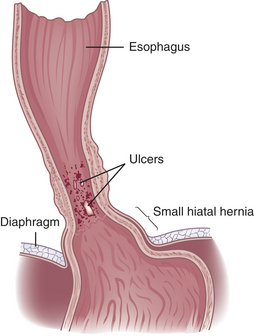
PATHOPHYSIOLOGY Hiatal hernia, a type of diaphragmatic hernia, is the protrusion (herniation) of the upper part of the stomach through the diaphragm and into the thorax. The two types of hiatal hernia are (1) sliding (direct) hiatal hernia, and (2) paraesophageal (rolling) hiatal hernia (Figure 39-3). In sliding hiatal hernia (the most common type, 90%) the stomach slides or moves into the thoracic cavity through the esophageal hiatus, an opening in the diaphragm for the esophagus and vagus nerves. A congenitally short esophagus, trauma, or weakening of the diaphragmatic muscles at the gastroesophageal junction contributes to the hernia. While the individual is in the supine position, the lower esophagus and stomach are pulled into the thorax. Standing causes the stomach to “slide” back into the abdomen. Sliding hiatal hernia is exacerbated by factors that increase intra-abdominal pressure. Therefore, coughing, bending, tight clothing, ascites, obesity, or pregnancy accentuates the hernia. This type of hernia is associated with gastroesophageal reflux and esophagitis because the hernia diminishes the resting pressure of the LES. In pregnant women with sliding hiatal hernia, progesterone and estrogen may lower the resting pressure of the LES further.
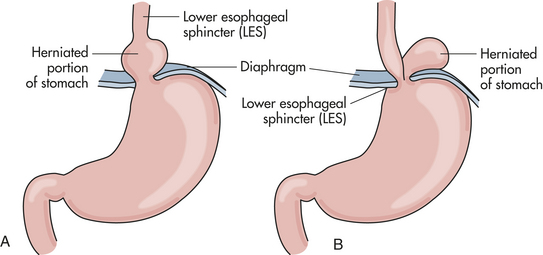
Figure 39-3 Types of hiatal hernia. A, In sliding hiatal hernia the visceral peritoneum remains intact and restrains the size of the hernia. B, In paraesophageal hernia the membrane becomes thinned out or defective, allowing a true peritoneal sac to protrude into the posterior mediastinum, where negative intrathoracic pressure causes it to enlarge. (From Monahan FD et al: Phipps’ Medical-surgical nursing: concepts and clinical practice, ed 8, St Louis, 2007, Mosby.)
Paraesophageal hiatal hernia (rolling hiatal hernia) is herniation of the greater curvature of the stomach through a secondary opening in the diaphragm (see Figure 39-3). The entire stomach can pass into the thorax. As the stomach protrudes through the opening into the thorax, it lies alongside the esophagus. The gastroesophageal junction remains below the diaphragm. With paraesophageal hernia, reflux is uncommon. The position of a portion of the stomach above the diaphragm, however, causes congestion of mucosal blood flow and can lead to gastritis and ulcer formation. A mechanical strangulation of the hernia is a major complication, and surgical correction is required. Strangulation occludes blood vessels and causes vascular engorgement, edema, ischemia, and hemorrhage. Hiatal hernias of both types tend to occur in conjunction with several other diseases, including reflux, peptic ulcer, cholecystitis (gallbladder inflammation), cholelithiasis (gallstones), chronic pancreatitis, and diverticulosis.
CLINICAL MANIFESTATIONS Hiatal hernias are often asymptomatic. Generally a wide variety of symptoms develop later in life and are associated with other gastrointestinal disorders as well. Manifestations of the various types of hiatal hernia are difficult to distinguish and include gastroesophageal reflux, dysphagia, heartburn, vomiting, and epigastric pain.42 Regurgitation and substernal discomfort after eating are common.
EVALUATION AND TREATMENT Diagnostic procedures include barium roentgenogram and endoscopy. A chest roentgenogram often will show the protrusion of the stomach into the thorax, indicating paraesophageal hiatal hernia.
Treatment for sliding hiatal hernia is usually conservative. The individual can diminish reflux by eating small, frequent meals and avoiding the recumbent position after eating. Abdominal supports and tight clothing are avoided, and weight control is recommended for obese individuals. Antacids alleviate reflux esophagitis. Anticholinergic drugs are contraindicated because they relax the LES and delay gastric emptying. Individuals who are uncomfortable at night benefit from sleeping in a semi-Fowler position. Surgery (i.e., fundoplication) may be performed for paraesophageal hiatal hernia or if medical management fails to control symptoms.43
Pyloric Obstruction
PATHOPHYSIOLOGY Pyloric obstruction is the narrowing or blocking of the opening between the stomach and the duodenum. This condition can be congenital (see Chapter 40) or acquired. Acquired obstruction is caused by peptic ulcer disease or carcinoma near the pylorus. Duodenal ulcers are more likely than gastric ulcers to obstruct the pylorus. Ulceration causes obstruction resulting from inflammation, edema, spasm, fibrosis, or scarring. Tumors cause obstruction by growing into the pylorus.
CLINICAL MANIFESTATIONS Early in the course of pyloric obstruction, the individual experiences vague epigastric fullness, which becomes more distressing after eating and later in the day. Nausea and epigastric pain may occur as the muscles of the stomach contract in attempts to force chyme past the obstruction. These symptoms disappear when the chyme finally moves into the duodenum. As obstruction progresses, anorexia develops, sometimes accompanied by weight loss. Severe obstruction causes gastric distention and atony (lack of muscle tone and gastric motility). Gastric distention stimulates gastric secretion, which increases the feeling of fullness. Rolling or jarring of the abdomen produces a sloshing sound called the succussion splash. At this stage, vomiting is a cardinal sign of obstruction. It is usually copious and occurs several hours after eating. The vomitus contains undigested food but no bile. Prolonged vomiting leads to dehydration, which is accompanied by a hypokalemic and hypochloremic metabolic alkalosis caused by loss of potassium and gastric acid. Because food does not enter the intestine, stools are infrequent and small. Prolonged pyloric obstruction causes malnutrition, dehydration, and extreme debilitation.
EVALUATION AND TREATMENT Diagnosis is based on clinical manifestations, a history of ulcer disease, and examination of residual gastric contents. Endoscopy is performed if gastric carcinoma is the suggested cause of pyloric obstruction. Barium studies are contraindicated because the barium may harden and be retained in the stomach.
Obstructions resulting from ulceration often resolve with conservative management. Gastric drainage is used to decompress the stomach and restore normal motility. Gastric secretions that contribute to inflammation and edema can be suppressed with omeprazole or cimetidine. Fluids and electrolytes (saline and potassium) are given intravenously to effect rehydration and correct hypokalemia and alkalosis (see Chapter 3). Severely malnourished individuals may require parenteral hyperalimentation (intravenous nutrition). Surgery or stenting may be required to treat gastric carcinoma or persistent obstruction caused by fibrosis and scarring.44
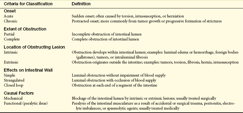
Intestinal Obstruction and Ileus
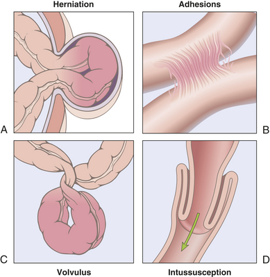
Intestinal obstruction can be caused by any condition that prevents the normal flow of chyme through the intestinal lumen or failure of normal intestinal motility in the absence of an obstructing lesion (ileus). The small intestine is more commonly obstructed because of its narrower lumen. Common causes of intestinal obstruction are summarized in Table 39-2. More specific causes of small and large bowel obstruction are summarized in Table 39-3. Criteria for classifying intestinal obstruction are summarized in Table 39-4. Intestinal obstruction is classified by cause as simple or functional. Simple obstruction is mechanical blockage of the lumen by a lesion; functional obstruction is a failure of motility (paralytic ileus) and is common after gastrointestinal or abdominal surgery. Anesthetic agents, local inflammatory reactions, use of opioid analgesia, and hyperactivity of the sympathetic nervous system contribute to postoperative ileus.45 Simple obstruction of the small intestine from fibrous adhesions is the most common type of intestinal obstruction.46 Acute obstructions usually have mechanical causes, such as adhesions or hernias (Figure 39-4). Chronic or partial obstructions are more often associated with tumors or inflammatory disorders, particularly of the large intestine. Intussusception is rare in adults compared with the more frequent occurrence in infants. Common causes of intestinal obstruction in children are presented in Chapter 40.
Table 39-2
Common Causes of Intestinal Obstruction
| Cause | Pathophysiology |
| Herniation | Protrusion of the intestine through a weakness in the abdominal muscles or through the inguinal ring |
| Intussusception | Telescoping of one part of the intestine into another; this usually causes strangulation of the blood supply; more common in the ileocecal area in infants 10 to 15 months of age than in adults |
| Torsion (volvulus) | Twisting of the intestine on its mesenteric pedicle, with occlusion of the blood supply; often associated with fibrous adhesions in the small intestine; occurs most often in the large intestine in older adults |
| Diverticulosis | Inflamed saccular herniations (diverticula) of the mucosa and submucosa through the tunica muscularis of the colon; diverticula are interspersed between thick, circular, fibrous bands; most common in obese individuals older than 60 years |
| Tumor | Tumor growth into the intestinal lumen; adenocarcinoma of the colon and rectum is the most common tumoral obstruction; most common in individuals older than 60 years |
| Paralytic (adynamic) ileus | Loss of peristaltic motor activity in the intestine; associated with abdominal surgery, peritonitis, hypokalemia, ischemic bowel, spinal trauma, pneumonia, neuropathies, or myopathies; affects small and large intestines |
| Fibrous adhesions | Peritoneal irritation from surgery or trauma leads to formation of fibrin and adhesions that attach to intestine, omentum, or peritoneum and can cause traction and obstruction; most common in small intestine |
Table 39-3
Large and Small Bowel Obstruction
| Cause | Pathogenesis |
| Small bowel obstruction | Adhesions: secondary to previous abdominal surgeries: 50%-70% |
| Hernia: inguinal, ventral, or femoral: 20%-25% | |
| Tumors: may be associated with intussusception: 10% | |
| Mesenteric ischemia 3%-5% | |
| Large bowel obstruction | Colon/rectal cancer 90% |
| Colonic volvulus 4%-5% | |
| Diverticular disease 3%-5% | |
| Other causes (inflammatory bowel disease, adhesions, hernia, adynamic ileus) |
Adapted from Feldman M et al: Sleisenger & Fordtran’s gastrointestinal liver disease, ed 8, Philadelphia, 2006, Saunders.
PATHOPHYSIOLOGY The consequences of intestinal obstruction are related to its onset and location, the length of intestinal tract proximal to the obstruction, and the presence and severity of ischemia. The major pathophysiologic alterations are presented in Figure 39-5. The most common causes of small intestine obstruction are intra-abdominal adhesions, hernias, and neoplasms.47 Obstruction leads to accumulation of fluid and gas inside the lumen proximal to the obstruction. Fluids accumulate from impaired water and electrolyte absorption and enhanced secretion with net movement of fluid from the vascular space to the intestinal lumen. Gas from swallowed air, and to a lesser extent from bacterial overgrowth, contributes to the distention. Distention begins almost immediately, as gases and fluids accumulate proximal to the obstruction. Distention decreases the intestine’s ability to absorb water and electrolytes and increases the net secretion of these substances into the lumen. Within 24 hours, up to 8 L of fluid and electrolytes enters the lumen in the form of saliva, gastric juice, bile, pancreatic juice, and intestinal secretions. Copious vomiting or sequestration of fluids in the intestinal lumen prevents their reabsorption and produces severe fluid and electrolyte disturbances. Extracellular fluid volume and plasma volume decrease, causing dehydration. Hemoconcentration (decreased plasma volume) elevates hematocrit, decreases central venous pressure, and causes tachycardia. Severe dehydration leads to hypovolemic shock.
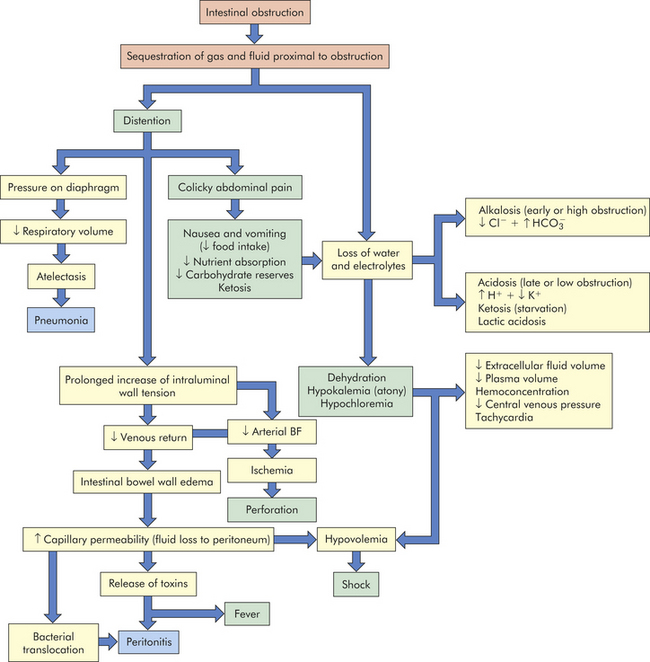
If the obstruction is at the pylorus or high in the small intestine, metabolic alkalosis develops initially as a result of excessive loss of hydrogen ions that normally would be reabsorbed from the gastric juice. With prolonged obstruction or obstruction lower in the intestine, metabolic acidosis is more likely to occur because bicarbonate from pancreatic secretions and bile cannot be reabsorbed. Hypokalemia can be extreme, promoting acidosis and atony of the intestinal wall. Metabolic acidosis also may be accentuated by ketosis, the result of declining carbohydrate stores caused by starvation. If pressure from the distention is severe enough, it occludes the arterial circulation and causes ischemia, necrosis, perforation, and peritonitis. Fever and leukocytosis are often associated with loss of intestinal motility, overgrowth of bacteria, strangulation, and bowel necrosis. Lack of circulation permits the buildup of significant amounts of lactic acid, which worsen the metabolic acidosis. Bacterial proliferation and translocation across the mucosa to the mesenteric lymph nodes or systemic circulation cause peritonitis or sepsis. The release of inflammatory mediators into the circulation causes remote organ failure.48
The most common causes of large bowel obstruction are malignancy, volvulus (twisting), and strictures related to diverticulitis. Consequences of colonic or large bowel obstruction are related to the competence of the ileocecal valve, which normally prevents reflux of colonic contents into the small intestine. When the ileocecal valve is competent, the cecum cannot decompress into the small intestine resulting in distention. Ischemia occurs when the intraluminal pressure exceeds capillary pressure in the lumen. Acute colonic pseudo-obstruction (Ogilvie syndrome) is a massive dilation of the large bowel that occurs in critically ill patients, and immobilized older adults. It is characterized by significant dilation of the cecum and absence of mechanical obstruction.
CLINICAL MANIFESTATIONS Signs and symptoms of small intestine obstruction are consistent with the pathophysiology. Colicky pains caused by distention followed by vomiting are the cardinal symptoms. Typically the pain occurs intermittently. Pain intensifies for seconds or minutes as a peristaltic wave of muscle contraction meets the obstruction. The passing of the wave is followed by a pain-free interval. Pain may be continuous with severe distention and then diminish in intensity. If strangulation occurs, the pain loses its colicky character, becoming more constant and severe as ischemia progresses to necrosis or perforation. Sweating, nausea, and hypotension occur as an autonomic nervous system response.
Vomiting and distention vary, depending on the level and completion of the obstruction. Obstruction at the pylorus causes early, profuse vomiting of clear gastric fluid. Obstruction in the proximal small intestine causes mild distention and vomiting of bile-stained fluid. Obstruction lower in the small intestine causes more pronounced distention because a greater length of intestine is proximal to the obstruction. In this case, vomiting may not occur or may occur later and contain fecal material. Partial obstruction can cause diarrhea or constipation, but complete obstruction usually causes constipation only. Complete obstruction increases the number of bowel sounds, which may be tinkly and accompanied by peristaltic rushes and crampy, abdominal pain. Signs of dehydration, hypovolemia, and metabolic acidosis may be observed as early as 24 hours after the occurrence of complete obstruction. Distention may be severe enough to push against the diaphragm and decrease lung volume. This can lead to atelectasis and pneumonia, particularly in debilitated individuals.
Colonic obstruction usually presents as hypogastric pain and abdominal distention. Pain can vary from vague to excruciating, depending on the degree of ischemia and the development of peritonitis. Colon cancer is the most common cause, followed by diverticular strictures or volvulus.
EVALUATION AND TREATMENT Evaluation is based on clinical manifestations and includes ultrasound and radiography.49,50 Successful management requires early identification of the site and type of obstruction. Replacement of fluid and electrolytes and decompression of the lumen with gastric or intestinal suction are essential forms of therapy. Immediate surgical intervention is required for strangulation and complete obstruction. Neostigmine is used for colonic pseudo-obstruction.51
Gastritis
Gastritis is an inflammatory disorder of the gastric mucosa. It can be acute or chronic and can affect the fundus or antrum or both. Acute gastritis erodes the surface epithelium in a diffuse or localized pattern. The erosions are usually superficial.
Acute gastritis is usually injury of the protective mucosal barrier by drugs, chemicals, or H. pylori infection (Figure 39-6). Nonsteroidal anti-inflammatory drugs (NSAIDs), such as aspirin, ibuprofen, naproxen, and indomethacin, are known to cause erosive gastritis because they inhibit prostaglandins, which normally stimulate the secretion of mucus.52 Alcohol, histamine, digitalis, and metabolic disorders such as uremia are contributing factors. H. pylori infection causes inflammation, pain, nausea, and vomiting.53 The clinical manifestations of acute gastritis can include vague abdominal discomfort, epigastric tenderness, and bleeding. Healing usually occurs spontaneously within a few days. Discontinuing injurious drugs, using antacids, or decreasing acid secretion with a histamine H2 receptor antagonist and proton pump inhibitor also promote healing.

Figure 39-6 Acute erosive gastritis. Acute erosive gastritis is shown in the opened stomach. The mucosa appears hyperemic, and the foci of superficial ulceration are manifested as scattered, small, red areas termed erosions. (From Kumar V et al: Pathologic basis of disease, ed 7, Philadelphia, 2006, Saunders.)
Chronic gastritis tends to occur in older adults and causes chronic inflammation, mucosal atrophy, and epithelial metaplasia. Chronic gastritis usually is classified as type A, or immune (fundal), or type B, nonimmune (antral), depending on the pathogenesis and location of the lesions. Chronic fundal gastritis is the most rare and severe type. The gastric mucosa degenerates extensively in the body and fundus of the stomach, leading to gastric atrophy. Loss of chief cells and parietal cells diminishes secretion of pepsinogen, hydrochloric acid, and intrinsic factor. Because acid secretion is insufficient, the feedback mechanism that normally inhibits gastrin secretion is impaired, causing elevated plasma levels of gastrin. Pernicious anemia can develop because intrinsic factor is less available to facilitate vitamin B12 absorption.
A significant number of individuals with chronic fundal gastritis have antibodies to parietal cells, intrinsic factor, and gastric cells in their sera, suggesting that an autoimmune mechanism is involved in the pathogenesis of the disease. The fact that chronic fundal gastritis occurs in association with other autoimmune diseases, such as diabetes, Addison disease, and thyroid disease, strengthens this association. Chronic fundal gastritis is a risk factor for gastric carcinoma, particularly in individuals who develop pernicious anemia.54,55
Chronic antral gastritis generally involves the antrum only and is approximately four times more common than fundal gastritis. It is not associated with decreased hydrochloric acid secretion, pernicious anemia, or presence of parietal cell antibodies. Several factors are associated with chronic antral gastritis, including use of alcohol, tobacco, and NSAIDs. H. pylori is a major causative factor associated with chronic atrophic antral gastritis and peptic ulcer disease. The host response to H. pylori infection is activation of T and B lymphocytes with infiltration of neutrophils. Release of inflammatory cytokines (e.g., tumor necrosis factor-α [TNF-α]; interleukin-1 [IL-1], IL-6, IL-8, IL-10; and leukotrienes) damage the gastric epithelium.56–58 An H. pylori gene (CagA) produces a vacuolating toxin (VacA) causing injury and promoting inflammation. In approximately 10% of cases, antibodies to gastrin-secreting cells are found in the serum. Chronic reflux of bile may contribute to the gastritis by persistently disrupting the mucosal barrier.
Signs and symptoms of chronic gastritis often do not correlate with the severity of the disease. Gastroscopic examination and biopsy may show a long-standing inflammatory process and gastric atrophy in an individual with no history of abdominal distress. The presence of antiparietal cell antibody is specific for type A gastritis. H. pylori infection is evidence for H. pylori gastritis. Failure to stimulate acid secretion confirms achlorhydria (diminished secretion of hydrochloric acid). The gastric secretions also can be evaluated for the presence of intrinsic factor. Individuals may report vague symptoms, including anorexia, fullness, nausea, vomiting, and epigastric pain. Gastric bleeding may be the only clinical manifestation of gastritis. There is increased risk for gastric carcinoma with chronic H. pylori infection.59
Symptoms usually can be managed with smaller meals; a soft, bland diet; and avoidance of alcohol and NSAIDs. Combination antibiotics are used to treat H. pylori, and the emergence of antimicrobial resistance is a concern.60 Vitamin B12 is administered to correct pernicious anemia (see Chapter 26).61
Peptic Ulcer Disease
A peptic ulcer is a break, or an ulceration, in the protective mucosal lining of the lower esophagus, stomach, or duodenum. Approximately 14.5 million people in the United States have peptic ulcer disease.62 Two major risk factors for peptic ulcer disease are H. pylori infection of the gastric mucosa (Box 39-2) and habitual use of NSAIDs. Alcohol and smoking may influence susceptibility to ulcer disease. Some chronic diseases, such as emphysema, rheumatoid arthritis, and cirrhosis, are associated with the development of peptic ulcers. Psychologic stress may be a risk factor for peptic ulcer disease, although studies of life stress and ulcer disease are inconclusive.63,64 The exact mechanism of causation is not known.65
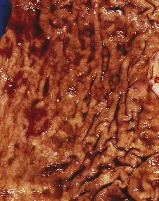
Peptic ulcers can be acute or chronic, and superficial or deep. Superficial ulcerations are called erosions because they erode the mucosa but do not penetrate the muscularis mucosae (Figure 39-7). True ulcers extend through the muscularis mucosae and damage blood vessels causing hemorrhage or perforate the gastrointestinal wall.
Figure 39-7 Chronic peptic ulcer. Gross photograph of a chronic peptic ulcer located in the lesser curvature, straddling the antrum and corpus of the stomach. (From Damjanov I, Linder J, editors: Anderson’s pathology, ed 10, St Louis, 1996, Mosby.)
Gastric mucosal infection with H. pylori is a major cause of peptic ulcers (Box 39-2).66 Chronic use of NSAIDs suppresses mucosal prostaglandin synthesis resulting in decreased bicarbonate secretion and mucin production and increased secretion of hydrochloric acid. The interaction of NSAIDs and H. pylori in the pathogenesis of peptic ulcer is not clear.67 Disruption of the mucosa exposes submucosal areas to gastric secretions and autodigestion causing erosion and ulceration.
Duodenal Ulcers
Duodenal ulcers occur with greater frequency than other types of peptic ulcers and affect 10% to 15% of the population.68 The incidence of duodenal ulcers is approximately the same among men and women in the United States.69 Duodenal ulcers tend to develop in younger persons, and there may be an association with type O blood.70,71
PATHOPHYSIOLOGY Factors other than H. pylori and use of NSAIDs that may be associated with duodenal ulcer include:
1. Increased mass of gastric parietal cells
2. Serum gastrin levels that remain high longer than normal after eating and continue to stimulate secretion of acid and pepsin (may be caused by H. pylori in gastric antrum)
3. Failure of the feedback mechanism whereby acid in the gastric antrum inhibits gastrin release
4. Rapid gastric emptying, which overwhelms the buffering capacity of the bicarbonate-rich pancreatic secretions
All these factors, singly or in combination, cause acid and pepsin concentrations in the duodenum to penetrate the mucosal barrier and lead to ulceration72,73 (Figure 39-8).
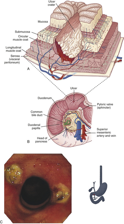
Figure 39-8 Duodenal ulcer. A, A deep ulceration in the duodenal wall extending as a crater through the entire mucosa and into the muscle layers. B, Duodenal ulcer. C, Bilateral (kissing) duodenal ulcers in a person using nonsteroidal anti-inflammatory drugs (NSAIDs). (C Courtesy David Bjorkman, MD, University of Utah School of Medicine, Department of Gastroenterology.)
CLINICAL MANIFESTATIONS The characteristic manifestation of a duodenal ulcer is chronic intermittent pain in the epigastric area. The pain begins 30 minutes to 2 hours after eating, when the stomach is empty. It is not unusual for pain to occur in the middle of the night and disappear by morning. The pain results from sensorineural stimulation by acid, muscle spasm, or both. Pain is relieved rapidly by ingestion of food or antacids, creating a typical “pain-food-relief” pattern. Some individuals with duodenal ulcer have no symptoms, particularly older adults; the first manifestation may be hemorrhage or perforation, particularly with a history of NSAID or anticoagulant use.
Duodenal ulcers often heal spontaneously but recur within months. Exacerbations tend to develop in the spring and fall. Healing is accompanied by relief of pain. Constant, unremitting pain may be caused by complications, such as intestinal obstruction or perforation. Bleeding from duodenal ulcers causes hematemesis or melena. It is not clear why individuals infected with H. pylori do not develop duodenal cancer.74
EVALUATION AND TREATMENT Several diagnostic approaches are used to differentiate duodenal ulcers from gastric ulcers or gastric carcinoma. Endoscopic evaluation allows visualization of lesions and biopsy. Radioimmune assays of gastrin levels are evaluated to identify ulcers associated with gastric carcinomas. The urea breath test, serum antibodies, stool and serum antigen, and positive findings from gastric biopsy detect H. pylori infection.75
Management of duodenal ulcers is aimed at relieving the causes and effects of hyperacidity. Antacids neutralize gastric contents, elevate pH, inactivate pepsin, and relieve pain. Acid secretion can be suppressed with drugs that block H2 receptors and inhibit the secretion of acid. Proton pump inhibitors inhibit acid production. Eradication of H. pylori with bismuth and combinations of antibiotics supplemented with vitamin C usually prevents relapse, although there is increasing drug resistance.76 Ulcer-coating agents, such as sucralfate and colloidal bismuth, promote healing. Anticholinergic drugs may be used to inhibit gastric secretion, suppress gastric motility, and delay gastric emptying. Surgical resection may be required for bleeding or perforating ulcers, obstruction, or peritonitis.77 Risk of duodenal ulcer may be reduced with a diet high in vitamin A and fiber.78 Clinical trials are in progress for a vaccine against H. pylori.79
Gastric Ulcers
Gastric ulcers are ulcers of the stomach. They occur with about equal frequency in males and females, usually between the ages of 55 and 65 years, and are about one fourth as common as duodenal ulcers (Table 39-5 and Figure 39-9).
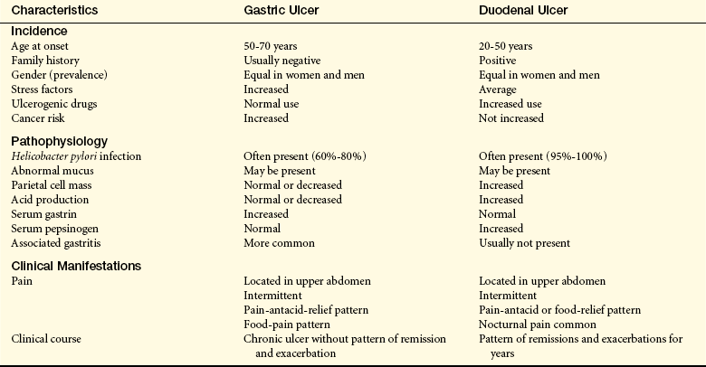
PATHOPHYSIOLOGY Generally gastric ulcers develop in the antral region, adjacent to the acid-secreting mucosa of the body, and are frequently caused by H. pylori (see Box 39-2).80 The primary defect is an abnormality that increases the mucosal barrier’s permeability to hydrogen ions. Gastric secretion may be normal or less than normal and there may be a decreased mass of parietal cells. Chronic pangastritis is often associated with development of gastric ulcers and may precipitate ulcer formation by limiting the mucosa’s ability to secrete a protective layer of mucus (Figure 39-10).
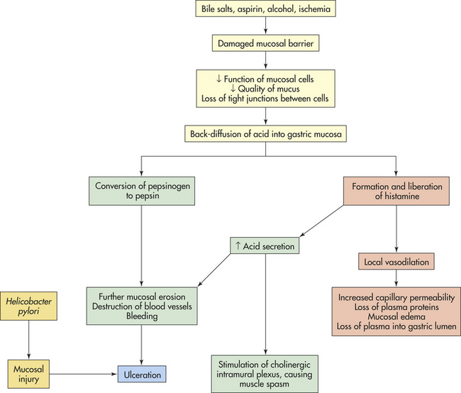
Duodenal reflux of bile is associated with gastric ulcer (alkaline reflux gastritis, p. 1469) and may occur after cholescystectomy, pyloroplasty, or gastrojejunostomy.81 The pyloric sphincter also may fail to respond to stimuli that normally increase resting tone, such as entry of acid, protein, and fat into the duodenum. An increased concentration of bile salts disrupts the gastric mucosa. The break damages the mucosal barrier by permitting hydrogen ions to diffuse into the mucosa, where they disrupt permeability and cellular structure. A vicious cycle can be established as the damaged mucosa liberates histamine, which stimulates the increase of acid and pepsinogen production, blood flow, and capillary permeability. The disrupted mucosa becomes edematous and loses plasma proteins. Destruction of small vessels causes bleeding.
Zollinger-Ellison syndrome is associated with peptic ulcers related to increased secretion of gastrin, which causes excess secretion of gastric acid. A gastrinoma (a gastrin-secreting neuroendocrine tumor or multiple tumors) of the pancreas or duodenum stimulates a proliferation of gastric parietal cells and chronic secretion of gastric acid. The resulting excess acid causes gastric and duodenal ulcers, gastroesophageal reflux with abdominal pain, and diarrhea. Diagnosis includes secretin- or calcium-stimulated measures of gastrin levels, gastric pH levels less than 2, and symptomatic evidence of peptic ulcer disease. Proton pump inhibitors reduce gastric acid secretion, and surgical removal of tumors limits metastasis.82,83
CLINICAL MANIFESTATIONS The clinical manifestations of gastric ulcers are similar to those of duodenal ulcers (see Table 39-5). The pattern of pain, food, and relief is common, but the pain of gastric ulcers also occurs immediately after eating. Gastric ulcers also tend to be chronic rather than alternate between periods of remission and exacerbation and cause more anorexia, vomiting, and weight loss than duodenal ulcers. The evaluation and treatment of gastric ulcers are similar to the evaluation and treatment of duodenal ulcers.
Stress-Related Mucosal Disease
A stress ulcer (stress-related mucosal disease) is an acute form of peptic ulcer that tends to accompany the physiologic stress of severe illness; multisystem organ failure; or major trauma, including severe burns or head injury. Usually, multiple sites of ulceration are distributed within the stomach or duodenum. Stress ulcers may be classified as ischemic ulcers or Cushing ulcers.
Ischemic ulcers develop within hours of an event—such as hemorrhage, multisystem trauma, severe burns, heart failure, or sepsis—that causes ischemia of the stomach and duodenal mucosa. Stress ulcers that develop as a result of burn injury are often called Curling ulcers.
The shock, anoxia, and sympathetic responses produced by the precipitating event decrease mucosal blood flow, leading to gastric ischemia. In intensive care units, use of positive-pressure mechanical ventilation can induce splanchnic hypoperfusion and contribute to stress-related mucosal injury.84 Because the metabolism of the mucosal cells declines as a result of ischemia, the mucosal lining degenerates. Acid diffuses back into the mucosa, causing inflammation, ulceration, hemorrhage, and necrosis. The ulcerative process is accelerated if bile or pancreatic enzymes are regurgitated from the duodenum. Bleeding occurs more readily with the presence of coagulopathy.85
Cushing ulcer is a stress ulcer associated with severe head trauma or brain surgery. This ulcer results from decreased mucosal blood flow and hypersecretion of acid caused by overstimulation of the vagal nuclei. Excessive acid damages the mucosal barrier, initiating the processes summarized in Figure 39-10.
The primary clinical manifestation of stress-related mucosal disease is bleeding. Other symptoms may not be present. The bleeding may be slight or, if a small vessel is perforated, amount to hundreds of milliliters. Prophylactic treatment regimens are used to prevent this disease.86 Stress ulcers seldom become chronic.
Surgical Treatment of Ulcer
Advances in the medical treatment of peptic ulcer disease with proton pump inhibitors and eradication of H. pylori, and laparoscopic and endoscopic repair techniques have significantly reduced the number of cases requiring surgery.87 The indications for ulcer surgery are recurrent or uncontrolled bleeding and complicated perforation of the stomach or duodenum.88 Different types of gastric resection may be performed to treat gastric cancer.
Acute complications of gastrectomy or anastomosis, such as poor wound healing, abscess formation, or suture failure, are relatively uncommon except in the debilitated person. Chronic complications, however, occur more often and are likely to develop if a large portion of the stomach has been removed. These complications and their pathophysiologic mechanisms are described in the next section.
Postgastrectomy Syndromes
Postgastrectomy syndromes are a group of signs and symptoms that occur after gastric resection. They are caused by changes in motor and control functions of the stomach and upper small intestine.89
Dumping Syndrome: Dumping syndrome is the rapid emptying of hypertonic chyme from the surgically created, residual stomach into the small intestine 10 to 20 minutes after eating (early dumping syndrome). It occurs with varying severity in 5% to 10% of individuals who have undergone partial gastrectomy or pyloroplasty.90 It is not common in individuals who have undergone a Billroth II anastomosis (gastrojejunostomy) accompanied by vagotomy. Factors that promote early dumping syndrome include (1) loss of gastric capacity, (2) loss of emptying control when the pylorus is removed, and (3) loss of feedback control by the duodenum when it is removed. Rapid gastric emptying and creation of a high osmotic gradient within the small intestine cause a sudden shift of fluid from the vascular compartment to the intestinal lumen. Plasma volume decreases, causing vasomotor responses, such as increased pulse rate, hypotension, weakness, pallor, sweating, and dizziness. Rapid distention of the intestine produces a feeling of epigastric fullness, cramping, nausea, vomiting, and diarrhea.91
A less common form of dumping syndrome, late dumping syndrome, occurs 1 to 3 hours after eating. The symptoms include weakness, diaphoresis, and confusion, but they cannot be explained by rapid gastric emptying. After a high-carbohydrate meal, individuals who have undergone gastrectomy may develop hypoglycemia, which causes the symptoms. The hypoglycemia is caused by an increase in insulin secretion stimulated by the hyperglycemia that follows eating. Other hormonal responses may also participate in the development of hypoglycemia.
Most cases of dumping syndrome respond well to dietary management.92 Frequent small meals that are high in protein and low in carbohydrates relieve symptoms. Other measures include drinking fluids between meals instead of at mealtime and reclining on the left side after eating. Some cases require surgical intervention, including reconstruction of the pylorus or a gastrojejunostomy.93 Octreotide reduces abdominal and vasomotor symptoms of dumping syndrome by unknown mechanisms.94
Alkaline Reflux Gastritis: Alkaline reflux gastritis is a stomach inflammation caused by reflux of bile and alkaline pancreatic secretions that contain proteolytic enzymes and disrupt the mucosal barrier. This form of gastritis occurs in 5% to 20% of individuals who have undergone gastrectomy or pyloroplasty. Clinical manifestations include nausea, bilious vomiting (vomiting in which the vomitus contains bile), and sustained epigastric pain that worsens after eating and is not relieved by antacids.95 Endoscopy shows a hemorrhagic and friable gastric mucosa. Conservative management is often difficult because antacids do not consistently improve symptoms. Avoidance of aspirin and alcohol may decrease gastric irritation, and a low-fat diet may limit bile secretion. Surgical correction may ultimately be required.
Afferent Loop Obstruction: Afferent loop obstruction is a rare problem that may occur after gastrojejunostomy (attachment of stomach remnant to jejunum [also known as Billroth II or Roux-en-Y procedures]). The problem is caused by recurring tumor growth, volvulus, hernia, adhesion, or stenosis in the duodenal stump on the proximal side of the gastrojejunostomy.96 Partial obstruction causes bile and pancreatic secretions to accumulate and distend the loop. Obstruction also causes delayed emptying. The symptoms of afferent loop obstruction include intermittent severe pain, epigastric fullness after eating, and vomiting. Conservative management consists of a low-fat diet. Surgical correction is required for complete obstruction.
Diarrhea: Diarrhea is one of the most common long-term alterations caused by gastric surgery. Diarrhea can accompany dumping syndrome or occur as a solitary symptom. Diarrhea can occur as frequent, persistent elimination of liquid stool or as intermittent, precipitous, and unpredictable elimination of a large volume of stool. Both types can be either mild or severe. Postgastrectomy diarrhea appears to be related to rapid gastric emptying, particularly after intake of large amounts of high-carbohydrate liquids, which increase the osmotic gradient and attract water into the intestinal lumen. Small, dry meals and anticholinergic drugs are effective control measures.
Weight Loss: Weight loss often follows gastric resection. Inadequate food intake is a common cause because many individuals cannot tolerate the osmotic effect of carbohydrates or a normal-size meal. Foods may be poorly absorbed because the stomach is less able to mix, churn, and break down food particles. Vomiting, diarrhea, and malabsorption of fats also contribute to weight loss.
Anemia: Anemia after gastrectomy results from iron, vitamin B12, or folate deficiency. Iron malabsorption may be caused by decreased acid secretion. Acid changes iron from a trivalent to a divalent molecule, making it easier to absorb. Iron absorption is also compromised in individuals who have undergone a Billroth II procedure because the duodenum is no longer available to absorb iron.
Vitamin B12 deficiency may occur several years after gastrectomy. Contributing factors include loss of parietal cells, which secrete intrinsic factor. (Intrinsic factor facilitates absorption of vitamin B12; see Chapter 38.) Vitamin B12 absorption is also compromised if gastric contents are not mixed adequately with pancreatic enzymes, such as may occur after a Billroth II anastomosis.
Folate deficiency is related to poor intake or malabsorption. Management of deficiencies consists of replacement of iron and folate with supplements. Vitamin B12 can be administered monthly by injection or oral supplements.97
Malabsorption Syndromes
Malabsorption syndromes interfere with nutrient absorption in the small intestine. Historically malabsorption disorders have been classified as maldigestion or malabsorption. Maldigestion is failure of the chemical processes of digestion that take place in the intestinal lumen or at the brush border of the intestinal mucosa. Malabsorption is the failure of the intestinal mucosa to absorb (transport) the digested nutrients. Often maldigestion and malabsorption are interrelated or occur together, making classification difficult. Generally, however, maldigestion is caused by deficiencies of enzymes, such as pancreatic lipase or intestinal lactase, which are necessary for digestion. Inadequate secretion of bile salts and inadequate reabsorption of bile in the ileum also contribute to maldigestion. Malabsorption is the result of mucosal disruption caused by gastric or intestinal resection, vascular disorders, or intestinal disease.
Pancreatic Insufficiency
The pancreatic enzymes (lipase, amylase, trypsin, chymotrypsin) are required for the digestion of proteins, carbohydrates, and fats. Pancreatic insufficiency is the deficient production of these enzymes by the pancreas. Causes of pancreatic insufficiency include chronic pancreatitis, pancreatic carcinoma, pancreatic resection, and cystic fibrosis. Significant damage to or loss of pancreatic tissue must occur before enzyme levels decrease sufficiently to cause maldigestion. Although pancreatic insufficiency causes poor digestion of all nutrients, fat maldigestion is the chief problem. Salivary amylase and enzymes secreted by the intestinal brush border assist in carbohydrate and protein digestion, but these enzymes do not digest fats. Absence of pancreatic bicarbonate in the duodenum and jejunum causes an acidic pH that worsens maldigestion by preventing activation of pancreatic enzymes that are present. Maldigestion, a large amount of fat in the stool (steatorrhea), and weight loss are the most common signs of pancreatic insufficiency. Lipase supplementation is usually successful.98
Lactase Deficiency
Deficiency of disaccharidase at the villus brush border of the small intestine is caused by a congenital defect in the lactase gene.99 Lactase deficiency inhibits the breakdown of lactose (milk sugar) into monosaccharides and therefore prevents lactose digestion and absorption across the intestinal wall. Lactase deficiency is most common in blacks. Congenital lactase deficiency causes watery diarrhea in breast milk or lactose-containing formulas in infants. Lactase expression is lost before adulthood in adult-type lactose intolerance and is genetically determined.100 Secondary (acquired) lactase deficiency can be caused by several diseases of the intestine, including gluten-sensitive enteropathy (see Chapter 40), enteritis, and bacterial overgrowth.
The undigested lactose remains in the intestine, where bacterial fermentation causes gases to form. Undigested lactose also increases the osmotic gradient in the intestine, causing irritation and osmotic diarrhea. Clinical manifestations of lactase deficiency are bloating, crampy pain, diarrhea, and flatulence. The disorder is diagnosed by a lactose-hydrogen breath test, dietary lactose withdrawal, or small intestinal biopsy.101 Avoiding milk products and adhering to a lactose-free diet relieve symptoms. Maintaining an adequate calcium intake with restricted intake of milk products decreases risk of osteoporosis.102
Bile Salt Deficiency
Conjugated bile acids (bile salts) are necessary for the digestion and absorption of fats. Bile salts are conjugated in the bile that is synthesized from cholesterol and secreted from the liver.103 When bile enters the duodenum, the bile salts aggregate with fatty acids and monoglycerides to form micelles. Micelle formation solubilizes fat molecules and allows them to pass through the unstirred layer at the brush border (see Chapter 38). A minimum concentration of bile salts, termed the critical micelle concentration, is required to allow micelles to form. Therefore, conditions that decrease the production or secretion of bile result in decreased micelle formation and fat malabsorption. These conditions include advanced liver disease, which decreases production of bile salts; obstruction of the common bile duct, which decreases flow of bile into the duodenum; intestinal stasis (lack of motility), which permits overgrowth of intestinal bacteria that deconjugate bile salts; and diseases of the ileum, which prevent the reabsorption and recycling of bile salts (enterohepatic circulation).
Clinical manifestations of bile salt deficiency are related to poor intestinal absorption of fat and fat-soluble vitamins (A, D, E, K). Increased fat in the stools (steatorrhea) leads to diarrhea and decreased plasma proteins. The losses of fat-soluble vitamins and their effects include the following:
1. Vitamin A deficiency results in night blindness.
2. Vitamin D deficiency results in decreased calcium absorption with bone demineralization (osteoporosis), bone pain, and fractures.
3. Vitamin K deficiency prolongs prothrombin time, leading to spontaneous development of purpura (bruising) and petechiae.
4. Vitamin E deficiency has uncertain effects but may cause testicular atrophy and neurologic defects in children.
The most effective treatment for fat-soluble vitamin deficiency is to increase medium-chain triglycerides in the diet, for example, by using coconut oil for cooking. Vitamins A, D, and K are given parenterally.
Inflammatory Bowel Disease
Ulcerative colitis and Crohn disease are chronic relapsing inflammatory bowel diseases (IBDs) of unknown origin that affect about 1 million people in the United States.104 Both diseases are associated with genetic factors, alterations in epithelial cell barrier functions, and immunopathology related to abnormal T-cell reactions to commensal microflora and other luminal antigens.105,106 Ulcerative colitis is limited to the mucosa of the colon and rectum. Crohn disease can involve any part of the gastrointestinal tract from the mouth to the anus and involves transmural granulomatous inflammatory lesions (Figure 39-11).
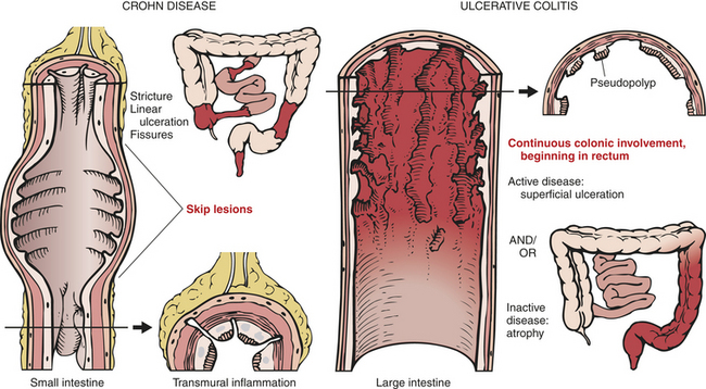
Figure 39-11 Distribution patterns of Crohn disease and ulcerative colitis. Comparison of distribution patterns of Crohn disease and ulcerative colitis as well as different conformations of ulcers and wall thickenings. (From Kumar V et al, editors: Robbins basic pathology, ed 7, St Louis, 2003, Mosby.)
Ulcerative Colitis
Ulcerative colitis (UC) is a chronic inflammatory disease that causes ulceration of the colonic mucosa and extends proximally from the rectum into the colon. The lesions appear in susceptible individuals between 20 and 40 years of age. Risk factors include family history of disease or Jewish descent, and the disease is more prevalent among white populations and Northern Europeans. UC is less common in smokers.107
Although the cause of UC is unknown, dietary, infectious, genetic, and immunologic factors are all suggested causes.108 Inflammation may be caused by commensal or pathogenic enteric microogansims with increased mucosal adherence and invasion and persistent activation of T cells. The familial tendency to develop ulcerative colitis and the occurrence of disease in identical twins supports a genetic theory of causation. Perhaps most significant are the humoral and cellular immunologic factors associated with the disease. Colonic epithelial antibodies of the immunoglobulin G (IgG) class have been identified in the sera of individuals with ulcerative colitis and a large number of plasma cells are found in the inflamed colon. Lymphocytes (T cells) in individuals with ulcerative colitis may have cytotoxic effects on the epithelial cells of the colon, as well as damage caused by inflammatory cytokines (IL-1, IL-2, IL-6, IL-8, IL-10, TNF-α), toxic oxygen radicals, and interferon-gamma (IFN-γ).109 Activated macrophages also contribute cytokines that cause fever and the acute phase response. Furthermore, autoimmune disorders, such as systemic lupus erythematosus and erythema nodosum, may accompany ulcerative colitis.
PATHOPHYSIOLOGY The primary lesions of UC are continuous with no skip lesions, limited to the mucosa, and not transmural. The mucous layer is thinner than normal and there is impairment of the epithelial barrier. The rectum is almost always involved. Inflammation begins at the base of the crypt of Lieberkühn in the large intestine, primarily the left colon, with infiltration and release of inflammatory cytokines from neutrophils, lymphocytes, plasma cells, macrophages, eosinophils, and mast cells.110 The disease is most severe in the rectum and sigmoid colon. With milder inflammation, the mucosa is hyperemic, edematous, and may appear dark red and velvety (Figure 39-12). In more severe inflammation, the mucosa becomes hemorrhagic, and small erosions form and coalesce into ulcers. Abscess formation occurs in the crypts. Necrosis and ragged ulceration of the mucosa ensue. Edema and thickening of the muscularis mucosae may narrow the lumen of the involved colon. In chronic disease, inflammatory polyps (pseudopolyps) develop in the colon from rapidly regenerating epithelium.
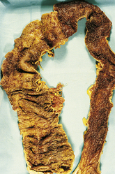
Figure 39-12 Acute ulcerative colitis. Colitis with extensive mucosal ulceration involving the entire colon. (From Damjanov I, Linder J, editors: Anderson’s pathology, ed 10, St. Louis, 1996, Mosby.)
CLINICAL MANIFESTATIONS The course of UC consists of intermittent periods of remission and exacerbation. Clinical manifestations vary with the severity and extent of disease. Loss of the absorptive mucosal surface and decreased colonic transit time can cause large volumes of watery diarrhea. Mucosal destruction causes bleeding, cramping pain, and an urge to defecate. Frequent diarrhea, with passage of small amounts of blood and purulent mucus, is common.111
Mild UC involves less mucosa and may be limited to proctitis, so that frequency of bowel movements, bleeding, and pain is minimal. Severe forms may involve the entire colon (pancolitis) and are characterized by fever; elevated pulse rate; frequent diarrhea (10 to 20 movements per day); urgency; obviously bloody stools; and continuous, crampy pain. Dehydration, weight loss, anemia, and fever result from fluid loss, bleeding, and inflammation. Complications include toxic megacolon, anal fissures, hemorrhoids, and perirectal abscess. Severe hemorrhage is rare, but chronic blood loss may precipitate hypotension and shock. Edema, strictures, or fibrosis can obstruct the colon. Perforation is an unusual but possible complication. The risk of left-sided colon cancer increases significantly after many years of ulcerative colitis and the presence of primary sclerosing cholangitis.112
Extraintestinal manifestations of UC and Crohn disease occur in 20% to 40% of cases and include cutaneous lesions (erythema nodosum and pyoderma gangrenosum), migratory polyarthritis and sacroiliitis, osteopenia and osteoporosis, mouth ulcers, episcleritis or anterior uveitis of the eye, and primary sclerosing colangitis in the liver.113 Gallstones are common. Alterations in coagulation can cause life-threatening microthrombi and deep vein thrombosis.114,115
EVALUATION AND TREATMENT Diagnosis of ulcerative colitis is based on the medical history, clinical manifestations, imaging procedures, and histologic criteria.116 Endoscopic evaluation shows an inflamed and hemorrhagic mucosa. Radiologic assessment may show loss of haustra, ulceration, and irregular mucosa. The laboratory data include low hemoglobin values, hypoalbuminemia, and low serum potassium levels. Infectious causes are ruled out by stool culture. The symptoms of ulcerative colitis can be very similar to those of Crohn disease, making differential diagnosis difficult.110
Treatment depends on the severity of symptoms and the extent of mucosal involvement. First line therapy is 5-aminosalicylic acid (mesalazine). Steroids and salicylates suppress the inflammatory response and help alleviate the cramping pain. Immunosuppressive agents (e.g., 6-mercaptopurine or azathioprine), cyclosporine, tacrolimus, and infliximab (a monoclonal anti-TNF-α antibody) are used for chronic active disease117. Broad-spectrum antibiotics or probiotics, or both, can modulate intestinal flora111 (see What’s New? Inflammatory Bowel Disease and Probiotics). For unknown reasons, nicotine may have a protective effect in ulcerative colitis but not in Crohn disease.118,119 Severe, unremitting disease can require hospital admission and administration of intravenous fluids. Extreme malnutrition may require intravenous hyperalimentation. Surgical resection of the colon or a colostomy may be performed if other forms of therapy are unsuccessful.120,121
Crohn Disease
Crohn disease (CD) (granulomatous colitis, ileocolitis, or regional enteritis) is an idiopathic inflammatory disorder that affects any part of the gastrointestinal tract from the mouth to the anus. The distal small intestine and proximal large colon are most commonly affected by the disease. In a small percentage of cases, CD is difficult to differentiate from ulcerative colitis (Table 39-6). Risk factors include family history, tobacco use, Jewish ethnicity, urban residency, and the CARD15/NOD2 (nucleotide-binding-oligomerization-domains) gene mutations (10% to 15% of cases).122 The CARD15/NOD2
WHAT’S NEW?
Inflammatory Bowel Disease and Probiotics
The etiology of inflammatory bowel disease (IBD) remains uncertain but the pathogenesis includes abnormal cell-mediated and humoral immune responses to commensal microflora in genetically susceptible individuals. There may be disturbances in mucosal permeability and the number and type of microflora. Probiotics are living microorganisms in food or dietary supplements that survive in stomach acid and bile and are safe for human ingestion. Their beneficial properties include altering the composition of bacterial flora, improvement of intestinal epithelial barrier function, and modulation of the mucosal immune system. The results of research using probiotics to treat IBD are controversial because there are few randomized controlled clinical trials. A multiagent mixture of probiotic strains appears to be more useful for treating pouchitis and ulcerative colitis than for Crohn disease and for maintaining remission in pediatric ulcerative colitis. There is a need for further research and controlled clinical trails to evaluate efficacy of probiotics for the remission of IBD.
Data from: Butterworth AD, Thomas AG, Akobeng AK: Cochrane Database Syst Rev (3):CD006634, 2008; Heilpern D, Szilagyi A: Rev Recent Clin Trials 3(3):167-184, 2008; Miele E et al: Am J Gastroenterol 104(2):437-443, 2009.
gene codes for a protein (a Toll-like receptor; see Chapter 9) involved in the recognition of gram-negative and gram-positive bacteria. Mutations in this gene are linked to the pathogenesis of CD, particularly ileal disease. Other candidate gene mutations are located on chromosomes 5 (IBD5), 6 (IBD3), and 10 (IBD10).123 The colony-stimulating factor IR gene, which is involved in monocyte to macrophage differentiation, also may be a susceptibility gene for CD.124 The pathogenesis of CD may be associated with an overly aggressive response to normal flora bacteria in genetically predisposed individuals.125,126 Th1-mediated inflammation with activation of leukocytes and cytokines (TNF-α, IFN-γ, and interleukins) causes injury. Recruited leukocytes release proinflammatory substances, including prostaglandins, leukotrienes, proteases, reactive oxygen species, and nitric oxide, which cause further injury and inflammation. Elevations in IgG are associated with severity of disease.127
PATHOPHYSIOLOGY The inflammatory process of CD begins in the intestinal submucosa and spreads across the intestinal wall to involve the mucosa and serosa in areas overlying lymphoid tissue. Progression of the disease involves neutrophil infiltration of the crypts resulting in abscess formation and crypt destruction. The most common site of the disease is the ileocolon, but both the large and small intestines may be involved. The inflammation can affect some haustral segments but not others, creating a pattern called skip lesions. One side of the intestinal wall may be affected but not the other.
The ulcerations of CD produce longitudinal and transverse fissures that extend inflammation into lymphoid tissue. The typical chronic lesion is a granuloma having cobblestone projections of inflamed tissue surrounded by areas of ulceration (Figure 39-13). (Granulomas are described in Chapter 6.) The lumen can narrow with inflammation, edema, and fibrotic strictures. Fistulae may form in the perianal area between loops of intestine or extend into the bladder.
Figure 39-13 Crohn disease. A, The mucosa in Crohn disease demonstrates a cobblestone pattern as a result of fissured ulcers (U) with intervening areas of edematous mucosa (M). B, Compared with normal small bowel wall (N), the Crohn segment (C) shows wall thickening that has caused a stenosis. (From Kumar V et al: Pathologic basis of disease, ed 7, Philadelphia, 2006, Saunders.)
CLINICAL MANIFESTATIONS Individuals with CD may have no specific symptoms other than an “irritable bowel” for several years. Symptoms vary and are associated with disease location. Abdominal pain and diarrhea are the most common signs (more than five stools per day), with passage of blood and mucus. Diarrhea can result from decreased colonic absorption, bypass fistulae, medications, bacterial overgrowth, and the presence of bile in the colon that inhibits water absorption.128 Other manifestations are related to the location and extent of intestinal involvement. Inflammation of the ileum, for example, causes tenderness in the lower right side of the abdomen. If the ileum is involved, the individual may be anemic as a result of malabsorption of vitamin B12. There also may be deficiencies in folic acid, vitamin D absorption, and calcium leading to bone disease. Proteins may be lost, leading to hypoalbuminemia. Weight loss is common. Anal manifestations occur in about 30% of cases, including anal fissure, perianal abscess, and fistula.129 Individuals with CD of long duration are also at risk for intestinal adenocarcinoma.130 Complications include obstruction, fistulae, abscess formation, and chronic blood loss. Extraintestinal manifestations are similar to those described for UC.
EVALUATION AND TREATMENT The diagnosis and treatment of CD are similar to the diagnosis and treatment of ulcerative colitis. Treatment with immunomodulatory agents can be effective. TNF-α–blocking agents are used for treatment of fistulas and to maintain remission.131 Surgery is generally performed to manage complications such as strictures, fistula, abscess, and perforation, or to relieve obstruction.132
When treatment involves surgical resection of small intestinal segments, complications related to short bowel syndrome can occur, including malabsorption, diarrhea and nutritional deficiencies. Symptoms are related to the extent and location of resection.
Diverticular Disease of the Colon
Diverticula are herniations or saclike outpouchings of mucosa through the muscle layers of the colon wall. Diverticulosis is asymptomatic diverticular disease. The cause is unknown but is associated with decreased dietary fiber and increased intracolonic pressure. Diverticulitis represents inflammation. Diverticular disease is most common in individuals older than 60 years of age, particularly those who live in developed countries where much of the diet consists of refined foods.133
PATHOPHYSIOLOGY Although diverticula can occur anywhere in the gastrointestinal tract, the most common site is the left colon.134 The diverticula form at weak points in the colon wall, usually where arteries penetrate the tunica muscularis to nourish the mucosal layer. Abnormal colonic motility with intraluminal hypertension also may be contributing factors. The colonic mucosa herniates through the smooth muscle layers (Figure 39-14). A common associated finding is thickening of the circular and longitudinal (teniae coli) muscles surrounding the diverticula. Hypertrophy and contraction of these muscles increase intraluminal pressure and degree of herniation. Habitual consumption of a low-residue diet reduces fecal bulk, thus reducing the diameter of the colon. According to Laplace’s law (see Chapter 29), wall pressure increases as the diameter of a cylindrical structure decreases. Therefore, pressure within the narrow lumen can increase enough to rupture the diverticula. Insoluble dietary fiber deficiency also may change the intestinal microflora, decreasing the immune response in the colon and permitting low-grade inflammation.135,136 Diverticulitis can cause abscess formation, fistula formation, peritonitis, or obstruction.137
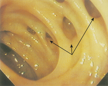
Figure 39-14 Diverticular disease. In diverticular disease, the outpouches (arrows) of mucosa seen in the sigmoid colon appear as slitlike openings from the mucosal surface of the opened bowel. (From Townsend C Sabiston textbook of surgery, ed 18, Philadelphia 2008, Saunders.)
CLINICAL MANIFESTATIONS Symptoms of diverticular disease are usually vague or absent. Cramping pain of the lower abdomen can accompany constriction of the hypertrophied colonic muscles. Diarrhea, constipation, distention, or flatulence may occur. Diverticula with an obstructed opening become inflamed or abscesses form, and the individual develops fever, leukocytosis (increased white blood cell count), and tenderness of the lower left quadrant. Right lower quadrant pain and severe complications, such as hemorrhage, perforation with peritonitis, bowel obstruction, and fistula formation, are rare.
EVALUATION AND TREATMENT Diverticula are often discovered during diagnostic procedures performed for other problems. Ultrasound, sigmoidoscopy, or barium enema is used for diagnosis of uncomplicated diverticula. Abdominal computed tomography (CT) is used for complicated cases.
An increase of dietary fiber intake increases stool weight, lowers colonic pressures, improves transit times, and often relieves symptoms (see Nutrition & Disease: Diverticular Disease and Diet). Probiotics combined with salicylates are effective in arresting infection. Diverticulitis with small abscesses is treated with antibiotics and reestablishing the microflora.134 Surgical resection may be required if there are severe complications.138
Appendicitis
Appendicitis is an inflammation of the vermiform appendix, which is a projection from the apex of the cecum. It is the most common surgical emergency of the abdomen and affects 7% to 12% of the population. The most common occurrence is between 20 and 30 years of age, although it may develop at any age.139
PATHOPHYSIOLOGY The exact cause of appendicitis is controversial. Obstruction of the lumen with stool, tumors, or foreign bodies with consequent, increased, intraluminal pressure, ischemia, bacterial infection, and inflammation is a common theory. The obstructed lumen does not allow drainage of the appendix, and as mucosal secretion continues, intraluminal pressure increases. The resultant increased pressure decreases mucosal blood flow, and the appendix becomes hypoxic. The mucosa ulcerates, promoting bacterial or other microbial invasion with further inflammation and edema. Inflammation may involve the distal or entire appendix. Gangrene develops from thrombosis of the luminal blood vessels, followed by perforation.140
CLINICAL MANIFESTATIONS Epigastric or periumbilical pain is the typical symptom of an inflamed appendix. The pain may be vague at first, increasing in intensity over 3 to 4 hours. It may subside and then recur with a shift of location to the right lower quadrant with rebound tenderness. Right lower quadrant pain is associated with extension of the inflammation to the surrounding tissues. Nausea, vomiting, and anorexia follow the onset of pain, and fever is common. Diarrhea occurs in some individuals, particularly children; others have a sensation of constipation. Perforation, peritonitis, and abscess formation are the most serious complications of appendicitis.
EVALUATION AND TREATMENT In addition to clinical manifestations, the clinician can usually locate the painful site with one finger. Rebound tenderness is usually referred to the right lower quadrant. The white blood cell count ranges from 10,000 to 16,000 cells/mm3, with increased neutrophils. C-reactive protein is elevated.141 Roentgenograms of the abdomen, CT scans, and ultrasound assist diagnostic accuracy. The combined information provides the best discriminating diagnosis.142
Antibiotics and appendectomy is the treatment for simple or perforated appendicitis. Laparoscopic surgery provides quick recovery for simple appendicitis. Recovery is more complicated in cases of perforation or abscess formation.143
Irritable Bowel Syndrome
Irritable bowel syndrome (IBS) is a functional gastrointestinal disorder characterized by abdominal pain and altered bowel habits that affects approximately 7 to 20 percent of individuals throughout the world and is more common in women, with a higher prevalence in youth and middle age. Individuals with IBS are more likely to have anxiety and depression. Subtypes of IBS are described on the basis of predominant symptoms—diarrhea, constipation, or pain. Symptoms of IBS can negatively affect quality of life and present a significant economic burden.
PATHOPHYSIOLOGY
There are no specific structural or biochemical alterations as a cause of IBS but there is increasing evidence to explain the varying symptom presentations and they are summarized below.144–146
Abnormal gastrointestinal motility and secretion: Individuals with diarrhea-type IBS have more rapid colonic transit times, whereas those with bloating and constipation have delayed transit times. The mechanism may be related to visceral hypersensitivity as well as dysregulation of the brain-gut axis or to the role of serotonin in the function of the enteric nervous system.
2. Visceral hypersensitivity or hyperalgesia particularly with distention of the rectum but also other areas of the gut: The mechanism may be related to a dysregulation of the “brain-gut-axis,” the role of serotonin in the enteric nervous system, infiltration and activation of mast cells and T lymphocytes, or alterations in autonomic or central nervous system processing of information in contributing to increased sensitivity to visceral pain.
3. Post infectious IBS: Intestinal infection(bacterial enteritis) has been associated with symptoms of IBS and may be related to ongoing low-grade inflammation and an abnormal immune response in gut tissues.
4. Overgrowth of intestinal flora: Overgrowth of normal gut bacteria may precipitate IBS symptoms and it is proposed that methane gas may slow intestinal transit time, resulting in constipation and bloating.
5. Food allergy or food intolerance: Food antigens may activate the mucosal immune system mediating hypersensitivity reactions and IBS symptoms. Food elimination approaches are helpful in some cases.
6. Psychosocial factors: Psychosocial factors including emotional stress influence brain-gut interaction including neuroendocrine, autonomic nervous system, and pain modulatory responses contributing to the symptoms of IBS.
CLINICAL MANIFESTATIONS
IBS is characterized by lower abdominal pain, diarrhea-predominant, constipation-predominant, or alternating diarrhea/constipation, gas, bloating, and nausea. Individuals may also describe fecal urgency and incomplete evacuation. Symptoms are usually relieved with defecation and usually to not interfere with sleep.
EVALUATION AND TREATMENT
The diagnosis of IBS is based on signs and symptoms and includes the exclusion of structural or biochemical causes of disease. In the absence of “alarm symptoms” such as fever, weight loss, gastrointestinal bleeding, anemia, or abdominal mass only limited diagnostic tests are needed. The individual may be evaluated for food allergies, lactose intolerance, parasites, or bacterial growth. The Rome III criteria for diagnosing IBS have been released to guide evaluation (Box 39-3).There is no cure for IBS, and treatment is individualized. Treatment of symptoms may include laxatives and fiber, antidiarrheals, antispasmodics, low-dose antidepressants, visceral analgesics, and serotonin agonists or antagonists. For more severe constipation, 5-hydroxytryptomine 4 agonist (e.g tegaserod) may be used (not approved for use in North America as of 2007) or CIC-2 chloride channel activators (e.g., lubiprostone). For more severe diarrhea 5-hydroxytryptomine 3 receptor antagonists (e.g., alosetron) may be used to normalize bowel habits. Alternative therapies including probiotics, hypnosis, and psychotherapy are treatment options. Research continues to advance the management of this complex syndrome.147–148
Vascular Insufficiency
The stomach and intestines are supplied by three branches of the abdominal aorta: the celiac axis and the superior and inferior mesenteric arteries. Because of the rich collateral circulation, at least two of the supplying vessels must be compromised to cause ischemia. Atherosclerotic lesions, thrombi, and emboli can develop in these vessels, occluding blood flow and causing ischemia or necrosis in the gastrointestinal tract.149
Mesenteric venous thrombosis is the least common of the causes of mesenteric vascular insufficiency. Malignancies, right-sided heart failure, and deep vein thrombosis are risk factors.
Acute occlusion of mesenteric artery blood flow (acute mesenteric ischemia) results in a significant reduction in mucosal blood flow from the arteries supplying the large and small intestine. Dissecting aortic aneurysms, thrombi, or emboli can be causes. Embolic obstruction is associated with atrial fibrillation, mitral valve disease, heart valve prostheses, or myocardial infarction. The superior mesenteric artery has a more direct line of flow from the aorta; therefore, emboli enter it more readily than the inferior branch, causing ischemia and necrosis of the small intestine.150 Ischemia and necrosis alter membrane permeability. There is initially increased motility, nausea and vomiting, urgent bowel evacuation, and severe abdominal pain. Ischemia leads to decreased motility and distention. The damaged intestinal mucosa cannot produce enough mucus to protect itself from digestive enzymes.151 Mucosal alteration causes fluid to move from the blood vessels into the bowel wall and peritoneum. Fluid loss causes hypovolemia and further decreases in intestinal blood flow. As intestinal infarction progresses, shock, fever, bloody diarrhea, and leukocytosis develop. Bacteria invade the necrotic intestinal wall, causing gangrene and peritonitis.
Chronic mesenteric insufficiency, or nonocculsive mesenteric insufficiency, can develop secondary to atherosclerosis, congestive heart failure, acute myocardial infarction, dysrhythmias, hemorrhage, stenosis, thrombus formation, aortic aneurysm, or any condition that decreases arterial blood flow. Older adults with arteriosclerosis are particularly susceptible. Chronic occlusion is often accompanied by formation of collateral circulation that may be able to nourish the resting intestine. After eating, however, when the intestine requires more blood, the arterial supply may be insufficient. Ischemia develops, causing a cramping abdominal pain, called abdominal angina, after meals. Progressive vascular obstruction eventually causes continuous abdominal pain and necrosis of the intestinal tissue. Reperfusion injury related to reactive oxygen metabolites and inflammatory mediators contributes to further tissue damage.
Colicky abdominal pain after eating is a cardinal symptom of chronic mesenteric insufficiency. Some individuals suffer significant weight loss because they stop eating to control the pain. Chronic segmental ischemia may lead to strictures and destruction.
Diagnosis of mesenteric artery occlusion is based on clinical manifestations, laboratory findings, mesenteric artery angiography, and abdominal radiography and ultrasonography. Bruit often can be heard over the occluded artery. With angiography a vasodilating agent may be injected into the vessels to improve the circulation. Heparin may be used if there are no contraindications. Medical management includes antibiotics, anticoagulation, vasodilators, and inhibitors of reperfusion injury. Surgery is required to remove necrotic tissue, repair sclerosed vessels, and for revascularization. Mortality is high (60% to 100%) for individuals with acute occlusion and compromised cardiac output. Early diagnosis and aggressive treatment result in the best survival rates.152
Disorders of Nutrition
Obesity is an increase in body fat mass and a metabolic disorder that has increased significantly over the past two decades. Obesity is an energy imbalance, with energy intake exceeding energy expenditure, and is defined as a body mass index (BMI) greater than 30.153 It is a major cause of morbidity, death, and high healthcare cost in the United States and worldwide.154 Three leading causes of death in the United States are associated with obesity: cardiovascular disease, type 2 diabetes mellitus, and cancer (colon, breast in postmenopausal women, endometrium, prostate, kidney, and esophagus.155 Obesity is also a risk factor for hypertension, stroke, hepatobiliary disease (gallstones and nonalcoholic steatohepatitis), osteoarthritis, and sleep apnea. Obesity is increasing in children and obese children tend to become obese adults.156
The causes and consequences of obesity are multiple and complex with rapidly advancing research regarding causal mechanisms and complications. Genotype and environmental-gene interactions are important predisposing factors.157 Single gene defects are rare and obesity is usually polygenic and associated with other phenotypes such as endocrine disorders (i.e., diabetes and hypothyroidism) and mental retardation (i.e., Down and Prader-Willi syndromes). Single gene defects include the melanocortin-receptor gene, leptin gene (also known as the obesity gene), and leptin-receptor-gene. All single-gene defects are directly or indirectly-related to leptin and melanocortin pathways.158 Metabolic abnormalities contributing to obesity include Cushing syndrome, Cushing disease, polycystic ovary syndrome, hypothyroidism, and hypothalamic injury. Environmental factors include culture, socioeconomic status, food intake, and exercise. Obesity is associated with adverse social and psychologic consequences.159
PATHOPHYSIOLOGY The pathophysiology of obesity is complex and involves the interaction of numerous cytokines, hormones, and neurotransmitters. Mechanisms contributing to the imbalance of energy intake in relation to energy expenditure and the multiple pathogenic effects of excess adipose tissue are not completely understood. Adipocytes secrete a number of hormones and cytokines known as adipokines (Box 39-4). These adipokines participate in regulation of food intake, lipid storage and metabolism, insulin sensitivity, the alternative complement system, vascular homeostasis, blood pressure regulation, angiogenesis, the inflammatory and immune responses, female reproduction, and regulation of energy metabolism.160 Visceral fat accumulation causes dysfunction of adipocytes and results in alterations in the regulation and interaction of these hormones and cytokines contributing to the causes and complications of obesity, particularly cardiovascular disease.161
Regulation of appetite and satiety occurs through neuroendocrine regulation of eating behavior, energy metabolism, and body fat mass. The system is complex and controlled by a dynamic circuit of signaling molecules from the periphery acting on central controls including the brain stem, hypothalamus, and autonomic nervous system. An imbalance in this system is usually associated with excessive caloric intake in relation to exercise with the consequence of weight gain and obesity.
The arcuate nucleus (ARC) in the hypothalamus has two sets of neurons with opposing effects that interact to regulate and balance food intake and energy metabolism. One set of neurons produces neuropeptide Y (NPY) and agouti-related protein (AGRP), which stimulates eating and decreases metabolism (anabolic). Another set of neurons synthesizes pro-opiomelanocortin (POMC)-producing peptide and cocaine-and-amphetamine-regulated transcript (CART), collectively known as POMC/CART neurons. They inhibit eating and increase metabolism (catabolic). Both sets of neurons express their effects by activating second-order neurons in the hypothalamus, which increases or decreases appetite and energy metabolism (Figure 39-15). Molecules that stimulate eating are called orexins (i.e., hypocretins [from the hypothalamus], a peptide family that acts as neurotransmitters for stimulating eating). Molecules that inhibit eating are called anorexins (Box 39-5). Peripheral effects of these signaling pathways are transmitted through the autonomic nervous and endocrine systems to regulate appetite, food intake, and energy metabolism.162
Many different hormones control appetite, satiety, and body weight. Their sources include ghrelin from the stomach; peptide YY, cholecystokinin (CCK), and glucagon-like peptide (GLP-1) from the intestines; insulin from pancreatic beta cells; and leptin, adiponectin, and resistin from adipose tissue. These hormones circulate in the blood at concentrations proportional to body fat mass, and serve as peripheral signals to the ARC in the hypothalamus, where appetite (food intake) and metabolism (energy expenditure) are regulated. Leptin and insulin normally decrease appetite by inhibiting NPY/AGRP neurons (anabolic circuits) and stimulating POMC/CART neurons (catabolic circuits) (Figure 39-15). Ghrelin, CCK, and other hormones stimulate appetite by activating NPY/AGRP-expressing neurons. Peptide YY (PYY) and other hormones inhibit these neurons and decrease appetite. Other peripheral hormones and neurotransmitters also can influence the hypothalamus and affect appetite and energy expenditure (see Box 39-4).
Figure 39-15 Neuroendocrine control of food intake and energy expenditure. Leptin and insulin normally decrease appetite, increase satiety, and increase energy expenditure (catabolism). Leptin/insulin inhibits NPY/AGRP gene expression resulting in decreased appetite and food intake; and stimulates POMC/CART gene expression with resulting α-MSH release and decrease in appetite and food intake. With leptin resistance as occurs in obesity, these effects are depressed and food intake increases in excess of energy expenditure. AGRP, agouti-related peptide; α-MSH, alpha-melanocyte stimulating hormone; NPY, neuropeptide Y; POMC/CART, proopiomelanocortin/cocaine-and-amphetamine-related transcript. (Reprinted withpermission from Schwartz MW et al, Central nervous system control of foodintake, Nature 404:661-671, 2000.)
Obesity is associated with increased circulating plasma levels of leptin, insulin, ghrelin, and PYY. Interaction among these hormones at the level of the hypothalamus may be an important determinant of excessive fat mass. Leptin receptors in the hypothalamus function to regulate satiety and body weight within a fairly narrow range or set point. Low leptin levels during fasting normally stimulate food intake and reduce energy expenditure and high leptin levels in the fed state inhibit food intake and increase energy expenditure. Leptin secretion increases as adipocytes increase in size or number (hyperleptinemia). The high levels of leptin in obesity are ineffective at decreasing appetite and increasing energy expenditure—this is known as leptin resistance. Leptin resistance disrupts hypothalamic satiety signaling and promotes overeating and excessive weight gain.163
The cause of leptin resistance is unknown. It may be related to a defect in leptin transport,164 an inability of leptin to cross the blood-brain barrier165; an alteration in the permissive effect of leptin on urocortin (a satiety-signaling molecule)166; or a defect in the leptin receptor.167 Hyperleptinemia also stimulates the sympathetic nervous system, chronic inflammation, oxidative stress, and ventricular hypertrophy and may contribute to the pathogenesis of hypertension, atherosclerosis, and cardiovascular disease associated with obesity.168,169
Ghrelin is produced by the stomach in response to hunger and stimulates food intake and induces metabolic changes leading to an increase in body weight and body fat mass. Ghrelin also stimulates release of growth hormone (GH) from anterior pituitary cells, the release of gastric acid and gastric motility, and affects pancreatic functions. It has vasodilatory, cardioprotective, and antiproliferative effects.170,171 Leptin and ghrelin are complementary, yet antagonistic signals reflecting acute and chronic changes in energy balance the effects of which are mediated by hypothalamic neuropeptides, such as NPY and AGRP. Plasma ghrelin is decreased in obesity, and its role in contributing to obesity is yet to be defined. Leptin may regulate ghrelin levels.172 Endocrine and vagal afferent pathways are also involved in the actions of ghrelin and leptin, adding to the complexity of mechanisms that can affect obesity.173
Adiponectin has insulin-sensitizing properties and plasma levels decrease with visceral obesity, contributing to insulin resistance, cardiovascular disease, and metabolic syndrome.174 Obese individuals, particularly those with expansion of visceral adipose tissue, are at increased risk for coronary artery disease resulting from hyperlipidemia, hypertension, and factors that promote thrombosis and inflammation (see Chapter 30). Decreased adiponectin levels are associated with increased levels of inflammatory markers, such as IL-6 and TNF-α. Adiponectin may serve as an anti-inflammatory and anti-atherogenic plasma protein and may have an important role in vascular remodeling that is limited with obesity.175
Obesity is associated with insulin resistance, which predisposes an individual to type 2 diabetes mellitus (see Chapter 21). The insulin resistance may be related to an insulin receptor defect or to postreceptor effects with alteration in glucose transporter functions. Excess insulin also may be a response to excessive caloric intake.176 Resistin is greatly increased in those with obesity and may be an antagonist to insulin action and a mediator of inflammation.177
CLINICAL MANIFESTATIONS Increased visceral fat is associated with metabolic syndrome (hypertriglyceridemia, reduced high-density lipoprotein, increased low-density lipoproteins, hypertension, and insulin resistance), a complex of traits that increase risk for ischemic heart disease and type 2 diabetes mellitus178,179 (see Chapters 21 and 30).
The hypertension of obesity is complex and may be related to insulin resistance, activation of the sympathetic nervous system, activation of renin-angiotensin, leptin resistance, physical compression of the kidneys, and alterations in vascular structure and function.180,181
Pulmonary function can be compromised by a large amount of adipose tissue overlying the chest cage. Work of breathing increases, and gas exchange, vital capacity, and expiratory volume all decrease causing low oxygen tension and high carbon dioxide tension. The hypoventilation causes daytime hypercapnia and sleep-disordered breathing. Hypoxemia causes pulmonary hypertension contributing to right ventricular hypertrophy and heart failure.182 Asthma is also associated with obesity, particularly among females, but the exact mechanisms of airway hyperresponsiveness are unknown. Obesity-related factors include airway narrowing, low-grade inflammation, gastroesophageal reflux, leptin resistance, and decreased adiponectin.183
Obstructive sleep apnea syndrome (OSAS) is a consequence of obesity and obesity hypoventilation syndrome. It involves episodic partial or complete obstruction of the upper airway, hypoventilation, and hypercapnia (see Chapters 15, 33, and 34). The hypoventilation also may be caused by leptin resistance, which has respiratory stimulant effects independent of the amount of visceral fat mass.184 OSAS results in fragmentation of sleep with daytime sleepiness and further complications of hypoxia and heart disease.
The development of osteoarthritis occurs as a function of mechanical stress and limb malalignment on weight-bearing joints. Exercise intolerance and pain in the weight-bearing joints, particularly the hips and knees, are common. Inflammation may cause erosion of cartilage.185
Obesity also increases the risk for cancer including adenocarcinoma of the esophagus and the gastric cardia, colorectal cancer, postmenopausal breast cancer, endometrial cancer, and renal-cell carcinoma. Less commonly associated cancers include thyroid, gallbladder, pancreatic, leukemia, multiple myeloma, and non-Hodgkin lymphoma. The pathophysiologic mechanisms that increase cancer risk are not clear, but insulin resistance and other factors, including insulin-like growth factors, sex steroids, adipokines, obesity-related inflammatory markers, the nuclear factor kappa beta system, and oxidative stresses, are being evaluated.186,187
EVALUATION AND TREATMENT There are several methods for measuring or estimating body fat mass, including CT and magnet resonance imaging (MRI) techniques; bioimpedance analysis; underwater weighing; and anthropometric measurements, such as skinfold thickness, circumferences, and various body diameters (i.e., waist-to-hip ratios and waist circumference, and BM-kg/m2 tables).188 The BMI and waist-to-hip ratios are most commonly used because they are the easiest to measure. Overweight is defined as a BMI greater than 25 and obesity is a BMI greater than 30. BMI charts are available for children ages 2 to 20 years; these can be used for comparison during adulthood because obese children generally become obese adults.153,189,190 No specific diagnostic criteria for obesity have been established.
Obesity is a chronic disease for which various treatment approaches have been used, including correction of metabolic abnormalities, individually tailored weight-reduction diets, and exercise programs.191–196 A combination of weight reduction and exercise is the most effective.197 Self-motivation and support systems are critical aspects of treatment. Additional treatments, such as psychotherapy, behavioral modification, medications, bariatric surgery (i.e., the Roux-en-Y gastric bypass or gastric banding), and liposuction are also prescribed and when successful result in a significant reduction in comorbidities and a decrease in insulin resistance.198–201 Unraveling the causes of obesity will lead to more specific prevention and pharmacotherapies.
Anorexia Nervosa and Bulimia Nervosa
Anorexia nervosa and bulimia nervosa are characterized by aberrant eating behavior and weight regulation, disturbed attitudes toward body weight and shape, and resistance to treatment.202 Many young adults and adolescents—as many as 1% of women and adolescent girls in the United States—are affected by these two complex and related eating disorders. They occur rarely in black women, and only 5% to 10% of cases are men.203 Risk factors include genetic, familial, biologic, psychologic, and social factors.204 There is an association between sexual assault history and eating disorders.205 An increasing number of children, young men, and older adult women are experiencing eating disorders, often with an associated depression, anxiety, or personality disorder.206
Alterations in hypothalamic and gut-related neuropeptides that effect eating behavior and neuroendocrine disturbances are associated with weight loss and malnutrition.207 Understanding these alterations may contribute to an explanation of the difficulty of reversing behaviors associated with these diseases.208
Anorexia nervosa (AN) is an obsessive-compulsive psychiatric disorder and physiologic syndrome that affects 1% to 3% of U.S. women. It has the highest mortality rate of any psychiatric disease and has a familial tendency.209 AN is characterized by the following210,211:
1. A fear of becoming obese despite progressive weight loss
2. A distorted body image: the perception that the body is fat when it is actually underweight
3. Body weight 15% less than normal for age and height because of refusal to eat
4. In women and girls, absence of three consecutive menstrual periods
Persons with AN frequently deny they have any eating problem. Two types of eating behavior are distinguished in AN and both involve subnormal body weight and ongoing malnutrition. Restrictive AN entails unremitting food avoidance. Binge eating/purging AN includes binge eating followed by self-induced vomiting or laxative abuse. AN usually emerges during adolescence and in females who have a history of childhood abuse.212
As the disease progresses, multiple organs are affected.213 Muscle and fat depletion gives the individual a skeleton-like appearance and increases the risk for osteopenia and osteoporosis. Iron deficiency anemia promotes fatigue, and low white blood cell count increases risk of infection. Reproductive functioning is affected, including ovarian function, menstruation, fertility, and pregnancy. Sodium, potassium, phosphate, and magnesium are depleted. Postural hypotension, edema, bradycardia, hypothermia, constipation, and sleep disturbances may ensue. The loss of 25% to 30% of ideal body weight can eventually lead to death caused by starvation-induced cardiac failure.214 Diagnosis of AN involves a thorough medical history, physical and psychologic examination, and ruling out other causes of anorexia and malnutrition.215
There are no universally accepted treatments. Treatment objectives for AN include reversing the compromised physical state, promoting insights and knowledge about the disorder, mutual goals, interaction with family members, restoring development growth, and modifying food habits.216–218 Correction of nutritional status may require intensive treatment, including total parenteral nutrition. When the individual demonstrates the willingness to eat food for nourishment, dietary protein, carbohydrate, and fat are introduced in tolerable amounts. Care must be taken to prevent refeeding syndrome, which can result in fluid and electrolyte, cardiac, neurologic, and hematologic complications during nutritional rehabilitation.219 Behavioral intervention begins as soon as physical symptoms are stabilized and may continue for several years. Family therapy is helpful for adolescents. Formal genetic studies are in progress and results may advance specificity of treatment.209
Bulimia nervosa is more common than anorexia, and body weight remains near normal but with aspirations for weight loss. The group at risk is the same as that for AN except that bulimia tends to occur in slightly older, less affluent women. Diagnosis of bulimia is based on the following findings220:
1. Recurrent episodes of binge eating during which the individual fears not being able to stop
2. Self-induced vomiting, use of laxatives (purging type)
3. Two binge-eating episodes per week for at least 3 months (purging type)
4. Fasting to oppose the effect of binge eating or excessive exercise (nonpurging type)
Because of negative connotations associated with self-stimulated vomiting and purging, individuals who have bulimia binge and purge secretly. Bulimic individuals may binge and purge as often as 20 times each day. Weight will fluctuate by about 10 pounds. Continual vomiting of acidic chyme can cause pitted teeth, pharyngeal and esophageal inflammation, and tracheoesophageal fistulae. Overuse of laxatives can cause rectal bleeding. Secret binge eating isolates the bulimic individual and leads to depression and anger that is turned inward. A vicious cycle of depression, overeating to try to feel better, vomiting and purging to maintain a normal weight, and returning depression perpetuates this eating disorder. Mood symptoms are often worse in the winter.221
Because persons with bulimia are usually older than individuals with AN and usually have separated from a family core, individual or group cognitive behavior change is the treatment focus. Fluoxetine may be beneficial.222 Individuals with bulimia rarely have physical problems requiring hospital care and respond to treatment more readily than those with AN.223
Starvation
Starvation is a reduction in energy intake leading to weight loss. Short-term and long-term starvation have different effects. Therapeutic short-term starvation is part of many weight-reduction programs because it causes an initial rapid weight loss that reinforces the individual’s motivation to diet. Therapeutic long-term starvation is used in medically controlled environments to facilitate rapid weight loss in morbidly obese individuals. Pathologic long-term starvation can be caused by poverty; chronic diseases of the cardiovascular, pulmonary, hepatic, and digestive systems; malabsorption syndromes; human immunodeficiency virus (HIV) infection; and cancer. In-hospital starvation primarily affects individuals with functional and cognitive deficits and inadequate caloric intake.224
Short-term starvation, or extended fasting, consists of several days of total dietary abstinence or deprivation. The body responds with protective mechanisms.225 For 4 to 6 hours after the last meal, the body is in a well-fed state and its energy requirements are supplied by glucose from recently ingested carbohydrates. Once all available energy has been absorbed from the intestine, glycogen in the liver is converted to glucose through glycogenolysis, the splitting of glycogen into glucose. This process peaks within 4 to 8 hours, and gluconeogenesis begins. Gluconeogenesis is the formation of glucose from noncarbohydrate molecules: lactate, pyruvate, amino acids, and the glycerol portion of fats. Like glycogenolysis, gluconeogenesis takes place within the liver. Both of these processes deplete stored nutrients and thus cannot meet the body’s energy needs indefinitely. Proteins continue to be catabolized to a minimal degree, providing carbon for the synthesis of glucose.225
Long-term starvation begins after several days of dietary abstinence and eventually causes death. Absolute deprivation of food causes marasmus or protein energy malnutrition. Protein deprivation in the presence of carbohydrate intake is called kwashiorkor. Marasmic kwashiorkor (edematous, severe childhood malnutrition) is a combination of chronic energy deficiency and chronic or acute protein deficiency.226 The major characteristic of long-term starvation is a decreased dependence on gluconeogenesis and an increased use of ketone bodies (products of lipid and pyruvate metabolism) as a cellular energy source. During long-term starvation, depressed insulin levels and increased glucagon, cortisone, epinephrine, and growth hormones promote lipolysis in adipose tissue. Lipolysis liberates fatty acids, which supply energy to cardiac and skeletal muscle cells, and ketone bodies, which sustain brain tissue. Fatty acid, or ketone body, oxidation meets most of the energy needs of the cells. (Some glucose is still needed as fuel for brain tissue.) Once the supply of adipose tissue is depleted, proteolysis begins. The breakdown of muscle and visceral protein is the last process the body engages to supply energy for life. Death results from severe alterations in electrolyte balance and loss of renal, pulmonary, and cardiac function.227
Adequate ingestion of appropriate nutrients is the obvious treatment for starvation. In medically induced starvation the body is maintained in a ketotic state until the desired amount of adipose tissue has been lysed. Starvation imposed by chronic disease, long-term illness, or malabsorption is treated with enteral or parenteral nutrition. Perioperative management of nutrition is necessary to prevent unnecessary starvation.228