Tubular and Interstitial Diseases

Most forms of tubular injury involve the interstitium as well; therefore, diseases affecting these two components are discussed together. Under this heading we consider two major groups of processes: (1) ischemic or toxic tubular injury, leading to acute kidney injury (AKI) and acute renal failure, and (2) inflammatory reactions of the tubules and interstitium (tubulointerstitial nephritis).

ACUTE KIDNEY INJURY (AKI) (ACUTE TUBULAR NECROSIS, ATN)

AKI, a term increasingly favored over the often synonymously used terms acute tubular necrosis (ATN) and acute tubular injury, is a clinicopathologic entity characterized clinically by acute diminution of renal function and often, but not invariably, morphologic evidence of tubular injury. It is the most common cause of acute renal failure,58,59 which signifies rapid reduction of renal function and urine flow, falling within 24 hours to less than 400 mL per day. It can be caused by a variety of conditions, including

Ischemia, due to decreased or interrupted blood flow, examples of which include diffuse involvement of the intrarenal blood vessels such as in microscopic polyangiitis, malignant hypertension, microangiopathies and systemic conditions associated with thrombosis (e.g., hemolytic uremic syndrome [HUS], thrombotic thrombocytopenic pupura [TTP], and disseminated intravascular coagulation [DIC]), or decreased effective circulating blood volume
Direct toxic injury to the tubules (e.g., by drugs, radiocontrast dyes, myoglobin, hemoglobin, radiation)
Acute tubulointerstitial nephritis, most commonly occurring as a hypersensitivity reaction to drugs
Urinary obstruction by tumors, prostatic hypertrophy, or blood clots (so-called postrenal acute renal failure)

AKI accounts for some 50% of cases of acute renal failure in hospitalized patients. Other causes of acute renal failure are discussed elsewhere in this chapter.

AKI is a reversible renal lesion that arises in a variety of clinical settings. Most of these, ranging from severe trauma to acute pancreatitis, have in common a period of inadequate blood flow to the peripheral organs, usually accompanied by marked hypotension and shock. This pattern of AKI is called ischemic AKI. The second pattern, called nephrotoxic AKI, is caused by a multitude of drugs, such as gentamicin and other antibiotics; radiographic contrast agents; poisons, including heavy metals (e.g., mercury); and organic solvents (e.g., carbon tetrachloride). Combinations of ischemic and nephrotoxic AKI also can occur, exemplified by mismatched blood transfusions and other hemolytic crises causing hemoglobinuria and skeletal muscle injuries causing myoglobinuria. Such injuries result in characteristic intratubular hemoglobin or myoglobin casts, respectively; the toxic iron content of these globin molecules contributes to the AKI. In addition to its frequency, the potential reversibility of AKI adds to its clinical importance. Proper management means the difference between full recovery and death.

Pathogenesis.

The critical events in both ischemic and nephrotoxic AKI are believed to be (1) tubular injury and (2) persistent and severe disturbances in blood flow60 (Fig. 20-23).

Tubule cell injury: Tubular epithelial cells are particularly sensitive to ischemia and are also vulnerable to toxins. Several factors predispose the tubules to toxic injury, including a vast charged surface for tubular reabsorption, active transport systems for ions and organic acids, a high metabolic rate and oxygen consumption requirement so as to perform these transport and reabsorption functions, and the capability for effective concentration.
image

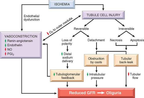
FIGURE 20-23 Postulated sequence in acute kidney injury. GFR, glomerular filtration rate; NO, nitric oxide; PGI2, prostaglandin I2 (prostacyclin).

(Modified from Brady HR et al.: Acute renal failure. In Brenner BM [ed]: Brenner and Rector’s The Kidney, 5th ed, Vol II. Philadelphia, WB Saunders, 1996, p 1210).

Ischemia causes numerous structural and functional alterations in epithelial cells, as discussed in Chapter 1. The structural changes include those of reversible injury (such as cellular swelling, loss of brush border and polarity, blebbing, and cell detachment) and those associated with lethal injury (necrosis and apoptosis). Biochemically there is depletion of ATP; accumulation of intracellular calcium; activation of proteases (e.g., calpain), which cause cytoskeletal disruption; activation of phospholipases, which damage membranes; generation of reactive oxygen species; and activation of caspases, which induce apoptotic cell death. One early reversible result of ischemia is loss of cell polarity due to redistribution of membrane proteins (e.g., the enzyme Na+, K+-ATPase) from the basolateral to the luminal surface of the tubular cells, resulting in abnormal ion transport across the cells, and increased sodium delivery to distal tubules. The latter incites vasoconstriction via tubuloglomerular feedback, which will be discussed below. In addition, ischemic tubular cells express cytokines (such as monocyte chemoattractant protein 1) and adhesion molecules (such as intercellular adhesion molecule 1), thus recruiting leukocytes that appear to participate in the subsequent injury. In time, injured cells detach from the basement membranes and cause luminal obstruction, increased intratubular pressure, and decreased GFR. In addition, fluid from the damaged tubules leaks into the interstitium, resulting in interstitial edema, increased interstitial pressure, and further damage to the tubule. All these effects, as shown in Figure 20-23, contribute to the decreased GFR.

Disturbances in blood flow: Ischemic renal injury is also characterized by hemodynamic alterations that cause reduced GFR. The major one is intrarenal vasoconstriction, which results in both reduced glomerular blood flow and reduced oxygen delivery to the functionally important tubules in the outer medulla (thick ascending limb and straight segment of the proximal tubule). Several vasoconstrictor pathways have been implicated, including the renin-angiotensin system, stimulated by increased distal sodium delivery (via tubuloglomerular feedback), and sublethal endothelial injury, leading to increased release of the vasoconstrictor endothelin and decreased production of the vasodilators nitric oxide and prostacyclin (prostaglandin I2). There is also some evidence of a direct effect of ischemia or toxins on the glomerulus, causing a reduced glomerular ultrafiltration coefficient, possibly due to mesangial contraction.

The patchiness of tubular necrosis and maintenance of the integrity of the basement membrane along many segments allow ready repair of the necrotic foci and recovery of function if the precipitating cause is removed. This repair is dependent on the capacity of reversibly injured epithelial cells to proliferate and differentiate. Re-epithelialization is mediated by a variety of growth factors and cytokines produced locally by the tubular cells themselves (autocrine stimulation) or by inflammatory cells in the vicinity of necrotic foci (paracrine stimulation).61 Of these, epidermal growth factor, TGF-α, insulin-like growth factor type 1, and hepatocyte growth factor have been shown to be particularly important in renal tubular repair. Growth factors, indeed, are being explored as possible therapeutic agents to enhance re-epithelialization in AKI, although clinical trials to date have been disappointing.61

Morphology. Ischemic AKI is characterized by focal tubular epithelial necrosis at multiple points along the nephron, with large skip areas in between, often accompanied by rupture of basement membranes (tubulorrhexis) and occlusion of tubular lumens by casts62 (Figs. 20-24 and 20-25). The straight portion of the proximal tubule and the ascending thick limb in the renal medulla are especially vulnerable, but focal lesions may also occur in the distal tubule, often in conjunction with casts. Paradoxically, the clinical syndrome of AKI is often associated with lesser degrees of tubular injury. This includes attenuation or loss of proximal tubule brush borders, simplification of cell structure, cell swelling and vacuolization, and sloughing of non-necrotic tubular cells into the tubular lumina (see Fig. 20-25). The severity of the morphologic findings often does not correlate well with the severity of the clinical manifestations.

image

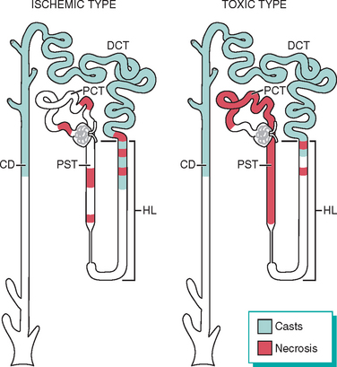
FIGURE 20-24 Patterns of tubular damage in ischemic and toxic acute kidney injury. In the ischemic type, tubular necrosis is patchy, relatively short lengths of tubules are affected, and straight segments of proximal tubules (PST) and ascending limbs of Henle’s loop (HL) are most vulnerable. In toxic acute kidney injury, extensive necrosis is present along the proximal convoluted tubule segments (PCT) with many toxins (e.g., mercury), but necrosis of the distal tubule, particularly ascending HL, also occurs. In both types, lumens of the distal convoluted tubules (DCT) and collecting ducts (CD) contain casts.

image

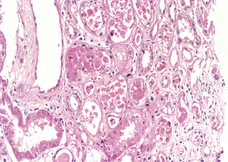
FIGURE 20-25 Acute kidney injury. Some of the tubular epithelial cells in the tubules are necrotic, and many have become detached (from their basement membranes) and been sloughed into the tubular lumens, whereas others are swollen, vacuolated, and regenerating.

(Courtesy of Dr. Agnes Fogo, Vanderbilt University, Nashville, TN.)

Eosinophilic hyaline casts, as well as pigmented granular casts, are common, particularly in distal tubules and collecting ducts. These casts consist principally of Tamm-Horsfall protein (a urinary glycoprotein normally secreted by the cells of ascending thick limb and distal tubules) in conjunction with other plasma proteins. Other findings in ischemic AKI are interstitial edema and accumulations of leukocytes within dilated vasa recta. There is also evidence of epithelial regeneration: flattened epithelial cells with hyperchromatic nuclei and mitotic figures are often present. In the course of time this regeneration repopulates the tubules so that, no residual evidence of damage is seen.

Toxic AKI is manifested by acute tubular injury, most obvious in the proximal convoluted tubules. On histologic examination the tubular necrosis may be entirely nonspecific, but it is somewhat distinctive in poisoning with certain agents. With mercuric chloride, for example, severely injured cells may contain large acidophilic inclusions. Later, these cells become totally necrotic, are desquamated into the lumen, and may undergo calcification. Carbon tetrachloride poisoning, in contrast, is characterized by the accumulation of neutral lipids in injured cells; again, such fatty change is followed by necrosis. Ethylene glycol produces marked ballooning and hydropic or vacuolar degeneration of proximal convoluted tubules. Calcium oxalate crystals are often found in the tubular lumens in such poisoning.

Clinical Course.

The clinical course of AKI is highly variable, but the classic case may be divided into initiation, maintenance, and recovery stages. The initiation phase, lasting for about 36 hours, is dominated by the inciting medical, surgical, or obstetric event in the ischemic form of AKI. The only indication of renal involvement is a slight decline in urine output with a rise in BUN. At this point, oliguria could be explained on the basis of a transient decrease in blood flow and declining GFR.

The maintenance phase is characterized by sustained decreases in urine output to between 40 and 400 mL/day (oliguria), salt and water overload, rising BUN concentrations, hyperkalemia, metabolic acidosis, and other manifestations of uremia. With appropriate attention to the balance of water and blood electrolytes, including dialysis, the patient can be supported through this oliguric crisis.

The recovery phase is ushered in by a steady increase in urine volume that may reach up to 3 L/day. The tubules are still damaged, so large amounts of water, sodium, and potassium are lost in the flood of urine. Hypokalemia, rather than hyperkalemia, becomes a clinical problem. There is a peculiar increased vulnerability to infection at this stage. Eventually, renal tubular function is restored and concentrating abilityimproves. At the same time, BUN and creatinine levels begin to return to normal. Subtle tubular functional impairment may persist for months, but most patients who reach this phase eventually recover completely.

The prognosis of AKI depends on the clinical setting. Recovery is expected with nephrotoxic AKI when the toxin has not caused serious damage to other organs, such as the liver or heart. With current supportive care, 95% of those who do not succumb to the precipitating cause recover. Conversely, in shock related to sepsis, extensive burns, or other causes of multi-organ failure, the mortality rate can rise to more than 50%.

Up to 50% of patients with AKI do not have oliguria and instead often have increased urine volumes. This so-called nonoliguric AKI occurs particularly often with nephrotoxins, and it generally tends to follow a more benign clinical course.

TUBULOINTERSTITIAL NEPHRITIS

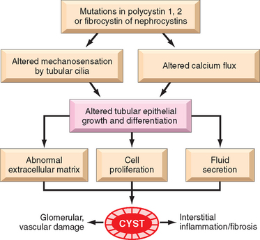
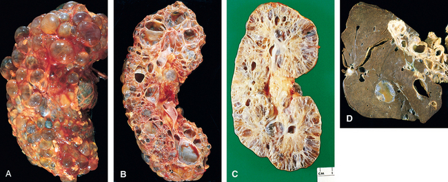
This group of renal diseases is characterized by histologic and functional alterations that involve predominantly the tubules and interstitium. We have previously seen that chronic tubulointerstitial injury may occur in diseases that primarily affect the glomerulus (see Fig. 20-22) and that such injury may be an important cause of progression in these diseases.18 Secondary tubulointerstitial nephritis is also present in a variety of vascular, cystic (polycystic kidney disease), and metabolic (diabetes) renal disorders, in which it may also contribute to progressive damage. Here we discuss disorders in which tubulointerstitial injury seems to be a primary event. These disorders have diverse causes and different pathogenetic mechanisms (Table 20-8). Glomerular and vascular abnormalities may also be present but either are mild or occur only in advanced stages of these diseases.

TABLE 20-8 Causes of Tubulointerstitial Nephritis

INFECTIONS
Acute bacterial pyelonephritis
Chronic pyelonephritis (including reflux nephropathy)
Other infections (e.g., viruses, parasites)
TOXINS
Drugs
Acute-hypersensitivity interstitial nephritis
Analgesics
Heavy metals
Lead, cadmium
METABOLIC DISEASES
Urate nephropathy
Nephrocalcinosis (hypercalcemic nephropathy)
Acute phosphate nephropathy
Hypokalemic nephropathy
Oxalate nephropathy
PHYSICAL FACTORS
Chronic urinary tract obstruction
NEOPLASMS
Multiple myeloma (light-chain cast nephropathy)
IMMUNOLOGIC REACTIONS
Transplant rejection
Sjögren syndrome
Sarcoidosis
VASCULAR DISEASES
MISCELLANEOUS
Balkan nephropathy
Nephronophthisis–medullary cystic disease complex
“Idiopathic” interstitial nephritis

Tubulointerstitial nephritis can be acute or chronic. Acute tubulointerstitial nephritis has a rapid clinical onset and is characterized histologically by interstitial edema, often accompanied by leukocytic infiltration of the interstitium and tubules, and focal tubular necrosis. In chronic interstitial nephritis there is infiltration with predominantly mononuclear leukocytes, prominent interstitial fibrosis, and widespread tubular atrophy. Morphologic features that are helpful in separating acute from chronic tubulointerstitial nephritis include edema and, when present, eosinophils and neutrophils in the acute form, while fibrosis and tubular atrophy characterize the chronic form.

These conditions are distinguished clinically from the glomerular diseases by the absence, in early stages, of such hallmarks of glomerular injury as nephritic or nephrotic syndrome and by the presence of defects in tubular function. The latter may be subtle and include impaired ability to concentrate urine, evidenced clinically by polyuria or nocturia; salt wasting; diminished ability to excrete acids (metabolic acidosis); and isolated defects in tubular reabsorption or secretion. The advanced forms, however, may be difficult to distinguish clinically from other causes of renal insufficiency.

Some of the specific conditions listed in Table 20-8 are discussed elsewhere in this book. In this section we deal principally with pyelonephritis and interstitial diseases induced by drugs.

Pyelonephritis and Urinary Tract Infection

Pyelonephritis is a renal disorder affecting the tubules, interstitium, and renal pelvis and is one of the most common diseases of the kidney. It occurs in two forms. Acute pyelonephritis is caused by bacterial infection and is the renal lesion associated with urinary tract infection. Chronic pyelonephritis is a more complex disorder; bacterial infection plays a dominant role, but other factors (vesicoureteral reflux, obstruction) are involved in its pathogenesis. Pyelonephritis is a serious complication of urinary tract infections that affect the bladder (cystitis), the kidneys and their collecting systems (pyelonephritis), or both. Bacterial infection of the lower urinary tract may be completely asymptomatic (asymptomatic bacteriuria) and most often remains localized to the bladder without the development of renal infection. However, lower urinary tract infection always carries the potential of spread to the kidney.

Etiology and Pathogenesis.

The dominant etiologic agents, accounting for more than 85% of cases of urinary tract infection, are the gram-negative bacilli that are normal inhabitants of the intestinal tract.63 By far the most common is Escherichia coli, followed by Proteus, Klebsiella, and Enterobacter. Streptococcus faecalis, also of enteric origin, staphylococci, and virtually every other bacterial and fungal agent can also cause lower urinary tract and renal infection. In immunocompromised persons, particularly those with transplanted organs, viruses such as Polyomavirus, cytomegalovirus, and adenovirus can also be a cause of renal infection.

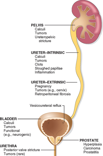
In most patients with urinary tract infection, the infecting organisms are derived from the patient’s own fecal flora. This is thus a form of endogenous infection. There are two routes by which bacteria can reach the kidneys: (1) through the bloodstream (hematogenous infection) and (2) from the lower urinary tract (ascending infection) (Fig. 20-26). The hematogenous route is the less common of the two and results from seeding of the kidneys by bacteria from distant foci in the course of septicemia or infective endocarditis. Hematogenous infection is more likely to occur in the presence of ureteral obstruction, in debilitated patients, in patients receiving immunosuppressive therapy, and with nonenteric organisms, such as staphylococci and certain fungi and viruses.

image

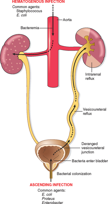
FIGURE 20-26 Schematic representation of pathways of renal infection. Hematogenous infection results from bacteremic spread. More common is ascending infection, which results from a combination of urinary bladder infection, vesicoureteral reflux, and intrarenal reflux.

Ascending infection is the most common cause of clinical pyelonephritis. Normal human bladder and bladder urine are sterile; therefore, a number of steps must occur for renal infection to occur:

The first step in ascending infection seems to be the colonization of the distal urethra and introitus (in the female) by coliform bacteria. This colonization is influenced by the ability of bacteria to adhere to urethral mucosal epithelial cells. Such bacterial adherence, as discussed in Chapter 8, involves adhesive molecules (adhesins) on the P-fimbriae (pili) of bacteria that interact with receptors on the surface of uroepithelial cells. Specific adhesins (e.g., that encoded by the pyelonephritis-associated pili [pap] gene64) are associated with infection. In addition, certain types of fimbriae promote renal tropism, persistence of infection, or an enhanced inflammatory response.64
From the urethra to the bladder, organisms gain entrance during urethral catheterization or other instrumentation. Long-term catheterization, in particular, carries a risk of infection. In the absence of instrumentation, urinary infections are much more common in females, and this has been ascribed to the shorter urethra in females, as well as the absence of antibacterial properties such as are found in prostatic fluid, hormonal changes affecting adherence of bacteria to the mucosa, and urethral trauma during sexual intercourse, or a combination of these factors.
Urinary tract obstruction and stasis of urine. Ordinarily, organisms introduced into the bladder are cleared by the continual flushing of voiding and by antibacterial mechanisms. However, outflow obstruction or bladder dysfunction results in incomplete emptying and increased residual volume of urine. In the presence of stasis, bacteria introduced into the bladder can multiply unhindered without being flushed out or destroyed. Accordingly, urinary tract infection is particularly frequent among patients with lower urinary tract obstruction, such as may occur with benign prostatic hypertrophy, tumors, or calculi, or with neurogenic bladder dysfunction caused by diabetes or spinal cord injury.
Vesicoureteral reflux. Although obstruction is an important predisposing factor in ascending infection, it is incompetence of the vesicoureteral valve that allows bacteria to ascend the ureter into the renal pelvis. The normal ureteral insertion into the bladder is a competent one-way valve that prevents retrograde flow of urine, especially during micturition, when the intravesical pressure rises. An incompetent vesicoureteral orifice allows the reflux of bladder urine into the ureters (vesicoureteral reflux) (Fig. 20-27). Reflux is most often due to a congenital absence or shortening of the intravesical portion of the ureter, such that the ureter is not compressed during micturition. In addition, bladder infection itself, probably as a result of the action of bacterial or inflammatory products on ureteral contractility, can cause or accentuate vesicoureteral reflux, particularly in children. Vesicoureteral reflux is not uncommon; it is estimated to affect 1% to 2% of otherwise normal children.65 Acquired vesicoureteral reflux in adults can result from persistent bladder atony caused by spinal cord injury. The effect of vesicoureteral reflux is similar to that of an obstruction in that there is residual urine in the urinary tract after voiding, which favors bacterial growth.
Intrarenal reflux. Vesicoureteral reflux also affords a ready mechanism by which the infected bladder urine can be propelled up to the renal pelvis and deep into the renal parenchyma through open ducts at the tips of the papillae (intrarenal reflux). Intrarenal reflux is most common in the upper and lower poles of the kidney, where papillae tend to have flattened or concave tips rather than the convex pointed type present in the midzones of the kidney (and depicted in most textbooks). Reflux can be demonstrated radiographically by a voiding cystourethrogram: The bladder is filled with a radiopaque dye, and films are taken during micturition. Vesicoureteral reflux can be demonstrated in about 30% of infants and children with urinary tract infection (see Fig. 20-27).
image

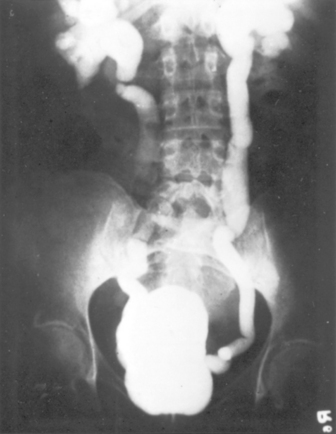
FIGURE 20-27 Vesicoureteral reflux demonstrated by a voiding cystourethrogram. Dye injected into the bladder refluxes into both dilated ureters, filling the pelvis and calyces.

In the absence of vesicoureteral reflux, infection usually remains localized in the bladder. Thus, the majority of individuals with repeated or persistent bacterial colonization of the urinary tract suffer from cystitis and urethritis (lower urinary tract infection) rather than pyelonephritis.

Acute Pyelonephritis

Acute pyelonephritis is an acute suppurative inflammation of the kidney caused by bacterial and sometimes viral (e.g., polyomavirus) infection, whether hematogenous and induced by septicemic spread or ascending and associated with vesicoureteral reflux.66

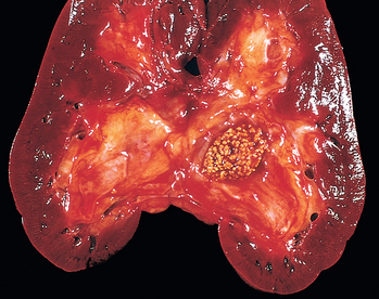
Morphology. The hallmarks of acute pyelonephritis are patchy interstitial suppurative inflammation, intratubular aggregates of neutrophils, and tubular necrosis. The suppuration may occur as discrete focal abscesses involving one or both kidneys, which can extend to large wedge-shaped areas of suppuration (Fig. 20-28). The distribution of these lesions is unpredictable and haphazard, but in pyelonephritis associated with reflux, damage occurs most commonly in the lower and upper poles.

image

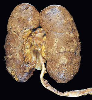
FIGURE 20-28 Acute pyelonephritis. Cortical surface shows grayish white areas of inflammation and abscess formation.

In the early stages, the neutrophilic infiltration is limited to the interstitial tissue. Soon, however, the reaction involves tubules and produces a characteristic abscess with the destruction of the engulfed tubules (Fig. 20-29). Since the tubular lumens present a ready pathway for the extension of the infection, large masses of intraluminal neutrophils frequently extend along the involved nephron into the collecting tubules. Characteristically, glomeruli seem to be relatively resistant to the infection. Large areas of severe necrosis, however, eventually destroy the glomeruli, and fungal pyelonephritis (e.g., Candida) often affects glomeruli.

image

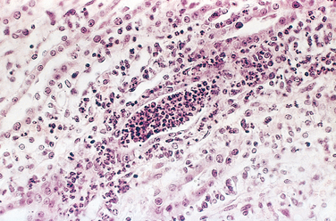
FIGURE 20-29 Acute pyelonephritis marked by an acute neutrophilic exudate within tubules and interstitial inflammation.

Three complications of acute pyelonephritis are encountered in special circumstances.

Papillary necrosis is seen mainly in diabetics and in those with urinary tract obstruction. Papillary necrosis is usually bilateral but may be unilateral. One or all of the pyramids of the affected kidney may be involved. On cut section, the tips or distal two thirds of the pyramids have areas of gray-white to yellow necrosis (Fig. 20-30). On microscopic examination the necrotic tissue shows characteristic coagulative necrosis, with preservation of outlines of tubules. The leukocytic response is limited to the junctions between preserved and destroyed tissue.
Pyonephrosis is seen when there is total or almost complete obstruction, particularly when it is high in the urinary tract. The suppurative exudate is unable to drain and thus fills the renal pelvis, calyces, and ureter with pus.
Perinephric abscess is an extension of suppurative inflammation through the renal capsule into the perinephric tissue.
image

FIGURE 20-30 Papillary necrosis. Areas of pale-gray necrosis involve the papillae (arrows).

After the acute phase of pyelonephritis, healing occurs. The neutrophilic infiltrate is replaced by one that is predominantly composed of macrophages, plasma cells, and (later) lymphocytes. The inflammatory foci are eventually replaced by irregular scars that can be seen on the cortical surface as fibrous depressions. Such scars are characterized microscopically by tubular atrophy, interstitial fibrosis, and a lymphocytic infiltrate in a characteristic patchy, jigsaw pattern with intervening preserved parenchyma. The pyelonephritic scar is almost always associated with inflammation, fibrosis, and deformation of the underlying calyx and pelvis, reflecting the role of ascending infection and vesicoureteral reflux in the pathogenesis of the disease.

Clinical Features.

Acute pyelonephritis is often associated with predisposing conditions, some of which were mentioned before. These include the following:

Urinary tract obstruction, either congenital or acquired
Instrumentation of the urinary tract, most commonly catheterization
Vesicoureteral reflux
Pregnancy. Between 4% and 6% of pregnant women develop bacteriuria sometime during pregnancy, and 20% to 40% of these eventually develop symptomatic urinary infection if not treated.
Gender and age. After the first year of life (when congenital anomalies in males commonly become evident) and up to around age 40 years, infections are much more frequent in females. With increasing age the incidence in males rises as a result of prostatic hypertrophy and instrumentation.
Preexisting renal lesions, causing intrarenal scarring and obstruction
Diabetes mellitus, in which increased susceptibility to infection, neurogenic bladder dysfunction, and more frequent instrumentation are predisposing factors
Immunosuppression and immunodeficiency

When acute pyelonephritis is clinically apparent, the onset is usually sudden, with pain at the costovertebral angle and systemic evidence of infection, such as fever and malaise. There are usually indications of bladder and urethral irritation, such as dysuria, frequency, and urgency. The urine contains many leukocytes (pyuria) derived from the inflammatory infiltrate, but pyuria does not differentiate upper from lower urinary tract infection. The finding of leukocyte casts, typically rich in neutrophils (pus casts), indicates renal involvement, because casts are formed only in tubules. The diagnosis of infection is established by quantitative urine culture.

Uncomplicated acute pyelonephritis usually follows a benign course, and the symptoms disappear within a few days after the institution of appropriate antibiotic therapy. Bacteria, however, may persist in the urine, or there may be recurrence of infection with new serologic types of E. coli or other organisms. Such bacteriuria then either disappears or may persist, sometimes for years. In the presence of unrelieved urinary obstruction, diabetes mellitus, or immunodeficiency, acute pyelonephritis may be more serious, leading to repeated septicemic episodes. The superimposition of papillary necrosis may lead to acute renal failure.

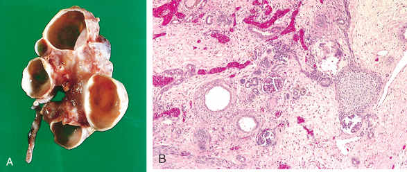
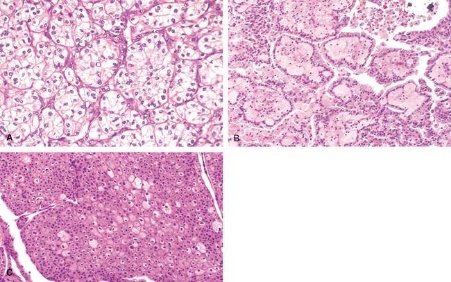
An emerging viral pathogen causing pyelonephritis in kidney allografts is polyomavirus. Latent infection with polyomavirus is widespread in the general population, but immunosuppression of the allograft recipient can lead to reactivation of latent infection and the development of a nephropathy resulting in allograft failure in as many as 1% to 5% of kidney transplant recipients.67 This form of pyelonephritis, now referred to as polyomavirus nephropathy, is characterized by viral infection of tubular epithelial cell nuclei, leading to nuclear enlargement and intranuclear inclusions visible by light microscopy (viral cytopathic effect). The inclusions are composed of virions arrayed in distinctive crystalline-like lattices when visualized by electron microscopy (Fig. 20-31). An interstitial inflammatory response is invariably present. Treatment consists of a reduction in immunosuppression.

image

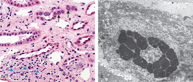
FIGURE 20-31 Polyomavirus nephropathy. A, The kidney shows enlarged tubular epithelial cells with nuclear inclusions (arrows) and interstitial inflammation (arrowheads). B, Intranuclear viral inclusions visualized by electron microscopy.

(Courtesy of Dr. Jean Olson, Department of Pathology, University of California San Francisco, San Francisco, CA.)

Chronic Pyelonephritis and Reflux Nephropathy

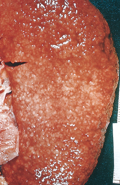
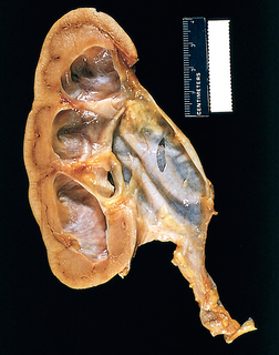
Chronic pyelonephritis is a disorder in which chronic tubulointerstitial inflammation and renal scarring are associated with pathologic involvement of the calyces and pelvis (Fig. 20-32). Pelvocalyceal damage is important in that virtually all the disease etiologies listed in Table 20-8 produce chronic tubulointerstitial alterations, but with the exception of chronic pyelonephritis and analgesic nephropathy, none affect the calyces. Chronic pyelonephritis is an important cause of end-stage kidney disease; at one time it accounted for as many as 10% to 20% of patients in renal transplant or dialysis units, until predisposing conditions such as reflux became better recognized. This condition remains an important cause of kidney destruction in children with severe lower urinary tract abnormalities.

image

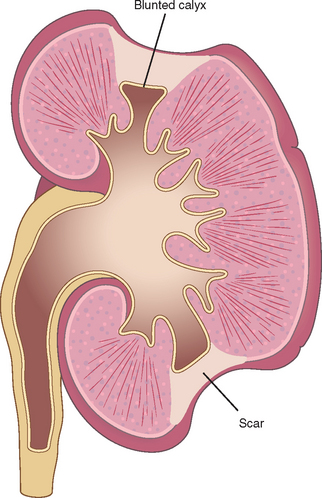
FIGURE 20-32 Typical coarse scars of chronic pyelonephritis associated with vesicoureteral reflux. The scars are usually polar and are associated with underlying blunted calyces.

Chronic pyelonephritis can be divided into two forms: chronic reflux-associated and chronic obstructive.

Reflux Nephropathy.

This is by far the more common form of chronic pyelonephritic scarring. Renal involvement in reflux nephropathy occurs early in childhood as a result of superimposition of a urinary infection on congenital vesicoureteral reflux and intrarenal reflux. Reflux may be unilateral or bilateral; thus, the resultant renal damage may cause scarring and atrophy of one kidney or involve both, leading to chronic renal insufficiency. Vesicoureteral reflux occasionally causes renal damage in the absence of infection (sterile reflux), but only when obstruction is severe.

Chronic Obstructive Pyelonephritis.

We have seen that obstruction predisposes the kidney to infection. Recurrent infections superimposed on diffuse or localized obstructive lesions lead to recurrent bouts of renal inflammation and scarring, resulting in a picture of chronic pyelonephritis. In this condition, the effects of obstruction contribute to the parenchymal atrophy; indeed, it is sometimes difficult to differentiate the effects of bacterial infection from those of obstruction alone. The disease can be bilateral, as with posterior urethral valves, resulting in renal insufficiency unless the anomaly is corrected, or unilateral, such as occurs with calculi and unilateral obstructive anomalies of the ureter.

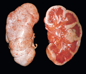
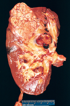
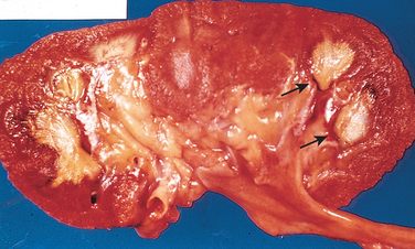
Morphology. The characteristic changes of chronic pyelonephritis are seen on gross examination (Figs. 20-32 and 20-33). The kidneys usually are irregularly scarred; if bilateral, the involvement is asymmetric. This contrasts with chronic glomerulonephritis, in which both kidneys are diffusely and symmetrically scarred. The hallmarks of chronic pyelonephritis are coarse, discrete, corticomedullary scars overlying dilated, blunted, or deformed calyces, and flattening of the papillae (see Fig. 20-33). The scars can vary from one to several in number and may affect one or both kidneys. Most are in the upper and lower poles, consistent with the frequency of reflux in these sites.

image

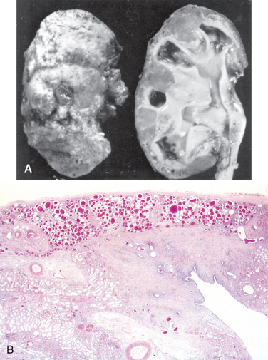
FIGURE 20-33 A, Chronic pyelonephritis. The surface (left) is irregularly scarred. The cut section (right) reveals characteristic dilation and blunting of calyces. The ureter is dilated and thickened, a finding that is consistent with chronic vesicoureteral reflux. B, Low-power view showing a corticomedullary renal scar with an underlying dilated deformed calyx. Note the thyroidization of tubules in the cortex.

The microscopic changes involve predominantly tubules and interstitium. The tubules show atrophy in some areas and hypertrophy or dilation in others. Dilated tubules with flattened epithelium may be filled with colloid casts (thyroidization). There are varying degrees of chronic interstitial inflammation and fibrosis in the cortex and medulla. In the presence of active infection there may be neutrophils in the interstitium and pus casts in the tubules. Arcuate and interlobular vessels demonstrate obliterative intimal sclerosis in the scarred areas; and in the presence of hypertension, hyaline arteriosclerosis is seen in the entire kidney. There is often fibrosis around the calyceal epithelium as well as a marked chronic inflammatory infiltrate. Glomeruli may appear normal except for periglomerular fibrosis, or exhibit a variety of changes, including ischemic fibrous obliteration and secondary changes related to hypertension. Individuals with chronic pyelonephritis and reflux nephropathy who develop proteinuria in advanced stages show secondary focal segmental glomerulosclerosis, as described later.

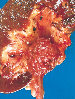
Xanthogranulomatous pyelonephritis is an unusual and relatively rare form of chronic pyelonephritis characterized by accumulation of foamy macrophages intermingled with plasma cells, lymphocytes, polymorphonuclear leukocytes, and occasional giant cells. Often associated with Proteus infections and obstruction, the lesions sometimes produce large, yellowish orange nodules that may be grossly confused with renal cell carcinoma.

Clinical Features.

Chronic obstructive pyelonephritis may be insidious in onset or present with clinical manifestations of acute recurrent pyelonephritis, such as back pain, fever, frequent pyuria, and bacteriuria. Chronic pyelonephritis associated with reflux may have a silent onset. These patients come to medical attention relatively late in the course of their disease because of the gradual onset of renal insufficiency and hypertension or because of the discovery of pyuria or bacteriuria on routine examination. Reflux nephropathy is often discovered when hypertension in children is investigated. Loss of tubular function—in particular of concentrating ability—gives rise to polyuria and nocturia. Radiographic studies show asymmetrically contracted kidneys with characteristic coarse scars and blunting and deformity of the calyceal system. Significant bacteriuria may be present, but it is often absent in the late stages.

Although proteinuria is usually mild, some individuals with pyelonephritic scars develop secondary focal segmental glomerulosclerosis with significant proteinuria, even in the nephrotic range, usually several years after the scarring has occurred and often in the absence of continued infection or persistent vesicoureteral reflux. The appearance of proteinuria and focal segmental glomerulosclerosis is a poor prognostic sign, and patients with these findings may proceed to chronic or end-stage renal failure. The glomerulosclerosis, as we have discussed, may be attributable to the adaptive glomerular alterations secondary to loss of renal mass caused by pyelonephritic scarring (renal ablation nephropathy).

Tubulointerstitial Nephritis Induced by Drugs and Toxins

Toxins and drugs can produce renal injury in at least three ways: (1) They may trigger an interstitial immunological reaction, exemplified by the acute hypersensitivity nephritis induced by such drugs as methicillin; (2) they may cause acute renal failure, as described earlier; and (3) they may cause subtle but cumulative injury to tubules that takes years to become manifest, resulting in chronic renal insufficiency.68 The last type of damage is especially treacherous, because it may be clinically unrecognized until significant renal damage has occurred. Such is the case with analgesic abuse nephropathy, which is usually detected only after the onset of chronic renal insufficiency.

Acute Drug-Induced Interstitial Nephritis

This is a well-recognized adverse reaction to a constantly increasing number of drugs. First reported after the use of sulfonamides, acute tubulointerstitial nephritis most frequently occurs with synthetic penicillins (methicillin, ampicillin), other synthetic antibiotics (rifampin), diuretics (thiazides), NSAIDs, and miscellaneous drugs (allopurinol, cimetidine). The disease begins about 15 days (range: 2–40) after exposure to the drug and is characterized by fever, eosinophilia (which may be transient), a rash in about 25% of patients, and renal abnormalities. The latter take the form of hematuria, mild proteinuria, and leukocyturia (often including eosinophils). A rising serum creatinine level or acute renal failure with oliguria develops in about 50% of cases, particularly in older patients.

Pathogenesis.

Many features of the disease suggest an immune mechanism. The immune response is idiosyncratic and not dose-related. Clinical evidence of hypersensitivity includes the latent period, the eosinophilia and rash, the fact that the onset of nephropathy is not dose-related, and the recurrence of hypersensitivity after re-exposure to the same or a cross-reactive drug. In some patients, serum IgE levels are increased, and IgE-containing plasma cells and basophils are present in the lesions, suggesting that the late-phase reaction of an IgE-mediated (type I) hypersensitivity may be involved in the pathogenesis (Chapter 6). In other cases, mononuclear or granulomatous infiltrate, together with positive results of skin tests to drug haptens, suggest a T cell–mediated delayedhypersensitivity reaction (type IV).

The most likely sequence of events is that the drugs act as haptens, which covalently bind to some cytoplasmic or extracellular component of tubular cells and become immunogenic. The resultant injury is then due to IgE and/or cell-mediated immune reactions to tubular cells or their basement membranes.

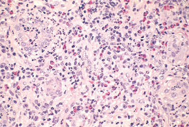
Morphology. On histologic examination the abnormalities are in the interstitium, which shows variable but frequently pronounced edema and infiltration by mononuclear cells, principally lymphocytes and macrophages. Eosinophils and neutrophils may be present (Fig. 20-34), often in clusters and large numbers, and plasma cells and basophils are sometimes found in small numbers. With some drugs (e.g., methicillin, thiazides), interstitial non-necrotizing granulomas containing giant cells may be seen. “Tubulitis,” the infiltration of tubules by lymphocytes, is common. Variable degrees of tubular necrosis and regeneration are present. The glomeruli are normal except in some cases caused by NSAIDs, when minimal-change disease and the nephrotic syndrome develop concurrently (see the discussion of NSAIDs later in the chapter).

image

FIGURE 20-34 Drug-induced interstitial nephritis, with prominent eosinophilic and mononuclear cell infiltrate.

(Courtesy of Dr. H. Rennke, Brigham and Women’s Hospital, Boston, MA.)

Clinical Features.

It is important to recognize drug-induced renal failure because withdrawal of the offending drug is followed by recovery, although it may take several months, and irreversible damage occurs occasionally in older subjects. It is also important to remember that while drugs are the leading identifiable cause of acute interstitial nephritis, in many affected patients (approximately 30% to 40%) an offending drug or mechanism cannot be identified.

Analgesic Nephropathy

This is a form of chronic renal disease caused by excessive intake of analgesic mixtures and characterized morphologically by chronic tubulointerstitial nephritis and renal papillary necrosis.69

The incidence of analgesic nephropathy reflects the consumption of analgesics in various populations throughout the world. In some parts of Australia, it ranked as one of the most common causes of chronic renal insufficiency until public health measures reduced its incidence. Its incidence in the United States is relatively low but varies among states, being highest in the southeast. Overall, it accounted for 9%, 3%, and 1% of patients undergoing dialysis in Australia, Europe, and the United States, respectively, before the recent surge in end-stage renal disease attributable to diabetes reduced these relative percentages. The renal damage was first ascribed to phenacetin, but the analgesic mixtures that are consumed often contain, in addition, aspirin, caffeine, acetaminophen (a metabolite of phenacetin), and codeine. Patients who develop this disease usually ingest large quantities of mixtures of at least two antipyretic analgesics. Most patients consume phenacetin-containing mixtures, and cases ascribed to ingestion of aspirin, phenacetin, or acetaminophen alone are uncommon. In most countries, restriction of over-the-counter sale of phenacetin or analgesic mixtures has reduced the incidence of the disorder but has not eradicated it, presumably because non-phenacetin-containing mixtures are available.

Pathogenesis.

Papillary necrosis is readily induced experimentally by a mixture of aspirin and phenacetin, usually combined with water depletion. It is now clear that in the sequence of events leading to renal damage, papillary necrosis occurs first, and cortical tubulointerstitial nephritis follows as a consequence of impeded urine outflow. The phenacetin metabolite acetaminophen, which can deplete cells of glutathione, then injures these cells by subsequent generation of oxidative metabolites. Aspirin induces its potentiating effect by inhibiting the vasodilatory effects of prostaglandins, predisposing the papillae to ischemia. Thus, the papillary damage may be due to a combination of direct toxic effects of phenacetin metabolites and ischemic injury to both tubular cells and vessels.

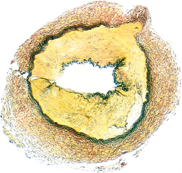
Morphology. In gross appearance the kidneys are either normal or slightly reduced in size, and the cortex shows depressed areas representing cortical atrophy overlying necrotic papillae. The papillae show various stages of necrosis, calcification, fragmentation, and sloughing. This gross appearance contrasts with the papillary necrosis seen in diabetic patients, in which all papillae are at the same stage of injury. On microscopic examination the papillary changes may take one of several forms. In early cases there is patchy necrosis, but in the advanced form the entire papilla is necrotic, often remaining in place as a structureless mass containing “ghosts” of tubules and foci of dystrophic calcification (Fig. 20-35). Segments of entire portions of the papilla may then be sloughed and excreted in the urine.

image

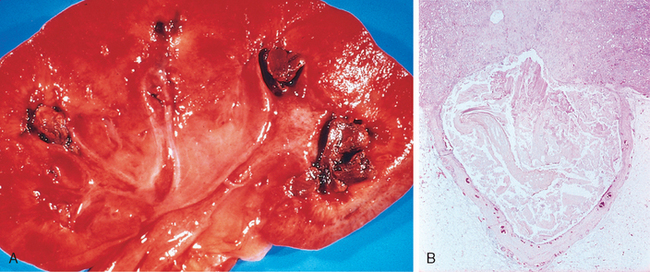
FIGURE 20-35 Analgesic nephropathy. A, The brownish necrotic papilla, transformed to a necrotic, structureless mass, fills the pelvis. B, Microscopic view. Note the fibrosis in the medulla.

(Courtesy of Dr. F.J. Gloor, Institut für Pathologie, Kantonsspital, St. Gallen, Switzerland.)

The cortical changes consist of loss and atrophy of tubules and interstitial fibrosis and inflammation. These changes are mainly due to obstructive atrophy caused by the tubular damage in the papillae. The cortical columns of Bertin are characteristically spared from this atrophy.

Clinical Features.

Analgesic nephropathy is more common in women than in men and is particularly prevalent in individuals with recurrent headaches and muscle pain, in psychoneurotic patients, and in factory workers. Early renal findings include inability to concentrate the urine (hyposthenuria), as would be expected for papillary lesions. Acquired distal renal tubular acidosis contributes to the development of renal stones. Headache, anemia, gastrointestinal symptoms, and hypertension are common accompaniments of analgesic nephropathy. Urinary tract infection complicates about 50% of cases. On occasion, entire tips of necrotic papillae are excreted, and these may cause gross hematuria or renal colic due to obstruction of the ureter by necrotic fragments. Magnetic resonance and computed tomographic imaging are helpful in detecting papillary necrosis and calcifications. Progressive impairment of renal function may lead to chronic renal failure, but with drug withdrawal, renal function may either stabilize or actually improve.

Unfortunately, a small percentage of patients with analgesic nephropathy develop transitional papillary carcinoma of the renal pelvis. Whether the carcinogenic effect is due to a metabolite of phenacetin or to some other component of the analgesic compounds is unsettled.

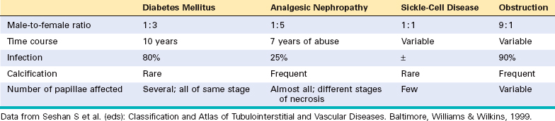
Papillary necrosis is not specific for analgesic nephropathy. It is also seen in diabetes mellitus, as was mentioned earlier, as well as in urinary tract obstruction, sickle cell disease or trait (described later), and focally in renal tuberculosis. Table 20-9 lists certain features of papillary necrosis in these conditions.

TABLE 20-9 Causes of Papillary Necrosis

image

Nephropathy Associated with NSAIDs

NSAIDs, one of the most common classes of drugs currently in use, produce several forms of renal injury. Although these complications are uncommon, they should be kept in mind since NSAIDs are frequently administered to patients with other potential causes of renal disease. Many NSAIDs are nonselective cyclooxygenase inhibitors, and their adverse renal effects are related to their ability to inhibit cyclooxygenase-dependent prostaglandin synthesis. The selective COX-2 inhibitors, while sparing the gastrointestinal tract, do affect the kidneys because COX-2 is expressed in human kidneys.70 NSAID-associated renal syndromes include

Hemodynamically induced acute renal failure, due to the decreased synthesis of vasodilatory prostaglandins. This is particularly likely to occur in the setting of other renal diseases or conditions causing volume depletion.
Acute hypersensitivity interstitial nephritis, resulting in acute renal failure, as described earlier.
Acute interstitial nephritis and minimal-change disease. This curious association of two diverse renal conditions, one leading to renal failure and the other to nephrotic syndrome, suggests a hypersensitivity reaction affecting the interstitium and possibly the glomeruli, but also is consistent with injury to podocytes mediated by cytokines released as part of the inflammatory process.
Membranous nephropathy, with the nephrotic syndrome, is a recently appreciated association, also of unclear pathogenesis.

Aristolochic Nephropathy

A syndrome of chronic tubulointerstitial nephritis caused by aristolochic acid, a supplement found in some herbal remedies, has been recognized recently. The drug forms covalent adducts with DNA and causes a distinctive picture of renal failure and interstitial fibrosis associated with a relative paucity of infiltrating leukocytes. As with analgesic nephropathy, there is an increased incidence of carcinoma in the kidney and urinary tract. Ingestion of aristolochic acid has also been identified as the cause of Balkan nephropathy, a chronic tubulointerstitial nephritis common in that part of the world.71

Other Tubulointerstitial Diseases

Urate Nephropathy

Three types of nephropathy can occur in persons with hyperuricemic disorders:

Acute uric acid nephropathy is caused by the precipitation of uric acid crystals in the renal tubules, principally in collecting ducts, leading to obstruction of nephrons and the development of acute renal failure. This type is particularly likely to occur in individuals with leukemias and lymphomas who are undergoing chemotherapy; the drugs increase the death of tumor cells, and uric acid is produced as released nucleic acids are broken down. Precipitation of uric acid is favored by the acidic pH in collecting tubules.
Chronic urate nephropathy, or gouty nephropathy, occurs in patients with more protracted forms of hyperuricemia. The lesions are ascribed to the deposition of monosodium urate crystals in the acidic milieu of the distal tubules and collecting ducts as well as in the interstitium. These deposits have a distinct histologic appearance and may form variably birefringent needle-like crystals either in the tubular lumens or in the interstitium (Fig. 20-36). The urates induce a tophus consisting of foreign-body giant cells, other mononuclear cells, and a fibrotic reaction (Chapter 26). Tubular obstruction by the urates causes cortical atrophy and scarring. Renal arterial and arteriolar thickening is common as a result of the relatively high frequency of hypertension in patients with gout. Clinically, urate nephropathy is a subtle disease associated with tubular defects that may progress slowly. Individuals with gout who actually develop a chronic nephropathy commonly have evidence of increased exposure to lead, sometimes by way of drinking “moonshine” whiskey contaminated with lead.
The third renal syndrome in hyperuricemia is nephrolithiasis; uric acid stones are present in 22% of individuals with gout and 42% of those with secondary hyperuricemia (see later discussion of renal stones).
image

FIGURE 20-36 Urate crystals in the renal medulla. Note the giant cells and fibrosis around the crystals.

Hypercalcemia and Nephrocalcinosis

Disorders associated with hypercalcemia, such as hyperparathyroidism, multiple myeloma, vitamin D intoxication, metastatic cancer, or excess calcium intake (milk-alkali syndrome), may induce the formation of calcium stones and deposition of calcium in the kidney (nephrocalcinosis). Extensive degrees of calcinosis, under certain conditions, may lead to chronic tubulointerstitial disease and renal insufficiency. The earliest damage induced by the hypercalcemia is to the tubular epithelial cells in the form of mitochondrial distortion and other signs of cell injury. Subsequently, calcium deposits appear within the mitochondria, cytoplasm, and basement membrane. Calcified cellular debris may obstruct tubular lumens and cause obstructive atrophy of nephrons and secondary interstitial fibrosis and inflammation. Atrophy of entire cortical areas drained by calcified tubules may occur, accounting for the alternating areas of normal and scarred parenchyma seen in such kidneys.

The earliest functional defect is an inability to concentrate the urine. Other tubular defects, such as tubular acidosis and salt-losing nephritis, may also occur. With further damage, a slowly progressive renal insufficiency develops. This is usually due to nephrocalcinosis, but many of these patients also have calcium stones and secondary pyelonephritis.

Acute Phosphate Nephropathy

Extensive accumulations of calcium phosphate crystals in tubules can occur in patients consuming high doses of oral phosphate solutions in preparation for colonoscopy.72 These patients are not hypercalcemic, but excess phosphate load, perhaps complicated by dehydration, causes marked precipitation of calcium phosphate, typically presenting as renal insufficiency several weeks after the exposure. Patients typically only partially recover renal function.

Light-Chain Cast Nephropathy (“Myeloma Kidney”)

Nonrenal malignant tumors, particularly those of hematopoietic origin, affect the kidneys in several ways (Table 20-10). The most common involvements are tubulointerstitial, caused by complications of the tumor (hypercalcemia, hyperuricemia, obstruction of ureters) or therapy (irradiation, hyperuricemia, chemotherapy, infections in immunosuppressed patients). As the survival rate of persons with malignant neoplasms increases, so do these renal complications. We limit the discussion here to the tubulointerstitial lesions in multiple myeloma that sometimes dominate the clinical picture in people with this disease.

TABLE 20-10 Renal Disease Related to Nonrenal Neoplasms

Direct tumor invasion of renal parenchyma
Ureters (obstruction)
Artery (renovascular hypertension)
Hypercalcemia
Hyperuricemia
Amyloidosis (AL, light-chain type)
Excretion of abnormal proteins (multiple myeloma)
Glomerulopathies
Membranous nephropathy, secondary (carcinomas)
Minimal-change disease (Hodgkin’s disease)
Membranoproliferative glomerulonephritis (leukemias and lymphomas)
Monoclonal immunoglobin/light-chain deposition disease (multiple myeloma)
Effects of radiation therapy, chemotherapy, secondary infection

Overt renal insufficiency occurs in half of those with multiple myeloma and related lymphoplasmacytic disorders. Several factors contribute to renal damage:

Bence Jones proteinuria and cast nephropathy. The main cause of renal dysfunction is related to Bence Jones (light-chain) proteinuria. Renal failure correlates well with the presence and amount of such proteinuria and is uncommon in its absence. Two mechanisms seem to account for the renal toxicity of Bence Jones proteins. First, some light chains are directly toxic to epithelial cells, apparently because of their intrinsic physicochemical properties. Second, Bence Jones proteins combine with the urinary glycoprotein (Tamm-Horsfall protein) under acidic conditions to form large, histologically distinct tubular casts that obstruct the tubular lumens and induce a characteristic inflammatory reaction around the casts (light-chain cast nephropathy).
Amyloidosis, of AL type formed from free light chains (usually of λ type), which occurs in 6% to 24% of individuals with myeloma.
Light-chain deposition disease. In some patients, light chains (usually of κ type) deposit in GBMs and mesangium in nonfibrillar forms, causing a glomerulopathy (described earlier), or in tubular basement membranes, which may cause tubulointerstitial nephritis.
Hypercalcemia and hyperuricemia are often present in these patients.

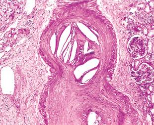
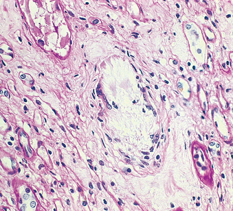
Morphology. The tubulointerstitial changes in light-chain cast nephropathy are fairly characteristic. The Bence Jones tubular casts appear as pink to blue amorphous masses, sometimes concentrically laminated and often fractured, which fill and distend the tubular lumens. Some of the casts are surrounded by multinucleate giant cells that are derived from mononuclear phagocytes (Fig. 20-37). The adjacent interstitial tissue usually shows a nonspecific inflammatory response and fibrosis. On occasion, the casts erode their way from the tubules into the interstitium and here evoke a granulomatous inflammatory reaction. Amyloidosis, light-chain deposition disease, nephrocalcinosis, and infection may also be present.

image

FIGURE 20-37 Light-chain cast nephropathy. Note the angulated and tubular casts, surrounded by macrophages, including multinucleate cells.

Clinical Features.

Clinically, the renal manifestations are of several types. In the most common form, chronic renal failure develops insidiously and usually progresses slowly during a period of several months to years. Another form occurs suddenly and is manifested by acute renal failure with oliguria. Precipitating factors in these patients include dehydration, hypercalcemia, acute infection, and treatment with nephrotoxic antibiotics. Bence Jones proteinuria occurs in 70% of individuals with multiple myeloma; the presence of significant non–light-chain proteinuria (e.g., albuminuria) suggests AL amyloidosis or light-chain deposition disease.

Vascular Diseases

Nearly all diseases of the kidney involve the renal blood vessels secondarily. Systemic vascular diseases, such as various forms of vasculitis, also affect renal vessels, and their effects on the kidney are often clinically important. Hypertension, as we discussed in Chapter 11, is intimately linked with the kidney, because kidney disease can be both the cause and consequence of increased blood pressure.73 In this chapter we discuss benign and malignant nephrosclerosis and renal artery stenosis, lesions associated with hypertension, and sundry lesions involving mostly smaller vessels of the kidney.

BENIGN NEPHROSCLEROSIS

Benign nephrosclerosis is the term used for the renal pathology associated with sclerosis of renal arterioles and small arteries. The resultant effect is focal ischemia of parenchyma supplied by vessels with thickened walls and consequent narrowed lumens. The parenchymal effects include glomerulosclerosis and chronic tubulointersititial injury, producing a reduction in functional renal mass. Nephrosclerosis at autopsy is associated with increasing age, more frequent in blacks than whites, and may be seen in the absence of hypertension.74,75 Hypertension and diabetes mellitus, however, increase the incidence and severity of the lesions.

Pathogenesis.

Two processes participate in the arterial lesions:

Medial and intimal thickening, as a response to hemodynamic changes, aging, genetic defects, or some combination of these
Hyaline deposition in arterioles, caused partly by extravasation of plasma proteins through injured endothelium and partly by increased deposition of basement membrane matrix

Morphology. The kidneys are either normal or moderately reduced in size, with average weights between 110 and 130 gm. The cortical surfaces have a fine, even granularity that resembles grain leather (Fig. 20-38). The loss of mass is due mainly to cortical scarring and shrinking.

image

FIGURE 20-38 Close-up of the gross appearance of the cortical surface in benign nephrosclerosis illustrating the fine, leathery granularity of the surface.

On histologic examination there is narrowing of the lumens of arterioles and small arteries, caused by thickening and hyalinization of the walls (hyaline arteriolosclerosis) (Fig. 20-39). Corresponding to the fine surface granulations are microscopic subcapsular scars with sclerotic glomeruli and tubular dropout, alternating with better preserved parenchyma. In addition, the interlobular and arcuate arteries show a characteristic lesion that consists of medial hypertrophy, reduplication of the elastic lamina, and increased myofibroblastic tissue in the intima, which combine to narrow the lumen. This change, called fibroelastic hyperplasia, often accompanies hyaline arteriolosclerosis and increases in severity with age and in the presence of hypertension.

image

FIGURE 20-39 Hyaline arteriolosclerosis. High-power view of two arterioles with hyaline deposition, marked thickening of the walls, and a narrowed lumen.

(Courtesy of Dr. M.A. Venkatachalam, Department of Pathology, University of Texas Health Sciences Center, San Antonio, TX.)

Consequent to the vascular narrowing, there is patchy ischemic atrophy, which consists of (1) foci of tubular atrophy and interstitial fibrosis and (2) a variety of glomerular alterations. The latter include collapse of the GBM, deposition of collagen within the Bowman space, periglomerular fibrosis, and total sclerosis of glomeruli. When the ischemic changes are pronounced and affect large areas of parenchyma, they can produce regional scars and histologic alterations that may resemble those seen in renal ablation injury, mentioned earlier.

Clinical Features.

It is unusual for uncomplicated benign nephrosclerosis to cause renal insufficiency or uremia. There are usually moderate reductions in renal blood flow, but the GFR is normal or only slightly reduced. On occasion, there is mild proteinuria. However, three groups of hypertensive patients with benign nephrosclerosis are at increased risk of developing renal failure: people of African descent, people with more severe blood pressure elevations, and persons with a second underlying disease, especially diabetes. In these groups renal insufficiency may supervene after prolonged benign hypertension, but more rapid renal failure results from the development of the malignant or accelerated phase of hypertension, discussed next.

MALIGNANT HYPERTENSION AND ACCELERATED NEPHROSCLEROSIS

Malignant nephrosclerosis is the form of renal disease associated with the malignant or accelerated phase of hypertension.76 This dramatic pattern of hypertension may occasionally develop in previously normotensive individuals but often is superimposed on preexisting essential benign hypertension, secondary forms of hypertension, or an underlying chronic renal disease, particularly glomerulonephritis or reflux nephropathy (Chapter 11). It is also a frequent cause of death from uremia in individuals with scleroderma. Malignant hypertension is relatively uncommon, occurring in 1% to 5% of all people with elevated blood pressure. In its pure form it usually affects younger individuals, and occurs more often in men and in blacks.

Pathogenesis.

The basis for this turn for the worse in hypertensive subjects is unclear, but the following sequence of events is suggested. The initial insult seems to be some form of vascular damage to the kidneys. This might result from long-standing benign hypertension, with eventual injury to the arteriolar walls, or the initiating injury may spring de novo from arteritis, a coagulopathy, or some injury causing acute exacerbation of the hypertension. In any case, the result is increased permeability of the small vessels to fibrinogen and other plasma proteins, endothelial injury, focal death of cells of the vascular wall, and platelet deposition. This leads to the appearance of fibrinoid necrosis of arterioles and small arteries, swelling of the vascular intima, and intravascular thrombosis. Mitogenic factors from platelets (e.g., PDGF), plasma, and other cells cause hyperplasia of intimal smooth muscle of vessels, resulting in the hyperplastic arteriolosclerosis that is typical of malignant hypertension and further narrowing of the lumens. The kidneys become markedly ischemic. With severe involvement of the renal afferent arterioles, the renin-angiotensin system receives a powerful stimulus; indeed, patients with malignant hypertension have markedly elevated levels of plasma renin. This sets up a self-perpetuating cycle in which angiotensin II causes intrarenal vasoconstriction, and the attendant renal ischemia perpetuates renin secretion. Other vasoconstrictors (e.g., endothelin) and loss of vasodilators (nitric oxide) may also contribute to vasoconstriction. Aldosterone levels are also elevated, and salt retention undoubtedly contributes to the elevation of blood pressure. The consequences of the markedly elevated blood pressure on the blood vessels throughout the body are known as malignant arteriosclerosis, and the renal disorder is malignant nephrosclerosis.

Morphology. On gross inspection the kidney size depends on the duration and severity of the hypertensive disease. Small, pinpoint petechial hemorrhages may appear on the cortical surface from rupture of arterioles or glomerular capillaries, giving the kidney a peculiar “flea-bitten” appearance.

Two histologic alterations characterize blood vessels in malignant hypertension (Fig. 20-40):

Fibrinoid necrosis of arterioles. This appears as an eosinophilic granular change in the blood vessel wall, which stains positively for fibrin by histochemical or immunofluorescence techniques. This change represents an acute event; it may be accompanied by limited inflammatory infiltrate within the wall, but prominent inflammation is not seen. Sometimes the glomeruli become necrotic and infiltrated with neutrophils, and the glomerular capillaries may thrombose.
In the interlobular arteries and arterioles, there is intimal thickening caused by a proliferation of elongated, concentrically arranged smooth muscle cells, together with fine concentric layering of collagen and accumulation of pale-staining material that probably represents accumulations of proteoglycans and plasma proteins. This alteration has been referred to as onion-skinning because of its concentric appearance. The lesion, also called hyperplastic arteriolitis, correlates well with renal failure in malignant hypertension. There may be superimposed intraluminal thrombosis. The arteriolar and arterial lesions result in considerable narrowing of all vascular lumens, ischemic atrophy and, at times, infarction distal to the abnormal vessels.
image

FIGURE 20-40 Malignant hypertension. A, Fibrinoid necrosis of afferent arteriole (PAS stain). B, Hyperplastic arteriolitis (onion-skin lesion).

(Courtesy of Dr. H. Rennke, Brigham and Women’s Hospital, Boston, MA.)

Clinical Features.

The full-blown syndrome of malignant hypertension is characterized by systolic pressures greater than 200 mm Hg and diastolic pressures greater than 120 mm Hg, papilledema, retinal hemorrhages, encephalopathy, cardiovascular abnormalities, and renal failure. Most often, the early symptoms are related to increased intracranial pressure and include headaches, nausea, vomiting, and visual impairments, particularly scotomas or spots before the eyes. “Hypertensive crises” are sometimes encountered, characterized by episodes of loss of consciousness or even convulsions. At the onset of rapidly mounting blood pressure, there is marked proteinuria and microscopic or sometimes macroscopic hematuria but no significant alteration in renal function. Soon, however, renal failure makes its appearance. The syndrome is a true medical emergency requiring the institution of aggressive and prompt antihypertensive therapy to prevent the development of irreversible renal lesions. Before the introduction of current antihypertensive drugs, malignant hypertension was associated with a 50% mortality rate within 3 months of onset, progressing to 90% within a year. At present, however, about 75% of patients survive 5 years, and 50% survive with restoration of pre-crisis renal function.

RENAL ARTERY STENOSIS

Unilateral renal artery stenosis is a relatively uncommon cause of hypertension, responsible for 2% to 5% of cases, but is important because it represents a potentially curable form of hypertension with surgical treatment. Furthermore, important insights into renal mechanisms of hypertension came from studies of experimental and human renal artery stenosis.

Pathogenesis.

The classic experiments of Goldblatt and colleagues78 showed that constriction of one renal artery in dogs results in hypertension and that the magnitude of the effect is roughly proportional to the amount of constriction. Later experiments in rats confirmed these results, and in time it was shown that the hypertensive effect, at least initially, is due to stimulation of renin secretion by cells of the juxtaglomerular apparatus and the subsequent production of the vasoconstrictor angiotensin II. A large proportion of individuals with renovascular hypertension have elevated plasma or renal vein renin levels, and almost all show a reduction of blood pressure when given drugs that block the activity of angiotensin II. Furthermore, unilateral renal renin hypersecretion can be normalized after renal revascularization, usually resulting in a decrease in blood pressure. Other factors, however, may contribute to the maintenance of renovascular hypertension after the renin-angiotensin system has initiated it, including sodium retention and possibly endothelin and loss of nitric oxide.

Morphology. The most common cause of renal artery stenosis (70% of cases) is occlusion by an atheromatous plaque at the origin of the renal artery. This lesion occurs more frequently in men, and the incidence increases with advancing age and diabetes mellitus. The plaque is usually concentrically placed, and superimposed thrombosis often occurs.

The second type of lesion leading to stenosis is so-called fibromuscular dysplasia of the renal artery. This is a heterogeneous group of lesions characterized by fibrous or fibromuscular thickening and may involve the intima, the media, or the adventitia of the artery. These lesions are thus subclassified into intimal, medial, and adventitial hyperplasia, the medial type being by far the most common (Fig. 20-41). The stenoses, as a whole, are more common in women and tend to occur in younger age groups (i.e., in the third and fourth decades). The lesions may consist of a single well-defined constriction or a series of narrowings, usually in the middle or distal portion of the renal artery. They may also involve the segmental branches and may be bilateral.

image

FIGURE 20-41 Fibromuscular dysplasia of the renal artery, medial type (elastic tissue stain). The media shows marked fibrous thickening, and the lumen is stenotic.

(Courtesy of Dr. Seymour Rosen, Beth Israel Hospital, Boston, MA.)

The ischemic kidney is usually reduced in size and shows signs of diffuse ischemic atrophy, with crowded glomeruli, atrophic tubules, interstitial fibrosis, and focal inflammatory infiltrates. The arterioles in the ischemic kidney are usually protected from the effects of high pressure, thus showing only mild arteriolosclerosis. In contrast, the contralateral nonischemic kidney may show more severe arteriolosclerosis, depending on the severity of the hypertension.

Clinical Course.

Few distinctive features suggest the presence of renal artery stenosis, and in general, these patients resemble those with essential hypertension. On occasion, a bruit can be heard on auscultation of the affected kidneys. Elevated plasma or renal vein renin, response to angiotensin-converting enzyme inhibitor, renal scans, and intravenous pyelography may aid with diagnosis, but arteriography is required to localize the stenotic lesion. The cure rate after surgery is 70% to 80% in well-selected cases.

THROMBOTIC MICROANGIOPATHIES

As was described in Chapter 14, this group of disorders is characterized clinically by microangiopathic hemolytic anemia, thrombocytopenia, and (in many cases) renal failure, and morphologically by thrombotic lesions in capillaries and arterioles in various tissue beds, including those of the kidney (Fig. 20-42).79,80 Schistocytes (fragmented red cells) in peripheral blood smears provide an important clue to the diagnosis. Unlike DIC, these disorders are generally associated with normal coagulation times and normal or only slightly elevated fibrin split products.

image

FIGURE 20-42 Fibrin stain showing platelet-fibrin thrombi (red) in the glomerular capillaries, characteristic of thrombotic microangiopathic disorders.

The classification of these disorders has been muddied by the fact that the two main forms, hemolytic-uremic syndrome (HUS) and thrombotic thrombocytopenic purpura (TTP), show considerable overlap in their clinical features.80,81 However, it is now evident that the category of HUS/TTP includes several entities with distinct causes, natural histories, and therapeutic approaches. We will thus classify these disorders according to our current understanding of their causes or associations, as follows:

1. Typical HUS (synonyms: epidemic, classic, diarrheapositive), most frequently associated with consumption of food contaminated by bacteria producing Shiga-like toxins
2. Atypical HUS (synonyms: non-epidemic, diarrheanegative), associated with:
a. Inherited mutations of complement-regulatory proteins
b. Diverse acquired causes of endothelial injury, including: antiphospholipid antibodies; complications of pregnancy and oral contraceptives; vascular renal diseases such as scleroderma and hypertension; chemotherapeutic and immunosuppressive drugs; and radiation
3. TTP, which is often associated with inherited or acquired deficiencies of ADAMTS13, a plasma metalloprotease that regulates the function of von Willebrand factor (vWF)
Pathogenesis.

Within the thrombotic microangiopathies, two pathogenetic triggers dominate: (1) endothelial injury, and (2) platelet activation and aggregation. As will be discussed, endothelial injury appears to be the primary cause of HUS, whereas platelet activation may be the inciting event in TTP.

Endothelial Injury.

In typical (epidemic, classic, diarrhea-positive) HUS, the trigger for endothelial injury and activation is usually a Shiga-like toxin, while in inherited forms of atypical HUS the cause of the endothelial injury appears to be excessive, inappropriate activation of complement. Many other exposures and conditions can occasionally precipitate a HUS-like picture, presumably also by injuring the endothelium. The endothelial injury in HUS appears to cause platelet activation and thrombosis within microvascular beds. There is evidence that reduced endothelial production of prostaglandin I2 and NO (both inhibitors of platelet aggregation) contributes to thrombosis. The reduction in these two factors and increased production of endothelium-derived endothelin may also promote vasoconstriction, exacerbating the hypoperfusion of tissues. Finally, adhesion molecules expressed on injured endothelium result in the recruitment of leukocytes, which may also contribute to thrombosis, as described in Chapter 4.

Platelet Aggregation.

In contrast to HUS, in TTP the initiating event appears to be platelet aggregation induced by very large multimers of vWF, which accumulate due to a deficiency of ADAMTS13, a plasma protease that cleaves vWF multimers into smaller sizes. The deficiency of ADAMTS13 is most often caused by autoantibodies that inhibit ADAMTS13 function. Less commonly, a chronic relapsing and remitting form of TTP is associated with inherited deficiencies of ADAMTS13. Very large vWF multimers can bind platelet surface glycoproteins and activate platelets spontaneously, providing a pathophysiologic explanation for the microthrombi that are observed in vascular beds.80,82

Regardless of the trigger, tissue dysfunction in all forms of HUS/TTP appears to result from the formation of microthrombi, vascular obstruction, and tissue ischemia.80 We will first describe the various subtypes of HUS/TTP, and then return to the morphologic features that are common to all.

Typical (epidemic, classic, diarrhea-positive) Hemolytic-Uremic Syndrome.

This is the best-characterized form of HUS. Most cases occur following intestinal infection with strains of E. coli (the most common being O157:H7) that produce Shiga-like toxins,83 so-called because they resemble those made by Shigella dysenteriae (Chapter 17). Epidemics have been traced to various sources, most commonly the ingestion of contaminated ground meat (as in hamburgers), but also drinking water, raw milk, and person-to-person transmission. However, most cases of typical HUS caused by E. coli are sporadic. Less commonly, infections by other agents, including Shigella dysenteriae, can give rise to a similar clinical picture.

Typical HUS can occur in adults, particularly the elderly, but it affects children preferentially, in whom it is one of the main causes of acute renal failure. Following a prodrome of influenza-like or diarrheal symptoms, there is a sudden onset of bleeding manifestations (especially hematemesis and melena), severe oliguria, and hematuria, associated with microangiopathic hemolytic anemia, thrombocytopenia, and (in some patients) prominent neurologic changes. Hypertension is present in about half the patients.

Shiga-like toxin injures endothelial cells, inducing increased expression of leukocyte adhesion molecules; increased endothelin and decreased nitric oxide production; and in the presence of cytokines such as TNF, endothelial apoptosis. These alterations lead to platelet activation and induce vasoconstriction, resulting in the characteristic microangiopathy. There is also some evidence that Shiga-like toxins bind and directly activate platelets.

In typical HUS, if the renal failure is managed properly with dialysis, most patients recover normal renal function in a matter of weeks. However, due to underlying renal damage the long-term (15 to 25 year) outlook is more guarded. In one study, only 10 of 25 patients with prior epidemic HUS had normal renal function, and 7 had chronic kidney disease.

Atypical (non-epidemic, diarrhea-negative) Hemolytic-Uremic Syndrome.

Atypical HUS occurs mainly in adults in a number of different settings. More than half of those affected have an inherited deficiency of complement-regulatory proteins, most commonly factor H, which normally breaks down the alternative pathway C3 convertase and protects cells from damage by uncontrolled complement activation (Chapter 2).82 A small number of patients have mutations in two other proteins that regulate complement, complement factor I and CD46 (membrane cofactor protein). Patients with genetic mutations in complement-regulatory proteins may develop HUS at any age. Roughly half of affected individuals have a course marked by multiple relapses and progression to end-stage renal disease. As the deficiencies in complement-regulatory factors are life-long, it is a mystery why the onset of HUS is delayed; additional unknown co-factors that trigger the development of HUS are suspected.

A variety of miscellaneous conditions or exposures are occasionally associated with atypical forms of HUS. These include:

1. The antiphospholipid syndrome, either primary or secondary to SLE (lupus anticoagulant). The syndrome is described in detail in Chapter 4. In this setting the microangiopathy tends to follow a chronic course.
2. Complications of pregnancy or the postpartum period. So-called postpartum renal failure is a form of HUS that usually occurs after an uneventful pregnancy, 1 day to several months after delivery. The condition has a grave prognosis, although recovery can occur in milder cases.
3. Vascular diseases affecting the kidney, such as systemic sclerosis and malignant hypertension.
4. Chemotherapeutic and immunosuppressive drugs, such as mitomycin, cyclosporine, cisplatin, and gemcitabine.
5. Irradiation of the kidney.

Patients with atypical HUS do not fare as well as those with typical HUS, in large part because the underlying conditions may be chronic and difficult to treat.80 As in typical HUS, some patients have neurologic symptoms; the disease in these patients can be distinguished from TTP by the presence of normal ADAMTS13 levels in the plasma (see below).

Thrombotic Thrombocytopenic Purpura.

TTP is classically manifested by the pentad of fever, neurologic symptoms, microangiopathic hemolytic anemia, thrombocytopenia, and renal failure.80 As discussed above, it is usually caused by antibodies (either autoimmune or drug-induced) or genetic defects that lead to functional deficits in ADAMTS13.82 The most common cause of deficient ADAMTS13 activity is inhibitory autoantibodies, and the majority of those with such antibodies are women. Regardless of cause, most patients present as adults at ages younger than 40.

In TTP, central nervous system involvement is the dominant feature, whereas renal involvement occurs in only about 50% of patients. The clinical findings are dictated by the distribution of the microthrombi, which are found in arterioles throughout the body. Untreated, the disease was once highly fatal, but in those with autoantibodies exchange transfusions and immunosuppressive therapy have reduced the mortality to less than 50%. As in HUS associated with inherited deficiencies of complement regulatory proteins, it is not understood why those with life-long genetic deficiency of ADAMTS13 present in adulthood. Such patients tend to follow a relapsing and remitting course.

Morphology. The morphological findings in the various forms of HUS/TTP show considerable overlap, and vary mainly according to chronicity rather than cause. In acute, active disease the kidney may show patchy or diffuse cortical necrosis (described later) and subcapsular petechiae. On microscopic examination, the glomerular capillaries are occluded by thrombi composed of aggregated platelets and to a lesser extent fibrin. The capillary walls are thickened due to endothelial cell swelling and subendothelial deposits of cell debris and fibrin. Disruption of the mesangial matrix and damage to the mesangial cells often results in mesangiolyis. Interlobular arteries and arterioles often show fibrinoid necrosis of the wall and occlusive thrombi. Chronic disease is confined to patients with atypical HUS or TTP, and has features that stem from continued injury and attempts at healing. The renal cortex reveals various degrees of scarring. By light microscopy the glomeruli are mildly hypercellular and have marked thickening of the capillary walls associated with splitting or reduplication of the basement membrane (so called double contours or tram tracks). The walls of arteries and arterioles often exhibit increased layers of cells and connective tissue (“onion-skinning”) that narrow the vessel lumens. These changes lead to persistent hypoperfusion and ischemic atrophy of the parenchyma, which manifests clinically as renal failure and hypertension.

OTHER VASCULAR DISORDERS

Atherosclerotic Ischemic Renal Disease

We have seen that atherosclerotic unilateral renal artery stenosis can lead to hypertension. Bilateral renal artery disease, usually diagnosed definitively by arteriography, now seems to be a fairly common cause of chronic ischemia with renal insufficiency in older individuals, sometimes in the absence of hypertension.84,85 The importance of recognizing this condition is that surgical revascularization is beneficial in reversing further decline in renal function.

Atheroembolic Renal Disease

Embolization of fragments of atheromatous plaques from the aorta or renal artery into intraparenchymal renal vessels occurs in elderly patients with severe atherosclerosis, especially after surgery on the abdominal aorta, aortography, or intra-aortic cannulization. These emboli can be recognized in the lumens and walls of arcuate and interlobular arteries by their content of cholesterol crystals, which appear as rhomboid clefts (Fig. 20-43). The clinical consequences of atheroemboli vary according to the number of emboli and the preexisting state of renal function. Frequently they have no functional significance. However, acute renal failure may develop in elderly patients in whom renal function is already compromised, principally after abdominal surgery on atherosclerotic aneurysms.

image

FIGURE 20-43 Atheroemboli with typical cholesterol clefts in an interlobar artery.

Sickle-Cell Disease Nephropathy

Sickle-cell disease (homozygous) or trait (heterozygous) may lead to a variety of alterations in renal morphology and function, some of which produce clinically significant abnormalities. The various manifestations are termed sickle-cell nephropathy.

The most common clinical and functional abnormalities are hematuria and a diminished concentrating ability (hyposthenuria). These are thought to be due largely to accelerated sickling in the hypertonic hypoxic milieu of the renal medulla; the hyperosmolarity dehydrates red cells and increases intracellular HbS concentrations, which likely explains why even those with sickle trait are affected. Patchy papillary necrosis may occur in both homozygotes and heterozygotes; this is sometimes associated with cortical scarring. Proteinuria is also common in sickle-cell disease, occurring in about 30% of patients. It is usually mild to moderate, but on occasion the overt nephrotic syndrome arises, associated with sclerosing glomerular lesions.

Diffuse Cortical Necrosis

This is an uncommon condition that occurs most frequently after an obstetric emergency, such as abruptio placentae (premature separation of the placenta), septic shock, or extensive surgery. When bilateral and symmetric, it is fatal in the absence of supportive therapy. The cortical destruction has the features of ischemic necrosis. Glomerular and arteriolar microthrombi are found in most cases, and clearly contribute to the necrosis and renal damage. The morphologic features have considerable overlap with thrombotic microangiopathy and disseminated intravascular coagulation, but the pathogenetic sequence of events in this injury remains obscure.

Morphology. The gross alterations of massive ischemic necrosis are sharply limited to the cortex (Fig. 20-44). The histologic appearance is that of acute ischemic infarction. The lesions may be patchy, with areas of coagulative necrosis and apparently better preserved cortex. Intravascular and intraglomerular thromboses may be prominent but are usually focal, and acute necroses of small arterioles and capillaries may occasionally be present. Hemorrhages occur into the glomeruli, together with the formation of fibrin plugs in the glomerular capillaries.

image

FIGURE 20-44 Diffuse cortical necrosis. The pale ischemic necrotic areas are confined to the cortex and columns of Bertin.

Massive acute cortical necrosis is of grave significance, since it gives rise to sudden anuria, terminating rapidly in uremic death. Instances of unilateral or patchy involvement are compatible with survival.

Renal Infarcts

The kidneys are favored sites for the development of infarcts. Contributing to this predisposition is the extensive blood flow to the kidneys (one fourth of the cardiac output), but probably more important is the “end-organ” nature of the arterial blood supply and the extremely limited collateral circulation from extrarenal sites (essentially small blood vessels penetrating from the renal capsule). Although thrombosis in advanced atherosclerosis and the acute vasculitis of polyarteritis nodosa may occlude arteries, most infarcts are due to embolism. A major source of such emboli is mural thrombosis in the left atrium and ventricle as a result of myocardial infarction. Vegetative endocarditis, aortic aneurysms, and aortic atherosclerosis are less frequent sources of emboli.

Morphology. Because of the end-organ type of arterial supply, most renal infarcts are of the “white” anemic variety. They may be solitary lesions or may be multiple and bilateral. Within 24 hours infarcts become sharply demarcated, pale, yellow-white areas that may contain small irregular foci of hemorrhagic discoloration. They are usually ringed by a zone of intense hyperemia.

On section the infarcts are wedge-shaped, with the base against the cortical surface and the apex pointing toward the medulla. There may be a narrow rim of preserved subcortical tissue that has been spared by the collateral capsular circulation. In time these acute areas of ischemic necrosis undergo progressive fibrous scarring, giving rise to depressed, pale, gray-white scars that assume a V-shape on section. The histologic changes in renal infarction are those of ischemic coagulative necrosis, described in Chapter 1.

Many renal infarcts are clinically silent. Sometimes, pain with tenderness localized to the costovertebral angle occurs, associated with showers of red cells in the urine. Large infarcts of one kidney are probably associated with narrowing of the renal artery or one of its major branches, which in turn may be a cause of hypertension.

Congenital Anomalies

About 10% of all people are born with potentially significant malformations of the urinary system. Renal dysplasias and hypoplasias account for 20% of chronic renal failure in children.

Congenital renal disease can be hereditary but is most often the result of an acquired developmental defect that arises during gestation. As was discussed in Chapter 10, defects in genes involved in development, including the Wilms tumor–associated genes, cause urogenital anomalies. As a rule, developmental abnormalities involve structural components of the kidney and urinary tract, causing syndromes termed CAKUT (congenital abnormalities of the kidney and urinary tract). However, genetic abnormalities also cause enzymatic or metabolic defects in tubular transport, such as cystinuria and renal tubular acidosis. Here, we restrict the discussion to structural anomalies involving primarily the kidney. All except horseshoe kidney are uncommon. Anomalies of the lower urinary tract are discussed in Chapter 21.

Agenesis of the Kidney.

Bilateral agenesis, which is incompatible with life, is usually encountered in stillborn infants. It is often associated with many other congenital disorders (e.g., limb defects, hypoplastic lungs) and leads to early death. Unilateral agenesis is an uncommon anomaly that is compatible with normal life if no other abnormalities exist. The opposite kidney is usually enlarged as a result of compensatory hypertrophy. Some patients eventually develop progressive glomerular sclerosis in the remaining kidney as a result of the adaptive changes in hypertrophied nephrons, discussed earlier in the chapter, and in time, chronic kidney disease ensues.

Hypoplasia.

Renal hypoplasia refers to failure of the kidneys to develop to a normal size. This anomaly may occur bilaterally, resulting in renal failure in early childhood, but it is more commonly encountered as a unilateral defect. True renal hypoplasia is extremely rare; most cases reported probably represent acquired scarring due to vascular, infectious, or other parenchymal diseases rather than an underlying developmental failure. Differentiation between congenital and acquired atrophic kidneys may be impossible, but a truly hypoplastic kidney shows no scars and has a reduced number of renal lobes and pyramids, usually six or fewer. In one form of hypoplastic kidney, oligomeganephronia, the kidney is small with fewer nephrons that are markedly hypertrophied.

Ectopic Kidneys.

The development of the definitive metanephros may occur in ectopic foci, usually at abnormally low levels. These kidneys lie either just above the pelvic brim or sometimes within the pelvis. They are usually normal or slightly small in size but otherwise are not remarkable. Because of their abnormal position, kinking or tortuosity of the ureters may cause some obstruction to urinary flow, which predisposes to bacterial infections.

Horseshoe Kidneys.

Fusion of the upper or lower poles of the kidneys produces a horseshoe-shaped structure that is continuous across the midline anterior to the great vessels. This anatomic anomaly is common and is found in about 1 in 500 to 1000 autopsies. Ninety percent of such kidneys are fused at the lower pole, and 10% are fused at the upper pole.

MULTICYSTIC RENAL DYSPLASIA

This sporadic disorder is due to an abnormality in metanephric differentiation characterized histologically by the persistence in the kidney of abnormal structures—cartilage, undifferentiated mesenchyme, and immature collecting ductules—and by abnormal lobar organization. Most cases are associated with ureteropelvic obstruction, ureteral agenesis or atresia, and other anomalies of the lower urinary tract.

Dysplasia can be unilateral or bilateral and is almost always cystic. The kidney is usually enlarged, extremely irregular, and multicystic (Fig. 20-45A). The cysts vary in size from microscopic structures to some that are several centimeters in diameter. On histologic examination, they are lined by flattened epithelium. Although normal nephrons are present, many have immature collecting ducts. The characteristic histologic feature is the presence of islands of undifferentiated mesenchyme, often with cartilage, and immature collecting ducts (Fig. 20-45B).

image

FIGURE 20-45 Multicystic renal dysplasia. A, Gross appearance. B, Histologic section showing disorganized architecture, dilated tubules with cuffs of primitive stroma, and an island of cartilage (H & E stain).

(A, Courtesy of Dr. D. Schofield, Children’s Hospital, Los Angeles, CA; B, courtesy of Dr. Laura Finn, Children’s Hospital, Seattle, WA.)

When unilateral, the dysplasia is discovered by the appearance of a flank mass that leads to surgical exploration and nephrectomy. The function of the opposite kidney is normal, and such patients have an excellent prognosis after surgical removal of the affected kidney. In bilateral multicystic renal dysplasia, renal failure may ultimately result.

Cystic Diseases of the Kidney

Cystic diseases of the kidney are heterogeneous, comprising hereditary, developmental, and acquired disorders. As a group, they are important for several reasons: (1) They are reasonably common and often represent diagnostic problems for clinicians, radiologists, and pathologists; (2) some forms, such as adult polycystic kidney disease, are major causes of chronic kidney disease; and (3) they can occasionally be confused with malignant tumors. A useful classification of renal cysts is as follows:86

1. Multicystic renal dysplasia
2. Polycystic kidney disease
a. Autosomal-dominant (adult) polycystic disease
b. Autosomal-recessive (childhood) polycystic disease
3. Medullary cystic disease
a. Medullary sponge kidney
b. Nephronophthisis
4. Acquired (dialysis-associated) cystic disease
5. Localized (simple) renal cysts
6. Renal cysts in hereditary malformation syndromes (e.g., tuberous sclerosis)
7. Glomerulocystic disease
8. Extraparenchymal renal cysts (pyelocalyceal cysts, hilar lymphangitic cysts)

Only the more important of the cystic diseases are discussed below. Table 20-11 summarizes the characteristic features of the principal renal cystic diseases.

TABLE 20-11 Summary of Renal Cystic Diseases

image

AUTOSOMAL-DOMINANT (ADULT) POLYCYSTIC KIDNEY DISEASE

Autosomal-dominant (adult) polycystic kidney disease (ADPKD) is a hereditary disorder characterized by multiple expanding cysts of both kidneys that ultimately destroy the renal parenchyma and cause renal failure.87 It is a common condition affecting roughly 1 of every 400 to 1000 live births and accounting for about 5% to 10% of cases of chronic renal failure requiring transplantation or dialysis. The pattern of inheritance is autosomal dominant, with high penetrance. Despite the autosomal dominant inheritance, as will be described later, the manifestation of the disease requires mutation of both alleles of either PKD gene. The disease is universally bilateral; reported unilateral cases probably represent multicystic dysplasia. The cysts initially involve only portions of the nephrons, so renal function is retained until about the fourth or fifth decade of life. ADPKD is genetically heterogeneous. Family studies show that the disease is caused by mutations in genes located on chromosome 16p13.3 (PKD1) and 4q21 (PKD2), and rare unlinked families point toward the presence of at least one additional disease-associated gene. Mutations of PKD1 account for about 85% of cases (most of the remainder involving PKD2) and are associated with a more severe disease, end-stage renal disease or death occurring at an average age of 53 years as compared with 69 years for PKD2.88 For PKD1 mutations, the likelihood of developing renal failure is less than 5% by 40 years of age, rising to more than 35% by 50 years, more than 70% at 60 years of age, and more than 95% by 70 years of age.89 Corresponding figures for PKD2 are less than 5% at 50 years of age, about 15% at 60 years of age, and about 45% at 70 years of age.87,90 Although the major pathologic process is in the kidneys, adult polycystic kidney disease is a systemic disorder in which cysts and other anomalies also arise in other organs (discussed later).

Genetics and Pathogenesis.

A wide range of different mutations in PKD1 and PKD2 has been described, and this allelic heterogeneity has complicated genetic diagnosis of this disorder.

The PKD1 gene encodes a large (460-kD) integral membrane protein named polycystin-1, which has a large extracellular region, multiple transmembrane domains, and a short cytoplasmic tail.90 It has been localized to tubular epithelial cells, particularly those of the distal nephron. At present its precise function is not known, but it contains domains that are usually involved in cell-cell and cell-matrix interactions.
The PKD2 gene product polycystin-2 is an integral membrane protein.90 It has been localized to all segments of the renal tubules and is also expressed in many extrarenal tissues. Polycystin-2 functions as a Ca2+-permeable cation channel, and a basic defect in ADPKD is a disruption in the regulation of intracellular Ca2+ levels.

The pathogenesis of polycystic disease is not established, but the hypothesis that is currently favored places the cilia-centrosome complex of tubular epithelial cells at the center of the disorder (Fig. 20-46).91-93 The epithelial cells of the kidney each contain a single nonmotile primary cilium, a 2–3 μm long hairlike organelle that projects into the tubular lumen from the apical surface of tubular cells. The cilium is made up of microtubules, and arises from and is attached to a basal body derived from the centriole. The cilia are part of a system of organelles and cellular structures that sense mechanical signals. It is believed that the apical cilia function in the kidney tubule as a mechanosensor to monitor changes in fluid flow and shear stress, while intercellular junctional complexes monitor forces between cells, and focal adhesions sense attachment to extracellular matrices. In response to external signals, these sensors regulate ion flux (cilia can induce Ca2+ flux in cultured kidney epithelial cells) and cellular behavior, including cell polarity and proliferation. The hypothesis that defects in mechanosensing, Ca2+ flux, and signal transduction underlie cyst formation is supported several observations.

Both polycystin-1 and polycystin-2 are localized to the primary cilium.91,93 Other genes that are mutated in cystic diseases (such as the NPHP genes described below) encode proteins that are also localized to cilia and/or basal bodies.
Knockout of the PKD1 gene in one model organism (the worm C. elegans) results in ciliary abnormalities and cyst formation.92
Tubular cells obtained from mice with a deletion of the PKD1 gene (which causes embryonic lethality in this species) have normal cilia architecture but not the flow-induced Ca2+ flux that occurs in normal tubular cells.92
image

FIGURE 20-46 Possible mechanisms of cyst formation in polycystic kidney disease (see text).

Polycystin-1 and polycystin-2 may form a protein complex that acts to regulate intracellular Ca2+ in response to fluid flow, perhaps because fluid moving through the kidney tubules causes ciliary bending that opens Ca2+ channels.91,93 Mutation of either of the PKD genes would lead to loss of the polycystin complex or the formation of an aberrant complex. The consequent disruption of normal polycystin activity then leads to changes in intracellular Ca2+ level and, given the second-messenger effects of Ca2+, to changes in cellular proliferation, basal levels of apoptosis, interactions with the ECM, and secretory function of the epithelia that together result in the characteristic feature of ADPKD. The interaction of PKD1 and PKD2 gene products probably accounts for the similar phenotype in the disease induced by mutations in either of the two genes.91 The increase in the number of cells caused by abnormal proliferation, and the expanding volume of intraluminal fluid caused by abnormal secretion from epithelial cells lining the cysts, result in progressive cyst enlargement. In addition, cyst fluids have been shown to harbor mediators, derived from epithelial cells, that enhance fluid secretion and induce inflammation. These abnormalities contribute to further enlargement of cysts and the interstitial fibrosis characteristic of progressive polycystic kidney disease.

Morphology. In gross appearance, the kidneys are usually bilaterally enlarged and may achieve enormous sizes; weights as high as 4 kg for each kidney have been reported. The external surface appears to be composed solely of a mass of cysts, up to 3 to 4 cm in diameter, with no intervening parenchyma (Fig. 20-47A and B). However, microscopic examination reveals functioning nephrons dispersed between the cysts. The cysts may be filled with a clear, serous fluid or, more usually, with turbid, red to brown, sometimes hemorrhagic fluid. As these cysts enlarge, they may encroach on the calyces and pelvis to produce pressure defects. The cysts arise from the tubules throughout the nephron and therefore have variable lining epithelia. On occasion, papillary epithelial formations and polyps project into the lumen. Bowman capsules are occasionally involved in cyst formation, and glomerular tufts may be seen within the cystic space.

image

FIGURE 20-47 A and B, Autosomal-dominant adult polycystic kidney disease (ADPKD) viewed from the external surface and bisected. The kidney is markedly enlarged and contains numerous dilated cysts. C, Autosomal-recessive childhood PKD, showing smaller cysts and dilated channels at right angles to the cortical surface. D, Liver cysts in adult PKD.

Clinical Features.

Many of these patients remain asymptomatic until renal insufficiency announces the presence of the disease. In others, hemorrhage or progressive dilation of cysts may produce pain. Excretion of blood clots causes renal colic. The enlarged kidneys, usually apparent on abdominal palpation, may induce a dragging sensation. The disease occasionally begins with the insidious onset of hematuria, followed by other features of progressive chronic kidney disease, such as proteinuria (rarely more than 2 gm/day), polyuria, and hypertension. Patients with PKD2 mutations tend to have an older age at onset and later development of renal failure. Both genetic and environmental factors influence disease severity. Progression is accelerated in blacks (largely correlated with sickle-cell trait), in males, and in the presence of hypertension.

Individuals with polycystic kidney disease also tend to have extrarenal congenital anomalies.87 About 40% have one to several cysts in the liver (polycystic liver disease) that are usually asymptomatic. The cysts are derived from biliary epithelium. Cysts occur much less frequently in the spleen, pancreas, and lungs. Intracranial berry aneurysms, presumably from altered expression of polycystin in vascular smooth muscle, arise in the circle of Willis, and subarachnoid hemorrhages from these account for death in about 4% to 10% of individuals. Mitral valve prolapse and other cardiac valvular anomalies occur in 20% to 25% of patients, but most are asymptomatic. The clinical diagnosis is made by radiologic imaging techniques.

This form of chronic renal failure is remarkable in that patients may survive for many years with azotemia slowly progressing to uremia. Ultimately, about 40% of adult patients die of coronary or hypertensive heart disease, 25% of infection, 15% of a ruptured berry aneurysm or hypertensive intracerebral hemorrhage, and the rest of other causes.

AUTOSOMAL-RECESSIVE (CHILDHOOD) POLYCYSTIC KIDNEY DISEASE

Autosomal-recessive (childhood) polycystic kidney disease (ARPKD) is genetically distinct from adult polycystic kidney disease. Perinatal, neonatal, infantile, and juvenile subcategories have been defined, depending on the time of presentation and presence of associated hepatic lesions. The first two are the most common; serious manifestations are usually present at birth, and the young infant might succumb rapidly to renal failure.

In most cases, the disease is caused by mutations of the PKHD1 gene, which maps to chromosome region 6p21–p23. The PKHD1 gene encodes a large novel protein, fibrocystin.94 The gene is highly expressed in adult and fetal kidney and also in liver and pancreas. Fibrocystin is a 447-kD integral membrane protein with a large extracellular region, a single transmembrane component, and a short cytoplasmic tail. The extracellular region contains multiple copies of a domain forming an Ig-like fold. Like polycystins 1 and 2, fibrocystin also has been localized to the primary cilium of tubular cells.93 The function of fibrocystin is unknown, but its putative conformational structure indicates it may be a cell surface receptor with a role in collecting-duct and biliary differentiation.

Analysis of ARPKD patients has revealed a wide range of different mutations. The vast majority of cases are compound heterozygotes (i.e. inherit a different mutant allele from each of the two parents). This complicates molecular diagnosis of ARPKD.

Morphology. The kidneys are enlarged and have a smooth external appearance. On cut section, numerous small cysts in the cortex and medulla give the kidney a spongelike appearance. Dilated elongated channels are present at right angles to the cortical surface, completely replacing the medulla and cortex (Fig. 20-47C). On microscopic examination, there is cylindrical or, less commonly, saccular dilation of all collecting tubules. The cysts have a uniform lining of cuboidal cells, reflecting their origin from the collecting ducts. In almost all cases the liver has cysts associated with portal fibrosis (Fig. 20-47D) and proliferation of portal bile ducts.

Patients who survive infancy (infantile and juvenile forms) may develop a peculiar type of hepatic fibrosis characterized by bland periportal fibrosis and the proliferation of welldifferentiated biliary ductules, a condition now termed congenital hepatic fibrosis. In older children the hepatic disease is the predominant clinical concern. Such patients may develop portal hypertension with splenomegaly. Curiously, congenital hepatic fibrosis sometimes occurs in the absence of polycystic kidneys and has been reported occasionally in the presence of adult polycystic kidney disease.

CYSTIC DISEASES OF RENAL MEDULLA

The three major types of medullary cystic disease are medullary sponge kidney, a relatively common and usually innocuous structural change, and nephronophthisis and adult-onset medullary cystic disease, which are almost always associated with renal dysfunction.

Medullary Sponge Kidney

The term medullary sponge kidney should be restricted to lesions consisting of multiple cystic dilations of the collecting ducts in the medulla. The condition occurs in adults and is usually discovered radiographically, either as an incidental finding or sometimes in relation to secondary complications. The latter include calcifications within the dilated ducts, hematuria, infection, and urinary calculi. Renal function is usually normal. On gross inspection the papillary ducts in the medulla are dilated, and small cysts may be present. The cysts are lined by cuboidal epithelium or occasionally by transitional epithelium. Unless there is superimposed pyelonephritis, cortical scarring is absent. The pathogenesis is unknown.

Nephronophthisis and Adult-Onset Medullary Cystic Disease

This is a group of progressive renal disorders. The common characteristic is the presence of a variable number of cysts in the medulla, usually concentrated at the corticomedullary junction. Initial injury probably involves the distal tubules with tubular basement membrane disruption, followed by chronic and progressive tubular atrophy involving both medulla and cortex and interstitial fibrosis. Although the presence of medullary cysts is important, the cortical tubulointerstitial damage is the cause of the eventual renal insufficiency.

Three variants of the nephronophthisis disease complex are recognized: (1) sporadic, nonfamilial; (2) familial juvenile nephronophthisis (most common); and (3) renal-retinal dysplasia (15%) in which the kidney disease is accompanied by ocular lesions. The familial forms are inherited as autosomal recessive traits and usually become manifest in childhood or adolescence. As a group, the nephronophthisis complex is now thought to be the most common genetic cause of end-stage renal disease in children and young adults. Adult-onset medullary cystic disease, at one time considered to be part of the nephronophthisis spectrum, has an autosomal dominant pattern of transmission and is now considered a distinct entity.

Affected children present first with polyuria and polydipsia, which reflect a marked defect in the concentrating ability of renal tubules. Sodium wasting and tubular acidosis are also prominent. Some variants of juvenile nephronophthisis can have extrarenal associations, including ocular motor abnormalities, retinal dystrophy, liver fibrosis, and cerebellar abnormalities. The expected course is progression to terminal renal failure during a period of 5 to 10 years.

Pathogenesis.

At least seven responsible gene loci have been identified. Three genes, NPH1, NPH2, and NPH3, are mutated in the juvenile forms of nephronophthisis.95 The protein products of NPH1 and NPH3–NPH6 have been identified (collectively called nephrocystins), but their functions are not yet known. As discussed earlier, these proteins are present in the primary cilia, basal bodies attached to these cilia, or the centrosome organelle from which the basal bodies originate. The NPHP2 gene product has been identified as inversin, which mediates left-right patterning during embryogenesis.91 Two genes (MCKD1 and MCKD2), with autosomal dominant transmission, have been identified as causing medullary cystic disease that is characterized by progression to end-stage kidney disease in adult life.87

Morphology. The kidneys are small, have contracted granular surfaces, and show cysts in the medulla, most prominently at the corticomedullary junction (Fig. 20-48). Small cysts are also seen in the cortex. The cysts are lined by flattened or cuboidal epithelium and are usually surrounded by either inflammatory cells or fibrous tissue. In the cortex there is widespread atrophy and thickening of the basement membranes of the proximal and distal tubules, together with interstitial fibrosis. Some glomeruli may be hyalinized, but in general, glomerular structure is preserved.

image

FIGURE 20-48 Medullary cystic disease. Cut section of kidney showing cysts at the corticomedullary junction and in the medulla.

There are few specific clues to diagnosis, because the medullary cysts might be too small to be visualized radiographically. The disease should be strongly considered in children or adolescents with otherwise unexplained chronic renal failure, a positive family history, and chronic tubulointerstitial nephritis on biopsy.

ACQUIRED (DIALYSIS-ASSOCIATED) CYSTIC DISEASE

The kidneys from patients with end-stage renal disease who have undergone prolonged dialysis sometimes show numerous cortical and medullary cysts. The cysts measure 0.5 to 2 cm in diameter, contain clear fluid, are lined by either hyperplastic or flattened tubular epithelium, and often contain calcium oxalate crystals. They probably form as a result of obstruction of tubules by interstitial fibrosis or by oxalate crystals.

Most are asymptomatic, but sometimes the cysts bleed, causing hematuria. The most ominous complication is the development of renal cell carcinoma in the walls of these cysts, occurring in 7% of dialyzed patients observed for 10 years.

SIMPLE CYSTS

These occur as multiple or single, usually cortical, cystic spaces that vary widely in diameter. They are commonly 1 to 5 cm but may reach 10 cm or more in size. They are translucent, lined by a gray, glistening, smooth membrane, and filled with clear fluid. On microscopic examination these membranes are composed of a single layer of cuboidal or flattened cuboidal epithelium, which in many instances may be completely atrophic.

Simple cysts are common postmortem findings without clinical significance. On occasion, hemorrhage into them may cause sudden distention and pain, and calcification of the hemorrhage may give rise to bizarre radiographic shadows. The main importance of cysts lies in their differentiation from kidney tumors when they are discovered either incidentally or because of hemorrhage and pain. Radiologic studies show that in contrast to renal tumors, renal cysts have smooth contours, are almost always avascular, and give fluid rather than solid signals on ultrasonography.

Urinary Tract Obstruction (Obstructive Uropathy)

Recognition of urinary obstruction is important because obstruction increases susceptibility to infection and to stone formation, and unrelieved obstruction almost always leads to permanent renal atrophy, termed hydronephrosis or obstructive uropathy. Fortunately, many causes of obstruction are surgically correctable or medically treatable.

Obstruction may be sudden or insidious, partial or complete, unilateral or bilateral; it may occur at any level of the urinary tract from the urethra to the renal pelvis. It can be caused by lesions that are intrinsic to the urinary tract or extrinsic lesions that compress the ureter.96 The common causes are as follows (Fig. 20-49):

1. Congenital anomalies: posterior urethral valves and urethral strictures, meatal stenosis, bladder neck obstruction; ureteropelvic junction narrowing or obstruction; severe vesicoureteral reflux
2. Urinary calculi
3. Benign prostatic hypertrophy
4. Tumors: carcinoma of the prostate, bladder tumors, contiguous malignant disease (retroperitoneal lymphoma), carcinoma of the cervix or uterus
5. Inflammation: prostatitis, ureteritis, urethritis, retroperitoneal fibrosis
6. Sloughed papillae or blood clots
7. Pregnancy
8. Uterine prolapse and cystocele
9. Functional disorders: neurogenic (spinal cord damage or diabetic nephropathy) and other functional abnormalities of the ureter or bladder (often termed dysfunctional obstruction)
image

FIGURE 20-49 Obstructive lesions of the urinary tract.

Hydronephrosis is the term used to describe dilation of the renal pelvis and calyces associated with progressive atrophy of the kidney due to obstruction to the outflow of urine. Even with complete obstruction, glomerular filtration persists for some time because the filtrate subsequently diffuses back into the renal interstitium and perirenal spaces, where it ultimately returns to the lymphatic and venous systems. Because of this continued filtration, the affected calyces and pelvis become dilated, often markedly so. The high pressure in the pelvis is transmitted back through the collecting ducts into the cortex, causing renal atrophy, but it also compresses the renal vasculature of the medulla, causing a diminution in inner medullary blood flow. The medullary vascular defects are initially reversible, but lead to medullary functional disturbances. Accordingly, the initial functional alterations caused by obstruction are largely tubular, manifested primarily by impaired concentrating ability. Only later does the GFR begin to fall. Obstruction also triggers an interstitial inflammatory reaction, leading eventually to interstitial fibrosis, by mechanisms similar to those discussed earlier (see Fig. 20-9).

Morphology. When the obstruction is sudden and complete, glomerular filtration is reduced. It leads to mild dilation of the pelvis and calyces and sometimes to atrophy of the renal parenchyma. When the obstruction is subtotal or intermittent, glomerular filtration is not suppressed, and progressive dilation ensues. Depending on the level of urinary block, the dilation may affect the bladder first, or the ureter and then the kidney.

The kidney may be slightly to massively enlarged, depending on the degree and the duration of the obstruction. The earlier features are those of simple dilation of the pelvis and calyces, but in addition there is often significant interstitial inflammation, even in the absence of infection. In chronic cases the picture is one of cortical tubular atrophy with marked diffuse interstitial fibrosis. Progressive blunting of the apices of the pyramids occurs, and these eventually become cupped. In far-advanced cases the kidney may become transformed into a thin-walled cystic structure having a diameter of up to 15 to 20 cm (Fig. 20-50) with striking parenchymal atrophy, total obliteration of the pyramids, and thinning of the cortex.

image

FIGURE 20-50 Hydronephrosis of the kidney, with marked dilation of the pelvis and calyces and thinning of the renal parenchyma.

Clinical Features.

Acute obstruction may provoke pain attributed to distention of the collecting system or renal capsule. Most of the early symptoms are produced by the underlying cause of the hydronephrosis. Thus, calculi lodged in the ureters may give rise to renal colic, and prostatic enlargements may give rise to bladder symptoms.

Unilateral complete or partial hydronephrosis may remain silent for long periods, since the unaffected kidney can maintain adequate renal function. Sometimes its existence first becomes apparent in the course of intravenous pyelography. It is regrettable that this disease tends to remain asymptomatic, because in its early stages, perhaps the first few weeks, relief of obstruction leads to reversion to normal function. Ultrasonography is a useful noninvasive technique in the diagnosis of obstructive uropathy.

In bilateral partial obstruction the earliest manifestation is inability to concentrate the urine, reflected by polyuria and nocturia. Some patients have acquired distal tubular acidosis, renal salt wasting, secondary renal calculi, and a typical picture of chronic tubulointerstitial nephritis with scarring and atrophy of the papilla and medulla. Hypertension is common in such patients.

Complete bilateral obstruction results in oliguria or anuria and is incompatible with survival unless the obstruction is relieved. Curiously, after relief of complete urinary tract obstruction, postobstructive diuresis occurs. This can often be massive, with the kidney excreting large amounts of urine that is rich in sodium chloride.

Urolithiasis (Renal Calculi, Stones)

Stones may form at any level in the urinary tract, but most arise in the kidney. Urolithiasis is a frequent clinical problem, affecting 5% to 10% of Americans in their lifetime.97 Men are affected more often than women, and the peak age at onset is between 20 and 30 years. Familial and hereditary predisposition to stone formation has long been known. Many inborn errors of metabolism, such as gout, cystinuria, and primary hyperoxaluria, provide examples of hereditary disease characterized by excessive production and excretion of stone-forming substances.

Cause and Pathogenesis.

There are four main types of calculi98 (Table 20-12): (1) calcium stones (about 70%), composed largely of calcium oxalate or calcium oxalate mixed with calcium phosphate; (2) another 15% are so-called triple stones or struvite stones, composed of magnesium ammonium phosphate; (3) 5% to 10% are uric acid stones; and (4) 1% to 2% are made up of cystine. An organic mucoprotein matrix, making up 1% to 5% of the stone by weight, is present in all calculi. Although there are many causes for the initiation and propagation of stones, the most important determinant is an increased urinary concentration of the stones’ constituents, such that it exceeds their solubility (supersaturation). A low urine volume in some metabolically normal patients may also favor supersaturation.

TABLE 20-12 Prevalence of Various Types of Renal Stones

Stone Type Percentage of All Stones
CALCIUM OXALATE AND PHOSPHATE 70
Idiopathic hypercalciuria (50%)
Hypercalciuria and hypercalcemia (10%)
Heperoxaluria (5%)
Enteric (4.5%)
Primary (0.5%)
Hyperuricosuria (20%)
Hypocitraturia
No known metabolic abnormality (15% to 20%)
MAGNESIUM AMMONIUM PHOSPHATE (STRUVITE) 15–20
URIC ACID 5–10
Associated with hyperuricemia
Associated with hyperuricosuria
Idiopathic (50% of uric stones)
CYSTINE 1–2
OTHERS OR UNKNOWN ±5

Calcium oxalate stones (Table 20-12) are associated in about 5% of patients with hypercalcemia and hypercalciuria, such as occurs with hyperparathyroidism, diffuse bone disease, sarcoidosis, and other hypercalcemic states. About 55% have hypercalciuria without hypercalcemia. This is caused by several factors, including hyperabsorption of calcium from the intestine (absorptive hypercalciuria), an intrinsic impairment in renal tubular reabsorption of calcium (renal hypercalciuria), or idiopathic fasting hypercalciuria with normal parathyroid function. As many as 20% of calcium oxalate stones are associated with increased uric acid secretion (hyperuricosuric calcium nephrolithiasis), with or without hypercalciuria. The mechanism of stone formation in this setting involves “nucleation” of calcium oxalate by uric acid crystals in the collecting ducts. Five percent are associated with hyperoxaluria, either hereditary (primary oxaluria) or, more commonly, acquired by intestinal overabsorption in patients with enteric diseases. The latter, so-called enteric hyperoxaluria, also occurs in vegetarians, because much of their diet is rich in oxalates. Hypocitraturia, associated with acidosis and chronic diarrhea of unknown cause, may produce calcium stones. In a variable proportion of individuals with calcium stones, no cause can be found (idiopathic calcium stone disease).

Magnesium ammonium phosphate stones are formed largely after infections by bacteria (e.g., Proteus and some staphylococci) that convert urea to ammonia. The resultant alkaline urine causes the precipitation of magnesium ammonium phosphate salts. These form some of the largest stones, as the amounts of urea excreted normally are huge. Indeed, so-called staghorn calculi occupying large portions of the renal pelvis are almost always a consequence of infection.

Uric acid stones are common in individuals with hyperuricemia, such as gout, and diseases involving rapid cell turnover, such as the leukemias. However, more than half of all patients with uric acid calculi have neither hyperuricemia nor increased urinary excretion of uric acid. In this group, it is thought that an unexplained tendency to excrete urine of pH below 5.5 may predispose to uric acid stones, because uric acid is insoluble in acidic urine. In contrast to the radiopaque calcium stones, uric acid stones are radiolucent.

Cystine stones are caused by genetic defects in the renal reabsorption of amino acids, including cystine, leading to cystinuria. Stones form at low urinary pH.

It can therefore be appreciated that increased concentration of stone constituents, changes in urinary pH, decreased urine volume, and the presence of bacteria influence the formation of calculi. However, many calculi occur in the absence of these factors; conversely, individuals with hypercalciuria, hyperoxaluria, and hyperuricosuria often do not form stones. It has therefore been postulated that stone formation is enhanced by a deficiency in inhibitors of crystal formation in urine. The list of such inhibitors is long, including pyrophosphate, diphosphonate, citrate, glycosaminoglycans, osteopontin, and a glycoprotein called nephrocalcin.

Morphology. Stones are unilateral in about 80% of patients. The favored sites for their formation are within the renal calyces and pelves (Fig. 20-51) and in the bladder. If formed in the renal pelvis they tend to remain small, having an average diameter of 2 to 3 mm. These may have smooth contours or may take the form of an irregular, jagged mass of spicules. Often many stones are found within one kidney. On occasion, progressive accretion of salts leads to the development of branching structures known as staghorn calculi, which create a cast of the pelvic and calyceal system.

image

FIGURE 20-51 Nephrolithiasis. A large stone impacted in the renal pelvis.

(Courtesy of Dr. E. Mosher, Brigham and Women’s Hospital, Boston, MA.)

Clinical Features.

Stones are of importance when they obstruct urinary flow or produce ulceration and bleeding. They may be present without producing any symptoms or they may cause significant renal damage. In general, smaller stones are most hazardous, because they may pass into the ureters, producing colic, one of the most intense forms of pain, and ureteral obstruction. Larger stones cannot enter the ureters and are more likely to remain silent within the renal pelvis. Commonly, these larger stones first manifest themselves by hematuria. Stones also predispose to superimposed infection, both by their obstructive nature and by the trauma they produce.

Tumors of the Kidney

Both benign and malignant tumors occur in the kidney. With the exception of oncocytoma, the benign tumors rarely cause clinical problems. Malignant tumors on the other hand, are of great importance clinically and deserve considerable emphasis. By far the most common of these malignant tumors is renal cell carcinoma, followed by Wilms tumor, which is found in children and is described in Chapter 10, and finally urothelial tumors of the calyces and pelves.

BENIGN TUMORS

Renal Papillary Adenoma

Small, discrete adenomas arising from the renal tubular epithelium are found commonly (7% to 22%) at autopsy. They are most frequently papillary and are therefore called papillary adenomas in the most recent classifications.99

Morphology. These are small tumors, usually less than 0.5 cm in diameter. They are present invariably within the cortex and appear grossly as pale yellow-gray, discrete, well-circumscribed nodules. On microscopic examination, they are composed of complex, branching, papillomatous structures with numerous complex fronds. Cells may also grow as tubules, glands, cords, and sheets of cells. The cells are cuboidal to polygonal in shape and have regular, small central nuclei, scanty cytoplasm, and no atypia.

By histologic criteria, these tumors do not differ from low-grade papillary renal cell adenocarcinoma and indeed share some immunohistochemical and cytogenetic features (trisomies 7 and 17) with papillary cancers, to be discussed later. The size of the tumor is used as a prognostic feature, with a cutoff of 3 cm separating those that metastasize from those that rarely do.99 However, because of occasional reports of small tumors that have metastasized, the current view is to regard all adenomas, regardless of size, as potentially malignant until an unequivocal marker of benignity is discovered.

Angiomyolipoma

This is a benign tumor consisting of vessels, smooth muscle, and fat. Angiomyolipomas are present in 25% to 50% of patients with tuberous sclerosis, a disease caused by loss-of-function mutations in the TSC1 or TSC2 tumor suppressor genes. It is characterized by lesions of the cerebral cortex that produce epilepsy and mental retardation, a variety of skin abnormalities, and unusual benign tumors at other sites, such as the heart (Chapters 12 and 28. The clinical importance of angiomyolipoma is due largely to their susceptibility to spontaneous hemorrhage.

Oncocytoma

This is an epithelial tumor composed of large eosinophilic cells having small, round, benign-appearing nuclei that have large nucleoli. It is thought to arise from the intercalated cells of collecting ducts. It is not an uncommon tumor, accounting for approximately 5% to 15% of surgically resected renal neoplasms. Ultrastructurally the eosinophilic cells have numerous mitochondria. In gross appearance the tumors are tan or mahogany brown, relatively homogeneous, and usually well encapsulated. However, they may achieve a large size (up to 12 cm in diameter). There are some familial cases in which these tumors are multicentric rather than solitary.

MALIGNANT TUMORS

Renal Cell Carcinoma (Adenocarcinoma of the Kidney)

Renal cell carcinomas represent about 3% of all newly diagnosed visceral cancers in the United States and account for 85% of renal cancers in adults. There are approximately 30,000 new cases per year and 12,000 deaths from the disease.100 The tumors occur most often in older individuals, usually in the sixth and seventh decades of life, and show a 2 : 1 male preponderance. Because of their gross yellow color and the resemblance of the tumor cells to clear cells of the adrenal cortex, they were at one time called hypernephroma. It is now clear that all these tumors arise from tubular epithelium and are therefore renal adenocarcinomas.

Epidemiology.

Tobacco is the most significant risk factor. Cigarette smokers have double the incidence of renal cell carcinoma, and pipe and cigar smokers are also more susceptible. An international study has identified additional risk factors, including obesity (particularly in women); hypertension; unopposed estrogen therapy; and exposure to asbestos, petroleum products, and heavy metals.101,102 There is also an increased incidence in patients with chronic renal failure and acquired cystic disease (see earlier) and in tuberous sclerosis.

Most renal cancer is sporadic, but unusual forms of autosomal dominant familial cancers occur, usually in younger individuals. Although they account for only 4% of renal cancers, familial variants have been enormously instructive in studying renal carcinogenesis.

Von Hippel-Lindau (VHL) syndrome: Half to two thirds of individuals with VHL (nearly all, if they live long enough) (Chapter 28) develop renal cysts and bilateral, often multiple, renal cell carcinomas. As we shall see, current studies implicate the VHL gene in the development of both familial and sporadic clear cell tumors.
Hereditary (familial) clear cell carcinoma, without the other manifestations of VHL but with abnormalities involving the same or a related gene, is another familial variant.
Hereditary papillary carcinoma. This autosomal dominant form is manifested by multiple bilateral tumors with papillary histology. These tumors show a series of cytogenetic abnormalities and, as will be described, mutations in the MET proto-oncogene.
Classification of renal cell carcinoma: histology, cytogenetics, and genetics.

The classification of renal cell carcinoma is based on correlative cytogenetic, genetic, and histologic studies of both familial and sporadic tumors.103,104 The major types of tumor are as follows (Fig. 20-52):

1. Clear cell carcinoma. This is the most common type, accounting for 70% to 80% of renal cell cancers. The tumors are made up of cells with clear or granular cytoplasm and are nonpapillary. They can be familial, but in most cases (95%) are sporadic. In 98% of these tumors, whether familial, sporadic, or associated with VHL, there is loss of sequences on the short arm of chromosome 3. This occurs by deletion (3p–) or by unbalanced chromosomal translocation (3;6, 3;8, 3;11) resulting in loss of chromosome 3 spanning 3p12 to 3p26. This region harbors the VHL gene (3p25.3).105 A second nondeleted allele of the VHL gene shows somatic mutations or hypermethylation-induced inactivation in up to 80% of clear cell cancers, indicating that the VHL gene acts as a tumor suppressor gene in both sporadic and familial cancers (Chapter 7).106 The VHL gene encodes a protein that is part of a ubiquitin ligase complex involved in targeting other proteins for degradation.106 Important among the targets of the VHL protein is hypoxia-inducible factor-1 (HIF-1). When VHL is mutated, HIF-1 levels remain high, and this constitutively active protein increases the transcription and production of hypoxia-inducible, pro-angiogenic proteins such as VEGF, PDGF, TGF-α, and TGF-β. In addition, insulin-like growth factor 1, another VHL target, is up-regulated. Thus, both cell growth and angiogenesis are stimulated. At least two other tumor suppressor genes have also been mapped to 3p.107
2. Papillary carcinoma accounts for 10% to 15% of renal cancers. It is characterized by a papillary growth pattern and also occurs in both familial and sporadic forms. These tumors are not associated with 3p deletions. The most common cytogenetic abnormalities are trisomies 7, 16, and 17 and loss of Y in male patients in the sporadic form, and trisomy 7 in the familial form. The gene for the familial form has been mapped to a locus on chromosome 7, encompassing the locus for MET, a proto-oncogene that serves as the tyrosine kinase receptor for hepatocyte growth factor.108 This gene has also been shown to be mutated in a proportion of the sporadic cases of papillary carcinoma. Described in Chapter 3, hepatocyte growth factor (also called scatter factor) mediates growth, cell mobility, invasion, and morphogenetic differentiation. Unlike clear cell carcinomas, papillary carcinomas are frequently multifocal in origin.
3. Chromophobe renal carcinoma represents 5% of renal cell cancers and is composed of cells with prominent cell membranes and pale eosinophilic cytoplasm, usually with a halo around the nucleus. On cytogenetic examination these tumors show multiple chromosome losses and extreme hypodiploidy. They are, like the benign oncocytoma, thought to grow from intercalated cells of collecting ducts and have an excellent prognosis compared with that of the clear cell and papillary cancers. Histologic distinction from oncocytoma can be difficult.
4. Collecting duct (Bellini duct) carcinoma represents approximately 1% or less of renal epithelial neoplasms. They arise from collecting duct cells in the medulla. Several chromosomal losses and deletions have been described for this tumor, but a distinct pattern has not been identified. Histologically these tumors are characterized by nests of malignant cells enmeshed within a prominent fibrotic stroma, typically in a medullary location.
image

FIGURE 20-52 Cytogenetics (blue) and genetics (red) of clear cell versus papillary renal cell carcinoma.

(Courtesy of Dr. Keith Ligon, Brigham and Women’s Hospital, Boston, MA.)

New variants of renal cell carcinoma that are distinctive (histologically, genetically, and clinically) are being recognized as a result of molecular profiling, illustrating how application of these techniques may improve our clinical understanding and management of these neoplasms.109

Morphology. Renal cell carcinomas may arise in any portion of the kidney, but more commonly affects the poles. Clear cell carcinomas arise most likely from proximal tubular epithelium, and usually occur as solitary unilateral lesions. They are spherical masses, which can vary in size, composed of bright yellow-gray-white tissue that distorts the renal outline. The yellow color is a consequence of the prominent lipid accumulations in tumor cells. There are commonly large areas of ischemic, opaque, gray-white necrosis, and foci of hemorrhagic discoloration. The margins are usually sharply defined and confined within the renal capsule (Fig. 20-53). Papillary tumors, thought to arise from distal convoluted tubules, can be multifocal and bilateral. They are typically hemorrhagic and cystic, especially when large. Papillary carcinomas are the most common type of renal cancer in patients who develop dialysis-associated cystic disease.

image

FIGURE 20-53 Renal cell carcinoma. Typical cross-section of yellowish, spherical neoplasm in one pole of the kidney. Note the tumor in the dilated thrombosed renal vein.

As tumors enlarge they may bulge into the calyces and pelvis and eventually may fungate through the walls of the collecting system to extend into the ureter. One of the striking characteristics of renal cell carcinoma is its tendency to invade the renal vein (see Fig. 20-53) and grow as a solid column of cells within this vessel. Further growth may produce a continuous cord of tumor in the inferior vena cava that may extend into the right side of the heart.

In clear cell carcinoma the growth pattern varies from solid to trabecular (cordlike) or tubular (resembling tubules). The tumor cells have a rounded or polygonal shape and abundant clear or granular cytoplasm, which contains glycogen and lipids (Fig. 20-54A). The tumors have delicate branching vasculature and may show cystic as well as solid areas. Most tumors are well differentiated, but some show marked nuclear atypia with formation of bizarre nuclei and giant cells. Papillary carcinoma is composed of cuboidal or low columnar cells arranged in papillary formations. Interstitial foam cells are common in the papillary cores (Fig. 20-54B). Psammoma bodies may be present. The stroma is usually scanty but highly vascularized. Chromophobe renal carcinoma is made up of pale eosinophilic cells, often with a perinuclear halo, arranged in solid sheets with a concentration of the largest cells around blood vessels (Fig. 20-54C). Collecting duct carcinoma is a rare variant showing irregular channels lined by highly atypical epithelium with a hobnail pattern. Sarcomatoid changes arise infrequently in all types of renal cell carcinoma and are a decidedly ominous feature.

image

FIGURE 20-54 Renal cell carcinoma. A, Clear cell type. B, Papillary type. Note the papillae and foamy macrophages in the stalk. C, Chromophobe type.

(Courtesy of Dr. A. Renshaw, Baptist Hospital, Miami, FL.)

Clinical Features.

The three classic diagnostic features of renal cell carcinoma are costovertebral pain, palpable mass, and hematuria, but these are seen in only 10% of cases. The most reliable of the three is hematuria, but it is usually intermittent and may be microscopic; thus, the tumor may remain silent until it attains a large size. At this time it is often associated with generalized constitutional symptoms, such as fever, malaise, weakness, and weight loss. This pattern of asymptomatic growth occurs in many patients, so the tumor may have reached a diameter of more than 10 cm when it is discovered. Currently, an increasing number of tumors are being discovered in the asymptomatic state by incidental radiologic studies (e.g., computed tomographic scan or magnetic resonance imaging) usually performed for nonrenal indications.

Renal cell carcinoma is classified as one of the great mimics in medicine, because it tends to produce a diversity of systemic symptoms not related to the kidney. In addition to fever and constitutional symptoms mentioned earlier, renal cell carcinomas produce a number of paraneoplastic syndromes (Chapter 7), ascribed to abnormal hormone production, including polycythemia, hypercalcemia, hypertension, hepatic dysfunction, feminization or masculinization, Cushing syndrome, eosinophilia, leukemoid reactions, and amyloidosis.

One of the common characteristics of this tumor is its tendency to metastasize widely before giving rise to any local symptoms or signs. In 25% of new patients with renal cell carcinoma, there is radiologic evidence of metastases at the time of presentation. The most common locations of metastasis are the lungs (more than 50%) and bones (33%), followed in frequency by the regional lymph nodes, liver, adrenal, and brain.

The average 5-year survival rate of persons with renal cell carcinoma is about 45% and as high as 70% in the absence of distant metastases. With renal vein invasion or extension into the perinephric fat, the figure is reduced to approximately 15% to 20%. Nephrectomy has been the treatment of choice, but partial nephrectomy to preserve renal function is being done with increasing frequency and similar outcome.

Urothelial Carcinomas of the Renal Pelvis

Approximately 5% to 10% of primary renal tumors originate from the urothelium of the renal pelvis (Fig. 20-55). These tumors span the range from apparently benign papillomas to invasive urothelial (transitional cell) carcinomas.

image

FIGURE 20-55 Urothelial carcinoma of the renal pelvis. The pelvis has been opened to expose the nodular irregular neoplasm, just proximal to the ureter.

Renal pelvic tumors usually become clinically apparent within a relatively short time, because they lie within the pelvis and, by fragmentation, produce noticeable hematuria. They are almost invariably small when discovered. These tumors may block the urinary outflow and lead to palpable hydronephrosis and flank pain. On histologic examination, pelvic tumors are the exact counterpart of those found in the urinary bladder; further details are in Chapter 21.

Urothelial tumors may occasionally be multiple, involving the pelvis, ureters, and bladder. In 50% of renal pelvic tumors there is a preexisting or concomitant bladder urothelial tumor. On histologic examination, there are also foci of atypia or carcinoma in situ in grossly normal urothelium remote from the pelvic tumor. As already mentioned, there is an increased incidence of urothelial carcinomas of the renal pelvis and bladder in individuals with analgesic nephropathy and Balkan nephropathy.

Infiltration of the wall of the pelvis and calyces is common. For this reason, despite their apparently small, deceptively benign appearance, the prognosis for these tumors is not good. Reported 5-year survival rates vary from 50% to 100% for low-grade noninvasive lesions to 10% with high-grade infiltrating tumors.

REFERENCES

1 Dinesen I. Seven Gothic Tales. New York: Modern Library, 1939.

2 National Center for Health Statistics: National Vital Statistics Report, 2002.

3 Miner JH. Renal basement membrane components. Kidney Int. 1999;56:2016.

4 Tryggvason K, Wartiovaara J. Molecular basis of glomerular permselectivity. Curr Opin Nephrol Hypertens. 2001;10:543.

5 Hudson BG, et al. Alport’s syndrome, Goodpasture’s syndrome, and type IV collagen. N Engl J Med. 2003;348:2543.

6 Kwoh C, et al. Pathogenesis of nonimmune glomerulopathies. Annu Rev Pathol. 2006;1:349.

7 Pavenstadt H, et al. Cell biology of the glomerular podocyte. Physiol Rev. 2003;83:253.

8 Tryggvason K, et al. Hereditary proteinuria syndromes and mechanisms of proteinuria. N Engl J Med. 2006;354:1387.

9 Drew AF, et al. Crescentic glomerulonephritis is diminished in fibrinogen-deficient mice. Am J Physiol Renal Physiol. 2001;281:F1157.

10 Couser WG. Glomerulonephritis. Lancet. 1999;353:1509.

11 Neilson EG, Couser WG, editors. Immunologic Renal Diseases, 2nd ed., Philadelphia: Lippincott-Raven, 2001.

12 Ronco P, Debiec H. Target antigens and nephritogenic antibodies in membranous nephropathy: of rats and men. Semin Immunopathol. 2007;29:445.

13 Nimmerjahn F, Ravetch JV. Fc-receptors as regulators of immunity. Adv Immunol. 2007;96:179.

14 Kurts C, Heymann F, et al. Role of T cells and dendritic cells in glomerular immunopathology. Semin Immunopathol. 2007;29:317.

15 Tipping PG, Holdsworth SR. T cells in glomerulonephritis. Springer Semin Immunopathol. 2003;24:377.

16 Floege J, et al. A new look at platelet-derived growth factor in renal disease. J Am Soc Nephrol. 2008;19:12.

17 Bottinger EP. TGF-beta in renal injury and disease. Semin Nephrol. 2007;27:309.

18 Eddy AA. Progression in chronic kidney disease. Adv Chronic Kidney Dis. 2005;12:353.

19 Fogo AB. Progression and potential regression of glomerulosclerosis. Kidney Int. 2001;59:804.

20 Remuzzi G, et al. Mechanisms of progression and regression of renal lesions of chronic nephropathies and diabetes. J Clin Invest. 2006;116:288.

21 Brenner BM. Remission of renal disease: recounting the challenge, acquiring the goal. J Clin Invest. 2002;110:1753.

22 Shankland SJ. The podocyte’s response to injury: role in proteinuria and glomerulosclerosis. Kidney Int. 2006;69:2131.

23 Abbate M, et al. How does proteinuria cause progressive renal damage? J Am Soc Nephrol. 2006;17:2974.

24 Couser WGJr, Alpers CE. Postinfectious glomerulonephritis. In: Neilson EG, Couser WG, editors. Immunologic Renal Diseases. 2nd ed. Philadelphia: Lippincott, Williams and Wilkins; 2001:899.

25 Rodriguez-Iturbe B, Batsford S. Pathogenesis of poststreptococcal glomerulonephritis a century after Clemens von Pirquet. Kidney Int. 2007;71:1094.

26 Jennette JC. Rapidly progressive crescentic glomerulonephritis. Kidney Int. 2003;63:1164.

27 Phelps RG, Rees AJ. The HLA complex in Goodpasture’s disease: a model for analyzing susceptibility to autoimmunity. Kidney Int. 1999;56:1638.

28 Jennette JC, et al. Pathogenesis of vascular inflammation by anti-neutrophil cytoplasmic antibodies. J Am Soc Nephrol. 2006;17:1235.

29 Kitiyakara C, et al. Trends in the epidemiology of focal segmental glomerulosclerosis. Semin Nephrol. 2003;23:172.

30 Cattran DC. Idiopathic membranous glomerulonephritis. Kidney Int. 2001;59:1983.

31 Ponticelli C. Membranous nephropathy. J Nephrol. 2007;20:268.

32 Couser WG, Nangaku M. Cellular and molecular biology of membranous nephropathy. J Nephrol. 2006;19:699.

33 Saha TC, Singh H. Minimal change disease: a review. South Med J. 2006;99:1264.

34 Grimbert P, et al. Recent approaches to the pathogenesis of minimal-change nephrotic syndrome. Nephrol Dial Transplant. 2003;18:245.

35 D’Agati V. The many masks of focal segmental glomerulosclerosis. Kidney Int. 1994;46:1223.

36 Sharma M, et al. “The FSGS factor:” enrichment and in vivo effect of activity from focal segmental glomerulosclerosis plasma. J Am Soc Nephrol. 1999;10:552.

37 Albaqumi M, et al. Collapsing glomerulopathy. J Am Soc Nephrol. 2006;17:2854.

38 Laurinavicius A, Rennke HG. Collapsing glomerulopathy—a new pattern of renal injury. Semin Diagn Pathol. 2002;19:106.

39 Pollak MR. Focal segmental glomerulosclerosis: recent advances. Curr Opin Nephrol Hypertens. 2008;17:138.

40 Wyatt CM, Klotman PE. HIV-1 and HIV-associated nephropathy 25 years later. Clin J Am Soc Nephrol. 2007;2(Suppl 1):S20.

41 Rennke HG. Secondary membranoproliferative glomerulonephritis. Kidney Int. 1995;47:643.

42 Smith KD, Alpers CE. Pathogenic mechanisms in membranopro-liferative glomerulonephritis. Curr Opin Nephrol Hypertens. 2005;14:396.

43 Appel GB, et al. Membranoproliferative glomerulonephritis type II (dense deposit disease): an update. J Am Soc Nephrol. 2005;16:1392.

44 Barratt J, Feehally J. IgA nephropathy. J Am Soc Nephrol. 2005;16:2088.

45 Barratt J, et al. Immunopathogenesis of IgAN. Semin Immunopathol. 2007;29:427.

46 Gubler MC. Inherited diseases of the glomerular basement membrane. Nat Clin Pract Nephrol. 2008;4:24.

47 Thorner PS. Alport syndrome and thin basement membrane nephropathy. Nephron Clin Pract. 2007;106:c82.

48 Kashtan CE. Alport syndromes: phenotypic heterogeneity of progressive hereditary nephritis. Pediatr Nephrol. 2000;14:502.

49 Donadio JV, Grande JP. IgA nephropathy. N Engl J Med. 2002;347:738.

50 Ibrahim HN, Hostetter TH. Diabetic nephropathy. J Am Soc Nephrol. 1997;8:487.

51 Sheetz MJ, King GL. Molecular understanding of hyperglycemia’s adverse effects for diabetic complications. JAMA. 2002;288:2579.

52 Bohlender JM, et al. Advanced glycation end products and the kidney. Am J Physiol Renal Physiol. 2005;289:F645.

53 Wolf G, Ziyadeh FN. Molecular mechanisms of diabetic renal hypertrophy. Kidney Int. 1999;56:393.

54 Dalla Vestra M, et al. Structural involvement in type 1 and type 2 diabetic nephropathy. Diabetes Metab. 2000;26(Suppl 4):8.

55 Wolf G, et al. From the periphery of the glomerular capillary wall toward the center of disease: podocyte injury comes of age in diabetic nephropathy. Diabetes. 2005;54:1626.

56 Fioretto P, Mauer M. Histopathology of diabetic nephropathy. Semin Nephrol. 2007;27:195.

57 Alpers CE, Kowalewska J. Fibrillary glomerulonephritis and immunotactoid glomerulopathy. J Am Soc Nephrol. 2008;19:34.

58 American Society of Nephrology. Renal Research Report. J Am Soc Nephrol. 2005;16:1886.

59 Schrier RW, et al. Acute renal failure: definitions, diagnosis, pathogenesis, and therapy. J Clin Invest. 2004;114:5.

60 Abuelo JG. Normotensive ischemic acute renal failure. N Engl J Med. 2007;357:797.

61 Jo SK, et al. Pharmacologic treatment of acute kidney injury: why drugs haven’t worked and what is on the horizon. Clin J Am Soc Nephrol. 2007;2:356.

62 Oliver J, et al. The pathogenesis of acute renal failure associated with traumatic and toxic injury; renal ischemia, nephrotoxic damage and the ischemic episode. J Clin Invest. 1951;30:1307.

63 Ronald A. The etiology of urinary tract infection: traditional and emerging pathogens. Am J Med. 2002;113(Suppl 1A):14S.

64 Lane MC, Mobley HL. Role of P-fimbrial-mediated adherence in pyelonephritis and persistence of uropathogenic Escherichia coli (UPEC) in the mammalian kidney. Kidney Int. 2007;72:19.

65 Gargollo PC, Diamond DA. Therapy insight: what nephrologists need to know about primary vesicoureteral reflux. Nat Clin Pract Nephrol. 2007;3:551.

66 Piccoli G, et al. Acute pyelonephritis: a new approach to an old clinical entity. J Nephrol. 2005;18:474.

67 Nickeleit V, Mihatsch MJ. Polyomavirus nephropathy in native kidneys and renal allografts: an update on an escalating threat. Transpl Int. 2006;19:960.

68 Choudhury D, Ahmed Z. Drug-associated renal dysfunction and injury. Nat Clin Pract Nephrol. 2006;2:80.

69 Fored CM, et al. Acetaminophen, aspirin, and chronic renal failure. N Engl J Med. 2001;345:1801.

70 Gambaro G, Perazella MA. Adverse renal effects of anti-inflammatory agents: evaluation of selective and nonselective cyclooxygenase inhibitors. J Intern Med. 2003;253:643.

71 Grollman AP, et al. Aristolochic acid and the etiology of ende-mic (Balkan) nephropathy. Proc Natl Acad Sci U S A. 2007;104:12129.

72 Markowitz GS, et al. Acute phosphate nephropathy following oral sodium phosphate bowel purgative: an underrecognized cause of chronic renal failure. J Am Soc Nephrol. 2005;16:3389.

73 Kurokawa K, et al. Hypertension: causes and consequences of renal injury. Kidney Int. 1997;49:S1.

74 Meyrier A, et al. Ischemic renal diseases: new insights into old entities. Kidney Int. 1998;54:2.

75 Marcantoni C, Fogo AB. A perspective on arterionephrosclerosis: from pathology to potential pathogenesis. J Nephrol. 2007;20:518.

76 Kitiyakara C, Guzman NJ. Malignant hypertension and hypertensive emergencies. J Am Soc Nephrol. 1998;9:133.

77 Safian RD, Textor SC. Renal-artery stenosis. N Engl J Med. 2001;344:431.

78 Goldblatt H, et al. Studies on experimental hypertension: I. Production of persistent elevation of systolic blood pressure by means of renal ischemia. J Exp Med. 1934;59:347.

79 Besbas N, et al. A classification of hemolytic uremic syndrome and thrombotic thrombocytopenic purpura and related disorders. Kidney Int. 2006;70:423.

80 Moake JL. Thrombotic microangiopathies. N Engl J Med. 2002;347:589.

81 Desch K, Motto D. Is there a shared pathophysiology for thrombotic thrombocytopenic purpura and hemolytic-uremic syndrome? J Am Soc Nephrol. 2007;18:2457.

82 Tsai HM. The molecular biology of thrombotic microangiopathy. Kidney Int. 2006;70:16.

83 Grabowski EF. The hemolytic-uremic syndrome—toxin, thrombin, and thrombosis. N Engl J Med. 2002;346:58.

84 Textor SC, Wilcox CS. Ischemic nephropathy/azotemic renovascular disease. Semin Nephrol. 2000;20:489.

85 Foley RN, Collins AJ. End-stage renal disease in the United States: an update from the United States Renal Data System. J Am Soc Nephrol. 2007;18:2644.

86 Gardner KDBJJr. The Cystic Kidney. Dordrecht: Kluwer Academic Publishers, 1990.

87 Torres VE, et al. Autosomal dominant polycystic kidney disease. Lancet. 2007;369:1287.

88 Rossetti S, Harris PC. Genotype-phenotype correlations in autosomal dominant and autosomal recessive polycystic kidney disease. J Am Soc Nephrol. 2007;18:1374.

89 Rossetti S, et al. The position of the polycystic kidney disease 1 (PKD1) gene mutation correlates with the severity of renal disease. J Am Soc Nephrol. 2002;13:1230.

90 Wilson PD, Goilav B. Cystic disease of the kidney. Annu Rev Pathol. 2007;2:341.

91 Guay-Woodford LM. Renal cystic diseases: diverse phenotypes converge on the cilium/centrosome complex. Pediatr Nephrol. 2006;21:1369.

92 Wilson PD. Polycystic kidney disease. N Engl J Med. 2004;350:151.

93 Yoder BK. Role of primary cilia in the pathogenesis of polycystic kidney disease. J Am Soc Nephrol. 2007;18:1381.

94 Ward CJ, et al. The gene mutated in autosomal recessive polycystic kidney disease encodes a large, receptor-like protein. Nat Genet. 2002;30:259.

95 Hildebrandt F, Zhou W. Nephronophthisis-associated ciliopathies. J Am Soc Nephrol. 2007;18:1855.

96 Chevalier RL. Obstructive nephropathy: towards biomarker discovery and gene therapy. Nat Clin Pract Nephrol. 2006;2:157.

97 Coe FL, et al. Kidney stone disease. J Clin Invest. 2005;115:2598.

98 Pak CY. Kidney stones. Lancet. 1998;351:1797.

99 Eble JN, Moch H. Papillary adenoma of the kidney. In: Eble JN, Sauter G, et al, editors. WHO Classification of Tumours: Pathology and Genetics of Tumours of the Urinary System and Male Genital Organs. Lyon, France: IARC Press; 2004:41.

100 Jemal A, et al. Cancer statistics, 2008. CA Cancer J Clin. 2008;58:71.

101 McLaughlin JK, Lipworth L. Epidemiologic aspects of renal cell cancer. Semin Oncol. 2000;27:115.

102 Moore LE, et al. Lifestyle factors, exposures, genetic susceptibility, and renal cell cancer risk: a review. Cancer Invest. 2005;23:240.

103 Eble JN, Sauter G, et al. WHO Classification of Tumours: Pathology and Genetics of Tumours of the Urinary System and Male Genital Organs. Lyon, France: IARC Press, 2004.

104 Cohen HT, McGovern FJ. Renal-cell carcinoma. N Engl J Med. 2005;353:2477.

105 Bodmer D, et al. Understanding familial and non-familial renal cell cancer. Hum Mol Genet. 2002;11:2489.

106 Kaelin WGJr. The von Hippel-Lindau tumor suppressor protein and clear cell renal carcinoma. Clin Cancer Res. 2007;13:680s.

107 Pavlovich CP, et al. The genetic basis of renal cell carcinoma. Urol Clin North Am. 2003;30:437.

108 Linehan WM, et al. Identification of the genes for kidney cancer: opportunity for disease-specific targeted therapeutics. Clin Cancer Res. 2007;13:671s.

109 Young AN, et al. Renal epithelial neoplasms: diagnostic applications of gene expression profiling. Adv Anat Pathol. 2008;15:28.