Hypoparathyroidism

Hypoparathyroidism is far less common than is hyperparathyroidism. Acquired hypoparathyroidism is almost always an inadvertent consequence of surgery; in addition, there are several genetic causes of hypoparathyroidism.

Surgically induced hypoparathyroidism occurs with inadvertent removal of all the parathyroid glands during thyroidectomy, excision of the parathyroid glands in the mistaken belief that they are lymph nodes during radical neck dissection for some form of malignant disease, or removal of too large a proportion of parathyroid tissue in the treatment of primary hyperparathyroidism.
Autoimmune hypoparathyroidism is often associated with chronic mucocutaneous candidiasis and primary adrenal insufficiency; this syndrome is known as autoimmune polyendocrine syndrome type 1 (APS1) and is caused by mutations in the autoimmune regulator (AIRE) gene. The syndrome typically presents in childhood with the onset of candidiasis, followed several years later by hypoparathyroidism and then adrenal insufficiency during adolescence. APS1 is discussed further under “Adrenal Glands.”
Autosomal-dominant hypoparathyroidism is caused by gain-of-function mutations in the calcium-sensing receptor (CASR) gene. Inappropriate CASR activity due to abnormal calcium sensing suppresses PTH, resulting in hypocalcemia and hypercalciuria.
Familial isolated hypoparathyroidism (FIH) is a rare condition with either autosomal dominant or autosomal recessive patterns of inheritance. Autosomal-dominant FIH is caused by a mutation in the gene encoding PTH precursor peptide, which impairs its processing to the mature hormone. Autosomal-recessive FIH is caused by loss-of-function mutations in the transcription factor gene glial cells missing-2 (GCM2), which is essential for development of the parathyroid.
Congenital absence of parathyroid glands can occur in conjunction with other malformations, such as thymic aplasia and cardiovascular defects, or as a component of the 22q11 deletion syndrome.28 As discussed in Chapter 5, when thymic defects are present, the condition is called DiGeorge syndrome.

The major clinical manifestations of hypoparathyroidism are related to the severity and chronicity of the hypocalcemia.

The hallmark of hypocalcemia is tetany, which is characterized by neuromuscular irritability, resulting from decreased serum ionized calcium concentration. These symptoms range from circumoral numbness or paresthesias (tingling) of the distal extremities and carpopedal spasm, to life-threatening laryngospasm and generalized seizures. The classic findings on physical examination are Chvostek sign and Trousseau sign. Chvostek sign is elicited in subclinical disease by tapping along the course of the facial nerve, which induces contractions of the muscles of the eye, mouth, or nose. Trousseau sign refers to carpal spasms produced by occlusion of the circulation to the forearm and hand with a blood pressure cuff for several minutes.
Mental status changes include emotional instability, anxiety and depression, confusional states, hallucinations, and frank psychosis.
Intracranial manifestations include calcifications of the basal ganglia, parkinsonian-like movement disorders, and increased intracranial pressure with resultant papilledema. The paradoxical association of hypocalcemia with calcifications may be because of an increase in phosphate levels, resulting in tissue deposits with locally produced calcium.
Ocular disease takes the form of calcification of the lens and cataract formation.
Cardiovascular manifestations include a conduction defect that produces a characteristic prolongation of the QT interval in the electrocardiogram.
Dental abnormalities occur when hypocalcemia is present during early development. These findings are highly characteristic of hypoparathyroidism and include dental hypoplasia, failure of eruption, defective enamel and root formation, and abraded carious teeth.

Pseudohypoparathyroidism

In this condition, hypoparathyroidism occurs because of end-organ resistance to the actions of PTH. Indeed, serum PTH levels are normal or elevated. In one form of pseudohypoparathyroidism, there is multi-hormone end-organ resistance to TSH and FSH/LH, besides PTH. All these hormones signal via G-protein–triggered second messengers, and the disorder results from genetic defects in this pathway. The PTH resistance is the most obvious clinical manifestation, presenting as hypocalcemia, hyperphosphatemia, and elevated circulating PTH. TSH resistance is generally mild, while LH/FSH resistance manifests as hypergonadotropic hypogonadism in females.

THE ENDOCRINE PANCREAS

The endocrine pancreas consists of about 1 million clusters of cells, the islets of Langerhans, which contain four major and two minor cell types. The four main types are β, α, δ, and PP (pancreatic polypeptide) cells. They can be differentiated by the ultrastructural characteristics of their granules, and by their hormone content (Fig. 24-26). The β cell produces insulin, as will be detailed in the discussion of diabetes. The insulin-containing intracellular granules contain a rectangular crystalline matrix, surrounded by a halo. The α cell secretes glucagon, inducing hyperglycemia by its glycogenolytic activity in the liver. α-cell granules are round, with closely applied membranes and a dense center. δ cells contain somatostatin, which suppresses both insulin and glucagon release; they have large, pale granules with closely applied membranes. PP cells contain a unique pancreatic polypeptide that exerts several gastrointestinal effects, such as stimulation of secretion of gastric and intestinal enzymes and inhibition of intestinal motility. These cells have small, dark granules and not only are present in islets but also are scattered in the exocrine pancreas. The two rare cell types are D1 cells and enterochromaffin cells. D1 cells elaborate vasoactive intestinal polypeptide (VIP), a hormone that induces glycogenolysis and hyperglycemia; it also stimulates gastrointestinal fluid secretion and causes secretory diarrhea. Enterochromaffin cells synthesize serotonin and are the source of pancreatic tumors that cause the carcinoid syndrome (Chapter 19).

image

FIGURE 24-26 Hormone production in pancreatic islet cells. Immunoperoxidase staining shows a dark reaction product for insulin in β cells (A), glucagon in α cells (B), and somatostatin in δ cells (C). D, Electron micrograph of a β cell shows the characteristic membrane-bound granules, each containing a dense, often rectangular core and distinct halo. E, Portions of an α cell (left) and a δ cell (right) also show granules, but with closely apportioned membranes. The α-cell granule shows a dense, round center.

(Electron micrographs courtesy of Dr. Arthur Like, University of Massachusetts Medical School, Worcester, MA.)

We now turn to the two main disorders of islet cells: diabetes mellitus and pancreatic endocrine tumors.

Diabetes Mellitus

Diabetes mellitus is not a single disease entity but rather a group of metabolic disorders sharing the common underlying feature of hyperglycemia. Hyperglycemia in diabetes results from defects in insulin secretion, insulin action, or, most commonly, both. The chronic hyperglycemia and attendant metabolic dysregulation may be associated with secondary damage in multiple organ systems, especially the kidneys, eyes, nerves, and blood vessels. According to the American Diabetes Association, diabetes affects over 20 million children and adults, or 7% of the population, in the United States, nearly a third of whom are currently unaware that they have hyperglycemia. Approximately 1.5 million new cases of diabetes are diagnosed each year in the United States, and diabetes is the leading cause of end-stage renal disease, adult-onset blindness, and nontraumatic lower extremity amputations. A staggering 54 million adults in this country have “pre-diabetes,” which is defined as elevated blood sugar that does not reach the criterion accepted for an outright diagnosis of diabetes (see below); individuals with pre-diabetes have an elevated risk for developing frank diabetes. Compared to non-Hispanic whites, Native Americans, African Americans, and Hispanics are 1.5 to 2 times more likely to develop diabetes over their lifetime. The total number of people with diabetes worldwide was estimated to be between 151 million and 171 million at the turn of the century, and is expected to rise to 366 million by 2030. The prevalence of diabetes is increasingly sharply in the developing world as people adopt more sedentary life styles, with India and China being the largest contributors to the world’s diabetic load.

DIAGNOSIS

Blood glucose values are normally maintained in a very narrow range, usually 70 to 120 mg/dL. The diagnosis of diabetes is established by noting elevation of blood glucose by any one of three criteria:

1. A random glucose concentration greater than 200 mg/dL, with classical signs and symptoms (discussed below)
2. A fasting glucose concentration greater than 126 mg/dL on more than one occasion
3. An abnormal oral glucose tolerance test (OGTT), in which the glucose concentration is greater than 200 mg/dL 2 hours after a standard carbohydrate load

Levels of blood glucose proceed along a continuum. Individuals with fasting glucose concentrations less than 100 mg/dL, or less than 140 mg/dL following an OGTT, are considered to be euglycemic. However, those with fasting glucose concentrations greater than 100 mg/dL but less than 126 mg/dL, or OGTT values greater than 140 mg/dL but less than 200 mg/dL, are considered to have impaired glucose tolerance, also known as “pre-diabetes.” Pre-diabetic individuals have a significant risk of progressing to overt diabetes over time, with as many as 5% to 10% advancing to diabetes mellitus per year. In addition, pre-diabetics are at risk for cardiovascular disease, as a result of the abnormal carbohydrate metabolism as well as the coexistence of other risk factors such as low levels of high-density lipoprotein, hypertriglyceridemia, and increased plasminogen activator inhibitor-1 (PAI-1) (see Chapter 11).

CLASSIFICATION

Although all forms of diabetes mellitus share hyperglycemia as a common feature, the underlying abnormalities involved in the development of hyperglycemia vary widely. The previous classification schemes of diabetes mellitus were based on the age at onset of the disease or on the mode of therapy; in contrast, the etiologic classification reflects our greater understanding of the pathogenesis of each variant (Table 24-6). The vast majority of cases of diabetes fall into one of two broad classes:

TABLE 24-6 Classification of Diabetes Mellitus

1. Type 1 diabetes (β-cell destruction, usually leading to absolute insulin deficiency)
Immune-mediated
Idiopathic
2. Type 2 diabetes (combination of insulin resistance and β-cell dysfunction)
3. Genetic defects of β-cell function
Maturity-onset diabetes of the young (MODY), caused by mutations in:
Hepatocyte nuclear factor 4α (HNF4A), MODY1
Glucokinase (GCK), MODY2
Hepatocyte nuclear factor 1α (HNF1A), MODY3
Pancreatic and duodenal homeobox 1 (PDX1), MODY4
Hepatocyte nuclear factor 1β (HNF1B), MODY5
Neurogenic differentiation factor 1 (NEUROD1), MODY6
Neonatal diabetes (activating mutations in KCNJ11 and ABCC8, encoding Kir6.2 and SUR1, respectively)
Maternally inherited diabetes and deafness (MIDD) due to mitochondrial DNA mutations (m.3243A→G)
Defects in proinsulin conversion
Insulin gene mutations
4. Genetic defects in insulin action
Type A insulin resistance
Lipoatrophic diabetes, including mutations in PPARG
5. Exocrine pancreatic defects
Chronic pancreatitis
Pancreatectomy/trauma
Neoplasia
Cystic fibrosis
Hemachromatosis
Fibrocalculous pancreatopathy
6. Endocrinopathies
Acromegaly
Cushing syndrome
Hyperthyroidism
Pheochromocytoma
Glucagonoma
7. Infections
Cytomegalovirus
Coxsackie B virus
Congenital rubella
8. Drugs
Glucocorticoids
Thyroid hormone
Interferon-α
Protease inhibitors
β-adrenergic agonists
Thiazides
Nicotinic acid
Phenytoin (Dilantin)
Vacor
9. Genetic syndromes associated with diabetes
Down syndrome
Kleinfelter syndrome
Turner syndrome
Prader-Willi syndrome
10. Gestational diabetes mellitus

American Diabetes Association: Position statement from the American Diabetes Association on the diagnosis and classification of diabetes mellitus. Diabetes Care 31 (Suppl. 1):S55–S60, 2008.

Type 1 diabetes is an autoimmune disease characterized by pancreatic β-cell destruction and an absolute deficiency of insulin. It accounts for approximately 5% to 10% of all cases, and is the most common subtype diagnosed in patients younger than 20 years of age.

Type 2 diabetes is caused by a combination of peripheral resistance to insulin action and an inadequate secretory response by the pancreatic β cells (“relative insulin deficiency”). Approximately 90% to 95% of diabetic patients have type 2 diabetes, and the vast majority of such individuals are overweight. Although classically considered “adult-onset,” the prevalence of type 2 diabetes in children and adolescents is increasing at an alarming pace.29

A variety of monogenic and secondary causes are responsible for the remaining cases, and these will be discussed later. It should be stressed that while the major types of diabetes have different pathogenic mechanisms, the long-term complications affecting the kidneys, eyes, nerves, and blood vessels are the same, as are the principal causes of morbidity and death. The pathogenesis of the two major types is discussed separately, but first we briefly review normal insulin secretion and the mechanism of insulin signaling, since these aspects are critical to understanding the pathogenesis of diabetes.

GLUCOSE HOMEOSTASIS

Normal glucose homeostasis is tightly regulated by three interrelated processes: glucose production in the liver; glucose uptake and utilization by peripheral tissues, chiefly skeletal muscle; and actions of insulin and counter-regulatory hormones, including glucagon, on glucose uptake and metabolism.

Insulin and glucagon have opposing regulatory effects on glucose homeostasis. During fasting states, low insulin and high glucagon levels facilitate hepatic gluconeogenesis and glycogenolysis (glycogen breakdown) while decreasing glycogen synthesis, thereby preventing hypoglycemia. Thus, fasting plasma glucose levels are determined primarily by hepatic glucose output. Following a meal, insulin levels rise and glucagon levels fall in response to the large glucose load. Insulin promotes glucose uptake and utilization in tissues (discussed later). The skeletal muscle is the major insulin-responsive site for postprandial glucose utilization, and is critical for preventing hyperglycemia and maintaining glucose homeostasis.

Regulation of Insulin Release

The insulin gene is expressed in the β cells of the pancreatic islets (see Fig. 24-26). Preproinsulin is synthesized in the rough endoplasmic reticulum from insulin mRNA and delivered to the Golgi apparatus. There, a series of proteolytic cleavage steps generate mature insulin and a cleavage peptide, C-peptide. Both insulin and C-peptide are then stored in secretory granules and secreted in equimolar quantities after physiologic stimulation; thus, C-peptide levels serve as a surrogate for β-cell function, decreasing with loss of β-cell mass in type 1 diabetes, or increasing with insulin resistance–associated hyperinsulinemia.

The most important stimulus for insulin synthesis and release is glucose itself.30 A rise in blood glucose levels results in glucose uptake into pancreatic β cells, facilitated by an insulinindependent glucose-transporter, GLUT-2 (Fig. 24-27). β cells express an ATP-sensitive K+ channel on the membrane, which comprises two subunits: an inward rectifying K+ channel (Kir6.2) and the sulfonylurea receptor (SUR1), the latter being the binding site for oral hypoglycemic agents (sulfonylureas) used in the treatment of diabetes (see below). Metabolism of glucose by glycolysis generates ATP, resulting in an increase in β-cell cytoplasmic ATP/ADP ratios. This inhibits the activity of the ATP-sensitive K+ channel, leading to membrane depolarization and the influx of extracellular Ca2+ through voltage-dependent Ca2+ channels. The resultant increase in intracellular Ca2+ stimulates secretion of insulin, presumably from stored hormone within the β-cell granules. This is the phase of immediate release of insulin. If the secretory stimulus persists, a delayed and protracted response follows that involves active synthesis of insulin. Other factors, including intestinal hormones and certain amino acids (leucine and arginine), also stimulate insulin release, but not its synthesis.

image

FIGURE 24-27 Insulin synthesis and secretion. Intracellular transport of glucose is mediated by GLUT-2, an insulin-independent glucose transporter in β cells. Glucose undergoes oxidative metabolism in the β cell to yield ATP. ATP inhibits an inward rectifying K+ channel receptor on the β-cell surface; the receptor itself is a dimeric complex of the sulfonylurea receptor (SUR1) and a K+-channel protein (Kir6.2). Inhibition of this receptor leads to membrane depolarization, influx of Ca2+ ions, and release of stored insulin from β cells. The sulfonylurea class of oral hypoglycemic agents bind to the SUR1 receptor protein.

Insulin Action and Insulin Signaling Pathways

Insulin is the most potent anabolic hormone known, with multiple synthetic and growth-promoting effects (Fig. 24-28).31 Its principal metabolic function is to increase the rate of glucose transport into certain cells in the body, thus providing an increased source of energy. These cells are the striated muscle cells (including myocardial cells) and to a lesser extent, adipocytes, which together represent about two thirds of the entire body weight. Glucose uptake in other peripheral tissues, most notably the brain, is insulin independent. In muscle cells, glucose is then either stored as glycogen or oxidized to generate ATP. In adipose tissue, glucose is primarily stored as lipid. Besides promoting lipid synthesis, insulin also inhibits lipid degradation in adipocytes. Similarly, insulin promotes amino acid uptake and protein synthesis, while inhibiting protein degradation. Thus, the anabolic effects of insulin are attributable to increased synthesis and reduced degradation of glycogen, lipids, and proteins. In addition, insulin has several mitogenic functions, including initiation of DNA synthesis in certain cells and stimulation of their growth and differentiation.

image

FIGURE 24-28 Metabolic actions of insulin in striated muscle, adipose tissue, and liver.

Elucidation of the insulin signaling pathway has been central to our understanding of the pathogenesis of diabetes. The complete description of this intricate network is beyond the scope of this book, and we will only summarize some of the more pertinent mediators (Fig. 24-29). The insulin receptor is a tetrameric protein composed of two α- and two βsubunits. The β-subunit cytosolic domain possesses tyrosine kinase activity. Insulin binding to the α-subunit extracellular domain activates the β-subunit tyrosine kinase, resulting in autophosphorylation of the receptor and the phosphorylation (activation) of several intracellular substrate proteins, such as the family of insulin receptor substrate (IRS) proteins, which includes IRS1–IRS4 and GAB1. The substrate proteins, in turn, activate multiple downstream signaling cascades, including the PI-3K and the MAP kinase pathways, which mediate the metabolic and mitogenic activities of insulin on the cell. Insulin signaling facilitates the trafficking and docking of vesicles containing the glucose transporter protein GLUT-4 to the plasma membrane, which promotes glucose uptake. This process is mediated by AKT, the principal effector of the PI-3K pathway, but also independently by the cytoplasmic protein CBL, which is a direct phosphorylation target of the insulin receptor. Insulin signaling is attenuated in vivo by several endogenous inhibitors that act along components of the pathway. For example, protein tyrosine phosphatase 1B (PTPN1B) dephosphorylates the insulin receptor and inhibits insulin signaling. The phosphatase PTEN can attenuate insulin signaling by blocking AKT activation by the PI-3K pathway.

image

FIGURE 24-29 Insulin action on a target cell. The metabolic actions of insulin include promoting glycogen synthesis by activating glycogen synthase, and enhancing protein synthesis and lipogenesis while inhibiting lipolysis (see text). Dashed arrows represent intermediate proteins and binding partners that are not shown in this overview diagram.

PATHOGENESIS OF TYPE 1 DIABETES MELLITUS

Type 1 diabetes is an autoimmune disease in which islet destruction is caused primarily by immune effector cells reacting against endogenous β-cell antigens. Type 1 diabetes most commonly develops in childhood, becomes manifest at puberty, and progresses with age. Since the disease can develop at any age, including late adulthood, the appellation “juvenile diabetes” is now considered obsolete. Similarly, the older moniker “insulin-dependent diabetes mellitus” has been excluded from the recent classification of diabetes because insulin dependence is not a consistent distinguishing feature. Nevertheless, most patients with type 1 diabetes depend on insulin for survival; without insulin they develop serious metabolic complications such as ketoacidosis and coma. A rare form of “idiopathic” type 1 diabetes has been described in which the evidence for autoimmunity is not definitive.32 Here we will focus on the typical immune-mediated type 1 diabetes.

As with most autoimmune diseases, the pathogenesis of type 1 diabetes represents interplay of genetic susceptibility and environmental factors.

Genetic Susceptibility.

Epidemiologic studies, such as those demonstrating higher concordance rates for disease in monozygotic vs dizygotic twins, have convincingly established a genetic basis for type 1 diabetes. More recently, genome-wide association studies have identified multiple genetic susceptibility loci for type 1 diabetes, as well as for type 2 diabetes (see below). Over a dozen susceptibility loci for type 1 diabetes are now known.33,34 Of these, by far the most important is the HLA locus on chromosome 6p21; according to some estimates, the HLA locus contributes as much as 50% of the genetic susceptibility to type 1 diabetes. Ninety to 95% of Caucasians with this disease have either a HLA-DR3 or HLA-DR4 haplotype, in contrast to about 40% of normal subjects; moreover, 40% to 50% of type 1 diabetics are combined DR3/DR4 heterozygotes, in contrast to 5% of normal subjects. Individuals who have either DR3 or DR4 concurrently with a DQ8 haplotype (which corresponds to DQA1*0301-DQB1*0302 alleles) demonstrate one of the highest inherited risks for type 1 diabetes in sibling studies.35 Predictably, the polymorphisms in the HLA molecules are located in or adjacent to the peptide-binding pockets, consistent with the notion that disease-associated alleles code for molecules that have particular features of antigen display. However, as we discussed in Chapter 6, it is still not known if these HLA-disease associations reflect the ability of specific HLA molecules to present self antigens or if they are related to T-cell selection and tolerance.

Several non-HLA genes also confer susceptibility to type 1 diabetes. The first disease-associated non-MHC gene to be identified was insulin, with variable number of tandem repeats (VNTRs) in the promoter region being associated with disease susceptibility.36 The mechanism underlying this association is unknown. It is possible that these polymorphisms influence the level of expression of insulin in the thymus, thus altering the negative selection of insulin-reactive T cells (Chapter 6). We have previously mentioned the association between polymorphisms in CTLA4 and PTPN22 and autoimmune thyroiditis (see above); both genes are also linked with susceptibility to type 1 diabetes. Both CTLA-4 and PTPN-22 are thought to inhibit T-cell responses, so polymorphisms that interfere with their functional activity are expected to set the stage for excessive T-cell activation. Whether this is the only mechanism of action of these proteins in the development of autoimmune diseases remains an open question. Another recently identified polymorphism is in CD25, which encodes the α chain of the IL-2 receptor. It is postulated that the polymorphism reduces the activity of this receptor, which is critical for the maintenance of functional regulatory T cells.37 Many of the other susceptibility loci identified in type 1 diabetes have been linked to various chromosomal regions but the involved genes are not defined.

Environmental Factors.

There is evidence that environmental factors, especially viral infections, may be involved in triggering islet cell destruction in type 1 diabetes. Epidemiologic associations have been reported between type 1 diabetes and infection with mumps, rubella, coxsackie B, or cytomegalovirus, among others. At least three different mechanisms have been proposed to explain the role of viruses in the induction of autoimmunity. The first is “bystander” damage, wherein viral infections induce islet injury and inflammation, leading to the release of sequestered β-cell antigens and the activation of autoreactive T cells. The second possibility is that the viruses produce proteins that mimic β-cell antigens, and the immune response to the viral protein cross-reacts with the self-tissue (“molecular mimicry”). The third hypothesis suggests that viral infections incurred early in life (“predisposing virus”) might persist in the tissue of interest, and subsequent re-infection with a related virus (“precipitating virus”) that shares antigenic epitopes leads to an immune response against the infected islet cells. This last mechanism, also known as “viral déj vu,” might explain the latency between infections and the onset of diabetes. It is unclear whether any of these mechanisms contribute to β-cell damage, and no causative viral infection is established. In fact, some epidemiologic data and studies of experimental models suggest that infections may be protective; the underlying mechanisms of such a protective effect are unknown. An epidemiologic study has also established no causal association between childhood vaccinations and the risk of developing type 1 diabetes.38

Mechanisms of β-Cell Destruction

Although the clinical onset of type 1 diabetes is often abrupt, the autoimmune process usually starts many years before the disease becomes evident, with progressive loss of insulin reserves over time39 (Fig. 24-30). The classic manifestations of the disease (hyperglycemia and ketosis) occur late in its course, after more than 90% of the β cells have been destroyed. Many of the advances in type 1 diabetes pathogenesis have emerged from studies of the nonobese diabetic (NOD) mouse model, which shares features of autoimmune islet destruction observed in the human disease. The fundamental immune abnormality in type 1 diabetes is a failure of self-tolerance in T-cells.40 This failure of tolerance may be a result of some combination of defective clonal deletion of self-reactive T cells in the thymus, as well as defects in the functions of regulatory T cells or resistance of effector T cells to suppression by regulatory cells. Thus, autoreactive T-cells not only survive but are poised to respond to self-antigens. The initial activation of these cells is thought to occur in the peripancreatic lymph nodes, perhaps in response to antigens that are released from damaged islets. The activated T cells then traffic to the pancreas, where they cause β cell injury. Multiple T-cell populations have been implicated in this damage, including TH1 cells (which may injure β cells by secreted cytokines, including IFN-γ and TNF), and CD8+ CTLs (which directly kill β cells). The islet auto-antigens that are the targets of immune attack may include insulin itself, as well as the β-cell enzyme glutamic acid decarboxylase (GAD), and islet cell autoantigen 512 (ICA512).41

image

FIGURE 24-30 Stages in the development of type 1 diabetes mellitus. The stages are listed from left to right, and hypothetical β-cell mass is plotted against age.

(From Eisenbarth GE: Type 1 diabetes: a chronic autoimmune disease. N Engl J Med 314:1360, 1986. Copyright © 1986, Massachusetts Medical Society. All rights reserved.)

A role for antibodies in type 1 diabetes is suspected because of the observation that autoantibodies against islet antigens are found in the vast majority of patients with type 1 diabetes, as well as in asymptomatic family members at risk for progression to overt disease; in fact, the presence of islet cell antibodies is used as a predictive marker for the disease.42 However, it is not clear if the autoantibodies are involved in causing injury or are produced as a consequence of islet injury.

PATHOGENESIS OF TYPE 2 DIABETES MELLITUS

Type 2 diabetes is a prototypic multifactorial complex disease. Environmental factors, such as a sedentary life style and dietary habits, unequivocally play a role, as will become evident when the association with obesity is considered. Genetic factors are also involved in the pathogenesis, as evidenced by the disease concordance rate of 35% to 60% in monozygotic twins compared with nearly half that in dizygotic twins. Such concordance is even greater than in type 1 diabetes, suggesting perhaps an even larger genetic component in type 2 diabetes. Furthermore, the lifetime risk for type 2 diabetes in an offspring is more than double if both parents are affected. Additional evidence for a genetic basis has emerged from recent large-scale genome-wide association studies, which have identified over a dozen susceptibility loci.43,44 The detailed description of these analyses is beyond the scope of this chapter, and only a few pertinent examples will be discussed here. Not surprisingly, polymorphisms in genes associated with β-cell function and insulin secretion seem to confer some of the strongest genetic risk for developing type 2 diabetes. The most reproducible association occurs with transcription factor 7–like-2 (TCF7L2) on chromosome 10q, which encodes a transcription factor in the WNT signaling pathway. Unlike type 1 diabetes, however, the disease is not linked to genes involved in immune tolerance and regulation (HLA, CTLA4, etc.), and there is no evidence of an autoimmune basis.

The two metabolic defects that characterize type 2 diabetes are (1) a decreased response of peripheral tissues to insulin (insulin resistance) and (2) β-cell dysfunction that is manifested as inadequate insulin secretion in the face of insulin resistance and hyperglycemia. Insulin resistance predates the development of hyperglycemia and is usually accompanied by compensatory β-cell hyperfunction and hyperinsulinemia in the early stages of the evolution of diabetes (Fig. 24-31).

image

FIGURE 24-31 Development of type 2 diabetes. Insulin resistance associated with obesity is induced by adipokines, free fatty acids, and chronic inflammation in adipose tissue. Pancreatic β cells compensate for insulin resistance by hypersecretion of insulin. However, at some point, β-cell compensation is followed by β-cell failure, and diabetes ensues.

(Reproduced with permission from Kasuga M: Insulin resistance and pancreatic β-cell failure. J Clin Invest 116:1756, 2006.)

Insulin Resistance

Insulin resistance is defined as the failure of target tissues to respond normally to insulin. It leads to decreased uptake of glucose in muscle, reduced glycolysis and fatty acid oxidation in the liver, and an inability to suppress hepatic gluconeogenesis. Studies in tissue-specific insulin receptor knockout mice suggest that loss of insulin sensitivity in the hepatocytes is likely to be the largest contributor to the pathogenesis of insulin resistance in vivo.45 A variety of functional defects have been reported in the insulin signaling pathway in states of insulin resistance (for example, reduced tyrosine phosphorylation and increased serine phosphorylation of the insulin receptor and IRS proteins), which attenuate signal transduction.46 Few factors play as important a role in the development of insulin resistance as obesity.

Obesity and Insulin Resistance.

The epidemiologic association of obesity with type 2 diabetes has been recognized for decades, with visceral obesity observed in greater than 80% of patients. Obesity has profound effects on sensitivity of tissues to insulin, and as consequence, on systemic glucose homeostasis. Insulin resistance is present even in simple obesity unaccompanied by hyperglycemia, indicating a fundamental abnormality of insulin signaling in states of fatty excess (see metabolic syndrome, below). The risk for diabetes increases as the body mass index (a measure of body fat content) increases. It is not only the absolute amount but also the distribution of body fat that has an effect on insulin sensitivity: central obesity (abdominal fat) is more likely to be linked with insulin resistance than are peripheral (gluteal/subcutaneous) fat depots. Obesity can adversely impact insulin sensitivity in numerous ways (see Fig. 24-31).47

Nonesterified fatty acids (NEFAs): Cross-sectional studies have demonstrated an inverse correlation between fasting plasma NEFAs and insulin sensitivity. The level of intracellular triglycerides is often markedly increased in muscle and liver tissues of obese individuals, presumably because excess circulating NEFAs are deposited in these organs. Central adipose tissue is more “lipolytic” than peripheral sites, which might explain the particularly deleterious consequences of this pattern of fat distribution. Excess intracellular NEFAs overwhelm the fatty acid oxidation pathways, leading to accumulation of cytoplasmic intermediates like diacylglycerol (DAG) and ceramide. These “toxic” intermediates can activate serine/threonine kinases, which cause aberrant serine phosphorylation of the insulin receptor and IRS proteins. Recall that, unlike tyrosine modification, phosphorylation at serine residues attenuates insulin signaling. Insulin normally inhibits hepatic gluconeogenesis by blocking the activity of phosphoenolpyruvate carboxykinase, the first enzymatic step in this process. Attenuated insulin signaling allows phosphoenolpyruvate carboxykinase to “ramp up” gluconeogenesis. Excess NEFAs also compete with glucose for substrate oxidation, leading to feedback inhibition of glycolytic enzymes, and thereby further exacerbating the existing glucose imbalance.
Adipokines: It is recognized that adipose tissue is not merely a passive storage depot for fat but is a functional endocrine organ that releases hormones in response to changes in the metabolic status. A variety of proteins secreted into the systemic circulation by adipose tissue have been identified, and these are collectively termed adipokines (or adipose cytokines). Both pro-hyperglycemic adipokines (e.g., resistin, retinol binding protein 4 [RBP4]) and anti-hyperglycemic adipokines (leptin, adiponectin) have been identified. Leptin and adiponectin improve insulin sensitivity by directly enhancing the activity of the AMP-activated protein kinase (AMPK), an enzyme that promotes fatty acid oxidation, in liver and skeletal muscle. Adiponectin levels are reduced in obesity, thus contributing to insulin resistance. Notably, AMPK is also the target for metformin, a commonly used oral antidiabetic medication.48
Inflammation: Adipose tissue also secretes a variety of pro-inflammatory cytokines like tumor necrosis factor, interleukin-6, and macrophage chemoattractant protein-1, the last attracting macrophages to fat deposits. Studies in experimental models have demonstrated that reducing the levels of pro-inflammatory cytokines enhances insulin sensitivity. These cytokines induce insulin resistance by increasing cellular “stress,” which in turn, activates multiple signaling cascades that antagonize insulin action on peripheral tissues.
Peroxisome proliferator-activated receptor γ (PPAR γ): PPARγ is a nuclear receptor and transcription factor expressed in adipose tissue, and plays a seminal role in adipocyte differentiation. A class of antidiabetic medications known as thiazolidinediones acts as agonist ligands for PPARγ and improves insulin sensitivity. Activation of PPARγ promotes secretion of anti-hyperglycemic adipokines like adiponectin, and shifts the deposition of NEFAs toward adipose tissue and away from liver and skeletal muscle. As discussed below, rare mutations of PPARG that cause profound loss of protein function can result in monogenic diabetes.

β-Cell Dysfunction

In type 2 diabetes, β cells seemingly exhaust their capacity to adapt to the long-term demands of peripheral insulin resistance. In states of insulin resistance like obesity, insulin secretion is initially higher for each level of glucose than in controls. This hyperinsulinemic state is a compensation for peripheral resistance and can often maintain normal plasma glucose for years. Eventually, however, β-cell compensation becomes inadequate, and there is progression to hyperglycemia. The observation that not all obese individuals with insulin resistance develop overt diabetes suggests that an intrinsic predisposition to β-cell failure must also exist. For example, recent studies have shown that allelic variants associated with the highest risk for type 2 diabetes in the diabetogenic gene TCF7L2 (see above) are associated with reduced insulin secretion from islets, indicating a preexisting propensity toward β-cell failure.49 The molecular mechanisms underlying β-cell dysfunction in type 2 diabetes are multifactorial and in many instances overlap with those implicated in insulin resistance. Thus, the excess NEFAs and attenuated insulin signaling (“lipotoxicity”) predispose to both insulin resistance and β-cell failure. Agents like metformin that enhance fatty acid oxidation through AMPK activation (see above) also improve β-cell function, further highlighting the shared pathogenetic mechanisms between insulin resistance and β-cell failure. Amyloid replacement of islets is a characteristic finding in individuals with long-standing type 2 diabetes and is present in more than 90% of diabetic islets examined. Some believe that the islet amyloid protein is directly cytotoxic to islets, analogous to the role played by amyloid plaques implicated in the pathogenesis of Alzheimer disease (Chapter 28).

MONOGENIC FORMS OF DIABETES

Although genetically defined causes of diabetes are uncommon, they have been intensively studied in the hope of gaining insights into the disease. As Table 24-6 illustrates, monogenic forms of diabetes are classified separately from types 1 and 2. These forms of diabetes result from either a primary defect in β-cell function or a defect in insulin–insulin receptor signaling, as described below.

Genetic Defects in β-Cell Function.

Approximately 1% to 2% of diabetics harbor a primary defect in β-cell function that occurs without β-cell loss, affecting either β-cell mass and/or insulin production. This form of monogenic diabetes is caused by a heterogeneous group of genetic defects, and is characterized by (1) autosomal-dominant inheritance, with high penetrance; (2) early onset, usually before age 25 and even in the neonatal period, as opposed to after age 40 for most patients with type 2 diabetes; (3) absence of obesity; and (4) absence of β-cell autoantibodies. Because of genetic heterogeneity, the clinical features vary widely from mild persistent hyperglycemia to severe diabetes requiring insulin for survival.

The largest subgroup of patients in this category was traditionally designated as having “maturity-onset diabetes of the young” (MODY) because of its superficial resemblance to type 2 diabetes and its occurrence in younger patients. MODY can result from hemizygous loss-of-function mutations in one of six genes (see Table 24-6). Glucokinase, implicated in MODY2, is an enzyme that catalyzes the transfer of phosphate from ATP to glucose, which is the first and rate-limiting step in glucose metabolism. β-cell glucokinase controls the entry of glucose into the glycolytic cycle, which, in turn, is coupled to insulin secretion. Mutations of the glucokinase (GCK) gene increase the glucose threshold that triggers insulin release, causing mild increases in fasting blood glucose (familial mild fasting hyperglycemia). As many as 50% of carriers of glucokinase mutations develop gestational diabetes mellitus, defined as any degree of glucose intolerance during pregnancy; conversely, approximately 2% to 5% of women with gestational diabetes mellitus and a first-degree relative with diabetes carry a mutation in the glucokinase gene. The other five genes mutated in MODY encode transcription factors that control insulin expression in β cells and β-cell mass; one such factor, IPF1 (also known as PDX1), plays a central role in the development of the pancreas.

Permanent neonatal diabetes (to be distinguished from transient neonatal hyperglycemic states) occurs as a result of mutations of KCNJ11 and ABCC8 genes, which encode the Kir6.2 and SUR1 subunits, respectively, of the ATP-sensitive K+ channel (see Fig. 24-27).50,51 You will recall that inactivation of this channel is required for membrane depolarization and physiologic insulin secretion from β cells. Gain-offunction KCNJ11 or ABCC8 mutations cause constitutive activation of the K+ channel, membrane hyperpolarization, and hypoinsulinemic diabetes. Permanent neonatal diabetes presents with severe hyperglycemia and ketoacidosis, and a fifth of such patients also demonstrate concurrent neurologic symptoms like epilepsy. Maternally inherited diabetes and deafness results from mitochondrial DNA mutations.52 Impairment of mitochondrial ATP synthesis in metabolically active islet cells results in decreased insulin secretion. Mitochondrial diabetes is associated with bilateral sensorineural deafness. Finally, mutations within the insulin gene itself have recently been described as a form of monogenic diabetes, most commonly presenting in the neonatal period, but also in childhood and adolescence.53

Genetic Defects in Insulin Action.

Rare instances of insulin receptor mutations that affect receptor synthesis, insulin binding, or receptor tyrosine kinase activity can cause severe insulin resistance, accompanied by hyperinsulinemia and diabetes (type A insulin resistance). Such patients often show a velvety hyperpigmentation of the skin, known as acanthosis nigricans. Females with type A insulin resistance frequently have polycystic ovaries and elevated androgen levels. Lipoatrophic diabetes, as the name suggests, is hyperglycemia accompanied by loss of adipose tissue, the latter occurring selectively in the subcutaneous fat. This rare group of genetic disorders has in common insulin resistance, diabetes, hypertriglyceridemia, acanthosis nigricans, and abnormal fat deposition in the liver (hepatic steatosis). Multiple subtypes of lipoatrophic diabetes, each ascribed to a different causal mutation, have been reported. Dominant-negative mutations in the DNA-binding domain of PPARG are found in a subset of patients, which interfere with the function of wild-type PPARγ in the nucleus, leading to severe insulin resistance.54 As discussed above, common PPARG polymorphisms are associated with susceptibility to type 2 diabetes, while PPARγ has emerged as a target for therapies that aim to improve insulin sensitivity in this disease.

PATHOGENESIS OF THE COMPLICATIONS OF DIABETES

The morbidity associated with long-standing diabetes of either type results from several serious complications, caused mainly by lesions involving both large- and medium-sized muscular arteries (macrovascular disease) and capillary dysfunction in target organs (microvascular disease). Macrovascular disease causes accelerated atherosclerosis among diabetics, resulting in increased risk of myocardial infarction, stroke, and lower extremity gangrene. The effects of microvascular disease are most profound in the retina, kidneys, and peripheral nerves, resulting in diabetic retinopathy, nephropathy, and neuropathy, respectively.

The pathogenesis of the long-term complications of diabetes is multifactorial, although persistent hyperglycemia (“glucotoxicity”) seems to be a key mediator. Much of the evidence supporting a role for glycemic control in ameliorating the long-term complications of diabetes has come from large randomized trials. The assessment of glycemic control in these trials has been based on the percentage of glycosylated hemoglobin, also known as HbA1C, which is formed by nonenzymatic covalent addition of glucose moieties to hemoglobin in red cells. Unlike blood glucose levels, HbA1C provides a measure of glycemic control over the lifespan of a red cell (120 days) and is little affected by day-to-day variations. The American Dietetic Association recommends that HbA1C be maintained below 7% in diabetic patients. It is important to stress that hyperglycemia is not the only factor responsible for the long-term complications of diabetes, and that other underlying abnormalities, such as insulin resistance, and co-morbidities like obesity, also play an important role.

At least three distinct metabolic pathways have been implicated in the deleterious effects of persistent hyperglycemia on peripheral tissues, although the primacy of any one over the others is unclear. The pathways are discussed below.

Formation of Advanced Glycation End Products.

Advanced glycation end products (AGEs) are formed as a result of nonenzymatic reactions between intracellular glucose-derived dicarbonyl precursors (glyoxal, methylglyoxal, and 3deoxyglucosone) with the amino groups of both intracellular and extracellular proteins. The natural rate of AGE formation is greatly accelerated in the presence of hyperglycemia. AGEs bind to a specific receptor (RAGE), which is expressed on inflammatory cells (macrophages and T cells), endothelium, and vascular smooth muscle. The detrimental effects of the AGE-RAGE signaling axis within the vascular compartment include (1) release of pro-inflammatory cytokines and growth factors from intimal macrophages; (2) generation of reactive oxygen species in endothelial cells; (3) increased procoagulant activity on endothelial cells and macrophages; and (4) enhanced proliferation of vascular smooth muscle cells and synthesis of extracellular matrix. Not surprisingly, endothelialspecific overexpression of RAGE in diabetic mice accelerates large vessel injury and microangiopathy, while RAGE-null mice show attenuation of these features.55,56 Antagonists of RAGE have emerged as a therapeutic strategy in diabetes and are being tested in clinical trials.

In addition to receptor-mediated effects, AGEs can directly cross-link extracellular matrix proteins. Cross-linking of collagen type I molecules in large vessels decreases their elasticity, which may predispose these vessels to shear stress and endothelial injury (Chapter 11). Similarly, AGE-induced cross-linking of type IV collagen in basement membrane decreases endothelial cell adhesion and increases extravasation of fluid. Proteins cross-linked by AGEs are resistant to proteolytic digestion. Thus, cross-linking decreases protein removal while enhancing protein deposition. AGE-modified matrix components also trap nonglycated plasma or interstitial proteins. In large vessels, trapping of LDL, for example, retards its efflux from the vessel wall and enhances the deposition of cholesterol in the intima, thus accelerating atherogenesis (Chapter 11). In capillaries, including those of renal glomeruli, plasma proteins such as albumin bind to the glycated basement membrane, accounting in part for the basement membrane thickening that is characteristic of diabetic microangiopathy.

Activation of Protein Kinase C.

Activation of intracellular protein kinase C (PKC) by Ca2+ ions and the second messenger diacyl glycerol (DAG) is an important signal transduction pathway in many cellular systems. Intracellular hyperglycemia stimulates the de novo synthesis of DAG from glycolytic intermediates, and hence causes activation of PKC. The downstream effects of PKC activation are numerous and include the following.

Production of proangiogenic vascular endothelial growth factor (VEGF), implicated in the neovascularization characterizing diabetic retinopathy (Chapter 29)
Elevated levels of the vasoconstrictor endothelin-1 and decreased levels of the vasodilator NO, due to decreased expression of endothelial nitric oxide synthase
Production of profibrogenic factors like TGF-β, leading to increased deposition of extracellular matrix and basement membrane material
Production of PAI-1, leading to reduced fibrinolysis and possible vascular occlusive episodes
Production of pro-inflammatory cytokines by the vascular endothelium

It should be evident that some effects of AGEs and activated PKC are overlapping, and both contribute to the long-term complications of diabetic microangiopathy. Clinical trials using a PKC inhibitor (ruboxistaurin) have yielded promising results in diabetic retinopathy,57 and this pathway is also under investigation as a therapeutic target in diabetic nephropathy.

Intracellular Hyperglycemia and Disturbances in Polyol Pathways.

In some tissues that do not require insulin for glucose transport (e.g., nerves, lenses, kidneys, blood vessels), persistent hyperglycemia in the extracellular milieu leads to an increase in intracellular glucose. This excess glucose is metabolized by the enzyme aldose reductase to sorbitol, a polyol, and eventually to fructose, in a reaction that uses NADPH (the reduced form of nicotinamide dinucleotide phosphate) as a cofactor. NADPH is also required by the enzyme glutathione reductase in a reaction that regenerates reduced glutathione (GSH). You will recall that GSH is one of the important antioxidant mechanisms in the cell (Chapter 1), and any reduction in GSH increases cellular susceptibility to oxidative stress. In the face of sustained hyperglycemia, progressive depletion of intracellular NADPH by aldol reductase compromises GSH regeneration, increasing cellular susceptibility to oxidative stress. In neurons, persistent hyperglycemia appears to be the major underlying cause of diabetic neuropathy (“glucose neurotoxicity”).58 Although clinical trials with aldose reductase inhibitors have been disappointing to date, targeting this pathway as a means for amelioration of diabetic complications remains on the horizon.

MORPHOLOGY OF DIABETES AND ITS LATE COMPLICATIONS

Pathologic findings in the pancreas are variable and not necessarily dramatic. The important morphologic changes are related to the many late systemic complications of diabetes. There is great variability among patients in the time of onset of these complications, their severity, and the particular organ or organs involved. In individuals with tight control of diabetes, the onset might be delayed. In most patients, however, morphologic changes are likely to be found in arteries (macrovascular disease), basement membranes of small vessels (microangiopathy), kidneys (diabetic nephropathy), retina (retinopathy), nerves (neuropathy), and other tissues (Fig. 24-32). These changes are seen in both type 1 and type 2 diabetes.

image

FIGURE 24-32 Long-term complications of diabetes.

Morphology

Pancreas. Lesions in the pancreas are inconstant and rarely of diagnostic value. Distinctive changes are more commonly associated with type 1 than with type 2 diabetes. One or more of the following alterations may be present:

Reduction in the number and size of islets. This is most often seen in type 1 diabetes, particularly with rapidly advancing disease. Most of the islets are small and inconspicuous.
Leukocytic infiltrates in the islets (insulitis) are principally composed of T lymphocytes, as is also seen in animal models of autoimmune diabetes (Fig. 24-33A). Lymphocytic infiltrates may be present in type 1 diabetics at the time of clinical presentation. The distribution of insulitis may be strikingly uneven. Eosinophilic infiltrates may also be found, particularly in diabetic infants who fail to survive the immediate postnatal period.
In type 2 diabetes there may be a subtle reduction in islet cell mass, demonstrated only by special morphometric studies.
Amyloid deposition within islets in type 2 diabetes begins in and around capillaries and between cells. At advanced stages, the islets may be virtually obliterated (Fig. 24-33B); fibrosis may also be observed. Similar lesions may be found in elderly nondiabetics, apparently as part of normal aging.
An increase in the number and size of islets is especially characteristic of nondiabetic newborns of diabetic mothers. Presumably, fetal islets undergo hyperplasia in response to the maternal hyperglycemia.
image

FIGURE 24-33 A, Insulitis, shown here from a rat (BB) model of autoimmune diabetes, also seen in type 1 human diabetes. B, Amyloidosis of a pancreatic islet in type 2 diabetes.

(A, Courtesy of Dr. Arthur Like, University of Massachusetts, Worchester, MA.)

Diabetic Macrovascular Disease. Diabetes exacts a heavy toll on the vascular system. Endothelial dysfunction (see Chapter 11), which predisposes to atherosclerosis and other cardiovascular morbidities, is widespread in diabetes, as a consequence of the deleterious effects of persistent hyperglycemia and insulin resistance on the vascular compartment. The hallmark of diabetic macrovascular disease is accelerated atherosclerosis involving the aorta and large- and medium-sized arteries. Except for its greater severity and earlier age at onset, atherosclerosis in diabetics is indistinguishable from that in nondiabetics (Chapter 11). Myocardial infarction, caused by atherosclerosis of the coronary arteries, is the most common cause of death in diabetics, and an elevated risk for cardiovascular disease is even observed in pre-diabetics. Significantly, myocardial infarction is almost as common in diabetic women as in diabetic men. In contrast, myocardial infarction is uncommon in nondiabetic women of reproductive age. Gangrene of the lower extremities, as a result of advanced vascular disease, is about 100 times more common in diabetics than in the general population. The larger renal arteries are also subject to severe atherosclerosis, but the most damaging effect of diabetes on the kidneys is exerted at the level of the glomeruli and the microcirculation. This is discussed later.

Hyaline arteriolosclerosis, the vascular lesion associated with hypertension (Chapters 11 and 20, is both more prevalent and more severe in diabetics than in nondiabetics, but it is not specific for diabetes and may be seen in elderly nondiabetics without hypertension. It takes the form of an amorphous, hyaline thickening of the wall of the arterioles, which causes narrowing of the lumen (Fig. 24-34). Not surprisingly, in diabetics it is related not only to the duration of the disease but also to the level of blood pressure.

image

FIGURE 24-34 Severe renal hyaline arteriolosclerosis. Note a markedly thickened, tortuous afferent arteriole. The amorphous nature of the thickened vascular wall is evident. (PAS stain).

(Courtesy of M.A. Venkatachalam, MD, Department of Pathology, University of Texas Health Science Center at San Antonio, TX.)

Diabetic Microangiopathy. One of the most consistent morphologic features of diabetes is diffuse thickening of basement membranes. The thickening is most evident in the capillaries of the skin, skeletal muscle, retina, renal glomeruli, and renal medulla. However, it may also be seen in such nonvascular structures as renal tubules, the Bowman capsule, peripheral nerves, and placenta. It should be noted that despite the increase in the thickness of basement membranes, diabetic capillaries are more leaky than normal to plasma proteins. The microangiopathy underlies the development of diabetic nephropathy, retinopathy, and some forms of neuropathy. An indistinguishable microangiopathy can be found in aged nondiabetic patients but rarely to the extent seen in patients with long-standing diabetes.

Diabetic Nephropathy. The kidneys are prime targets of diabetes. Renal failure is second only to myocardial infarction as a cause of death from this disease. Three lesions are encountered: (1) glomerular lesions; (2) renal vascular lesions, principally arteriolosclerosis; and (3) pyelonephritis, including necrotizing papillitis.

The most important glomerular lesions are capillary basement membrane thickening, diffuse mesangial sclerosis, and nodular glomerulosclerosis.

Capillary Basement Membrane Thickening. Widespread thickening of the glomerular capillary basement membrane (GBM) occurs in virtually all cases of diabetic nephropathy and is part and parcel of the diabetic microangiopathy. Pure capillary basement membrane thickening can be detected only by electron microscopy (Fig. 24-35). Careful morphometric studies demonstrate that this thickening begins as early as 2 years after the onset of type 1 diabetes and by 5 years amounts to about a 30% increase. The thickening continues progressively and usually concurrently with mesangial widening. Simultaneously, there is thickening of the tubular basement membranes (Fig 24-36).

image

FIGURE 24-35 Electron micrograph of a renal glomerulus showing markedly thickened glomerular basement membrane (B) in a diabetic. L, glomerular capillary lumen; U, urinary space.

(Courtesy of Dr. Michael Kashgarian, Department of Pathology, Yale University School of Medicine, New Haven, CT.)

image

FIGURE 24-36 Renal cortex showing thickening of tubular basement membranes in a diabetic patient (PAS stain).

Diffuse Mesangial Sclerosis. This lesion consists of diffuse increase in mesangial matrix. There can be mild proliferation of mesangial cells early in the disease process, but cell proliferation is not a prominent part of this injury. The mesangial increase is typically associated with the overall thickening of the GBM. The matrix depositions are PAS-positive (Fig. 24-37). As the disease progresses, the expansion of mesangial areas can extend to nodular configurations. The progressive expansion of the mesangium has been shown to correlate well with measures of deteriorating renal function such as increasing proteinuria.

image

FIGURE 24-37 Diffuse and nodular diabetic glomerulosclerosis (PAS stain). Note the diffuse increase in mesangial matrix and characteristic acellular PAS-positive nodules.

Nodular Glomerulosclerosis. This is also known as intercapillary glomerulosclerosis or Kimmelstiel-Wilson disease. The glomerular lesions take the form of ovoid or spherical, often laminated, nodules of matrix situated in the periphery of the glomerulus. The nodules are PAS-positive. They lie within the mesangial core of the glomerular lobules and can be surrounded by patent peripheral capillary loops (Fig 24-37) or loops that are markedly dilated. The nodules often show features of mesangiolysis with fraying of the mesangial/capillary lumen interface, disruption of sites at which the capillaries are anchored into the mesangial stalks, and resultant capillary microaneurysm formation as the untethered capillaries distend outward as a result of intracapillary pressures and flows. Usually, not all the lobules in the individual glomerulus are involved by nodular lesions, but even uninvolved lobules and glomeruli show striking diffuse mesangial sclerosis. As the disease advances, the individual nodules enlarge and may eventually compress and engulf capillaries, obliterating the glomerular tuft. These nodular lesions are frequently accompanied by prominent accumulations of hyaline material in capillary loops (“fibrin caps”) or adherent to Bowman’s capsules (“capsular drops”). Both afferent and efferent glomerular hilar arterioles show hyalinosis. As a consequence of the glomerular and arteriolar lesions, the kidney suffers from ischemia, develops tubular atrophy and interstitial fibrosis, and usually undergoes overall contraction in size (Fig. 24-38). Approximately 15% to 30% of individuals with long-term diabetes develop nodular glomerulosclerosis, and in most instances it is associated with renal failure.

image

FIGURE 24-38 Nephrosclerosis in a patient with long-standing diabetes. The kidney has been bisected to demonstrate both diffuse granular transformation of the surface (left) and marked thinning of the cortical tissue (right). Additional features include some irregular depressions, the result of pyelonephritis, and an incidental cortical cyst (far right).

Renal atherosclerosis and arteriolosclerosis constitute part of the macrovascular disease in diabetics. The kidney is one of the most frequently and severely affected organs; however, the changes in the arteries and arterioles are similar to those found throughout the body. Hyaline arteriolosclerosis affects not only the afferent but also the efferent arteriole. Such efferent arteriolosclerosis is rarely, if ever, encountered in individuals who do not have diabetes.

Pyelonephritis is an acute or chronic inflammation of the kidneys that usually begins in the interstitial tissue and then spreads to affect the tubules. Both the acute and chronic forms of this disease occur in nondiabetics as well as in diabetics but are more common in diabetics than in the general population, and, once affected, diabetics tend to have more severe involvement. One special pattern of acute pyelonephritis, necrotizing papillitis (or papillary necrosis), is much more prevalent in diabetics than in nondiabetics.

Diabetic Ocular Complications.

The ocular involvement may take the form of retinopathy, cataract formation, or glaucoma. The morphologic features are discussed further in Chapter 29.

Diabetic Neuropathy.

The central and peripheral nervous systems are not spared by diabetes. The morphology of diabetes in the nervous system is described further in Chapter 27.

CLINICAL FEATURES OF DIABETES

It is difficult to sketch with brevity the diverse clinical presentations of diabetes mellitus. Only a few characteristic patterns will be presented.

Type 1 diabetes was formerly thought to occur primarily in those under age 18 but is now known to occur at any age. In the initial 1 or 2 years following the onset of overt type 1 diabetes, the exogenous insulin requirements may be minimal because of ongoing endogenous insulin secretion (referred to as the honeymoon period). Thereafter, any residual β-cell reserve is exhausted and insulin requirements increase dramatically. Although β-cell destruction is a prolonged process, the transition from impaired glucose tolerance to overt diabetes may be abrupt, and is often brought on by an event, such as infection, that is also associated with increased insulin requirements.

The onset is marked by polyuria, polydipsia, polyphagia, and, when severe, ketoacidosis, all resulting from metabolic derangements. Since insulin is a major anabolic hormone in the body, deficiency of insulin results in a catabolic state that affects not only glucose metabolism but also fat and protein metabolism. Unopposed secretion of counter-regulatory hormones (glucagon, growth hormone, epinephrine) also plays a role in these metabolic derangements. The assimilation of glucose into muscle and adipose tissue is sharply diminished or abolished. Not only does storage of glycogen in liver and muscle cease, but also reserves are depleted by glycogenolysis. The resultant hyperglycemia exceeds the renal threshold for reabsorption, and glycosuria ensues. The glycosuria induces an osmotic diuresis and thus polyuria, causing a profound loss of water and electrolytes (Fig. 24-39). The obligatory renal water loss combined with the hyperosmolarity resulting from the increased levels of glucose in the blood tends to deplete intracellular water, triggering the osmoreceptors of the thirst centers of the brain. In this manner, intense thirst (polydipsia) appears. With a deficiency of insulin the scales swing from insulin-promoted anabolism to catabolism of proteins and fats. Proteolysis follows, and the gluconeogenic amino acids are removed by the liver and used as building blocks for glucose. The catabolism of proteins and fats tends to induce a negative energy balance, which in turn leads to increasing appetite (polyphagia), thus completing the classic triad of diabetes: polyuria, polydipsia, and polyphagia. Despite the increased appetite, catabolic effects prevail, resulting in weight loss and muscle weakness. The combination of polyphagia and weight loss is paradoxical and should always raise the suspicion of diabetes.

image

FIGURE 24-39 Sequence of metabolic derangements underlying the clinical manifestations of diabetes. An absolute insulin deficiency leads to a catabolic state, culminating in ketoacidosis and severe volume depletion. These cause sufficient central nervous system compromise to lead to coma and eventual death if left untreated.

Diabetic ketoacidosis is a serious complication of type 1 diabetes but may also occur in type 2 diabetes, though not as commonly and not to as marked an extent. These patients have marked insulin deficiency, and the release of the catecholamine hormone epinephrine blocks any residual insulin action and stimulates the secretion of glucagon. The insulin deficiency coupled with glucagon excess decreases peripheral utilization of glucose while increasing gluconeogenesis, severely exacerbating hyperglycemia (the plasma glucose levels are usually in the range of 500 to 700 mg/dL). The hyperglycemia causes an osmotic diuresis and dehydration characteristic of the ketoacidotic state. The second major effect of an alteration in the insulin-to-glucagon ratio is activation of the ketogenic machinery. Insulin deficiency stimulates lipoprotein lipase, with resultant breakdown of adipose stores, and an increase in levels of free fatty acids. When these free fatty acids reach the liver, they are esterified to fatty acyl coenzyme A. Oxidation of fatty acyl coenzyme A molecules within the hepatic mitochondria produces ketone bodies (acetoacetic acid and β-hydroxybutyric acid). The rate at which ketone bodies are formed may exceed the rate at which acetoacetic acid and β-hydroxybutyric acid can be utilized by peripheral tissues, leading to ketonemia and ketonuria. If the urinary excretion of ketones is compromised by dehydration, systemic metabolic ketoacidosis results. Release of ketogenic amino acids by protein catabolism aggravates the ketotic state.

Type 2 diabetes mellitus may also present with polyuria and polydipsia, but unlike in type 1 diabetes, patients are often older (over 40 years) and frequently obese. However, with the increase in obesity and sedentary life style in our society, type 2 diabetes is now seen in children and adolescents with increasing frequency. In some cases medical attention is sought because of unexplained weakness or weight loss. Most frequently, however, the diagnosis is made after routine blood or urine testing in asymptomatic persons. The infrequency of ketoacidosis and milder presentation in type 2 diabetes is presumably because of higher portal vein insulin levels in these patients than in type 1 diabetics, which prevents unrestricted hepatic fatty acid oxidation and keeps the formation of ketone bodies in check. In the decompensated state, these patients may develop hyperosmolar nonketotic coma due to severe dehydration resulting from sustained osmotic diuresis (particularly in patients who do not drink enough water to compensate for urinary losses from chronic hyperglycemia). Typically, the patient is an elderly diabetic who is disabled by a stroke or an infection and is unable to maintain adequate water intake. Furthermore, the absence of ketoacidosis and its symptoms (nausea, vomiting, respiratory difficulties) delays the seeking of medical attention until severe dehydration and coma occur. Table 24-7 summarizes some of the pertinent clinical, genetic, and histopathologic features that distinguish type 1 and type 2 diabetes.

TABLE 24-7 Type 1 Versus Type 2 Diabetes Mellitus

  Type 1 Diabetes Mellitus Type 2 Diabetes Mellitus
CLINICAL
  Onset: usually childhood and adolescence Onset: usually adult; increasing incidence in childhood and adolescence
Normal weight or weight loss preceding diagnosis Vast majority are obese (80%)
Progressive decrease in insulin levels Increased blood insulin (early); normal or moderate decrease in insulin (late)
Circulating islet autoantibodies (anti-insulin, anti-GAD, anti-ICA512) No islet auto-antibodies
Diabetic ketoacidosis in absence of insulin therapy Nonketotic hyperosmolar coma more common
GENETICS
  Major linkage to MHC class I and II genes; also linked to polymorphisms in CTLA4 and PTPN22, and insulin gene VNTRs No HLA linkage; linkage to candidate diabetogenic and obesity-related genes (TCF7L2, PPARG, FTO, etc.)
PATHOGENESIS
  Dysfunction in regulatory T cells (Tregs) leading to breakdown in self-tolerance to islet auto-antigens Insulin resistance in peripheral tissues, failure of compensation by β-cells
Multiple obesity-associated factors (circulating nonesterified fatty acids, inflammatory mediators, adipocytokines) linked to pathogenesis of insulin resistance
PATHOLOGY
  Insulitis (inflammatory infiltrate of T cells and macrophages) No insulitis; amyloid deposition in islets
β-cell depletion, islet atrophy Mild β-cell depletion

HLA, human leukocyte antigen; MHC, major histocompatibility complex; VNTRs, variable number of tandem repeats.

In both types it is the long-term effects of diabetes, more than the acute metabolic complications, that are responsible for the overwhelming majority of the morbidity and mortality.59,60 In most instances these complications appear approximately 15 to 20 years after the onset of hyperglycemia.

Macrovascular complications such as myocardial infarction, renal vascular insufficiency, and cerebrovascular accidents are the most common causes of mortality in long-standing diabetes.61 Diabetics have a two to four times greater incidence of coronary artery disease, and a fourfold higher risk of dying from cardiovascular complications than nondiabetics. Diabetes is often accompanied by underlying conditions that favor the development of adverse cardiovascular events. For example, hypertension is found in approximately 75% of individuals with type 2 diabetes and potentiates the effects of hyperglycemia and insulin resistance on endothelial dysfunction and atherosclerosis. Another cardiovascular risk frequently seen in diabetics is dyslipidemia, which includes both increased triglycerides and LDL levels and decreased levels of the “protective” lipoprotein, high-density lipoprotein (Chapter 11). Insulin resistance is believed to contribute to “diabetic dyslipidemia” by favoring the hepatic production of atherogenic lipoproteins and by suppressing the uptake of circulating lipids in peripheral tissues. Finally, diabetics have elevated levels of PAI-1, which is an inhibitor of fibrinolysis and therefore acts as a procoagulant in the formation of atherosclerotic plaques.
Diabetic nephropathy is a leading cause of end-stage renal disease in the United States. Approximately 30% to 40% of all diabetics develop clinical evidence of nephropathy, but a considerably smaller fraction of patients with type 2 diabetes progress to end-stage renal disease. However, because of the much greater prevalence of type 2 diabetes, these patients constitute slightly over half the diabetic patients starting dialysis each year. The frequency of diabetic nephropathy is greatly influenced by the genetic makeup of the population in question; for example, Native Americans, Hispanics, and African Americans have a greater risk of developing end-stage renal disease than do non-Hispanic whites with type 2 diabetes. The earliest manifestation of diabetic nephropathy is the appearance of low amounts of albumin in the urine (>30 mg/day, but <300 mg/day), that is, microalbuminuria. Notably, microalbuminuria is also a marker for greatly increased cardiovascular morbidity and mortality for persons with either type 1 or type 2 diabetes. Therefore, all patients with microalbuminuria should be screened for macrovascular disease, and aggressive intervention should be undertaken to reduce cardiovascular risk factors. Without specific interventions, approximately 80% of type 1 diabetics and 20% to 40% of type 2 diabetics will develop overt nephropathy with macroalbuminuria (>300 mg of urinary albumin per day) over 10 to 15 years, usually accompanied by the appearance of hypertension. The progression from overt nephropathy to end-stage renal disease can be highly variable. By 20 years, more than 75% of type 1 diabetics and approximately 20% of type 2 diabetics with overt nephropathy will develop end-stage renal disease, requiring dialysis or renal transplantation. Diabetic nephropathy is also discussed in Chapter 20.
Visual impairment, sometimes even total blindness, is one of the more feared consequences of long-standing diabetes. Approximately 60% to 80% of patients develop some form of diabetic retinopathy approximately 15 to 20 years after diagnosis. The fundamental lesion of retinopathy—neovascularization—is attributable to hypoxia-induced overexpression of VEGF in the retina. Indeed, current treatment for this condition includes intravitreous injection of anti-angiogenic agents. Diabetic retinopathy, described in Chapter 29, consists of a constellation of changes that together are considered by many ophthalmologists to be virtually diagnostic of the disease. In addition to retinopathy, diabetics also have an increased propensity for glaucoma and cataract formation, both of which contribute to visual impairment in diabetes.
Diabetic neuropathy can elicit a variety of clinical syndromes, afflicting the central nervous system, peripheral sensorimotor nerves, and the autonomic nervous system (Chapter 27). The most frequent pattern of involvement is a distal symmetric polyneuropathy of the lower extremities that affects both motor and sensory function, but particularly the latter. Over time the upper extremities may be involved as well, thus approximating a “glove and stocking” pattern of polyneuropathy. Other forms include autonomic neuropathy, which produces disturbances in bowel and bladder function and sometimes sexual impotence, and diabetic mononeuropathy, which may manifest as sudden footdrop, wristdrop, or isolated cranial nerve palsies.
Diabetics are plagued by enhanced susceptibility to infections of the skin and to tuberculosis, pneumonia, and pyelonephritis. Such infections cause the deaths of about 5% of diabetics. In an individual with diabetic neuropathy, a trivial infection in a toe may be the first event in a long succession of complications (gangrene, bacteremia, pneumonia) that may ultimately lead to death. The basis of enhanced susceptibility is multifactorial, and includes decreased neutrophil functions (chemotaxis, adherence to the endothelium, phagocytosis, and microbicidal activity), and impaired cytokine production by macrophages. The vascular compromise also reduces delivery of circulating cells and molecules that are required for host defense.

In recent years increasingly sedentary life styles and poor eating habits have contributed to the simultaneous escalation of diabetes and obesity worldwide, which some have termed as the diabesity epidemic.62 Sadly, obesity and diabetes have now percolated even to children exposed to “junk” food and lacking adequate exercise. The term metabolic syndrome (previously called “syndrome X”) has been applied to an increasingly common condition wherein abdominal obesity and insulin resistance are accompanied by a constellation of risk factors for cardiovascular disease like abnormal lipid profiles.63 Persons with metabolic syndrome benefit greatly from changes in their life style, including dietary modification and weight reduction; a similar benefit is observed in individuals with frank type 2 diabetes.64 As the incidence of communicable diseases has declined and expected life span has increased, diabetes has become a major public health problem, and it continues to be one of the top 10 “killers” in the United States. The American Diabetes Association estimates that the total costs from diabetes to the United States economy is an astounding $132 billion dollars, including $92 billion from direct medical costs and the additional $40 billion from indirect costs such as disability, work loss, and premature mortality. There is hope, however, since the role of primary prevention of type 2 diabetes by life-style and dietary alterations, and secondary prevention of diabetic complications by strict glycemic control, has become increasingly recognized. It is also hoped that islet cell transplantation, stem cell therapies, and immune modulators may result in a cure for those afflicted with type 1 diabetes.

Pancreatic Endocrine Neoplasms

The preferred term for tumors of the pancreatic islet cells (“islet cell tumors”) is pancreatic endocrine neoplasms. They are rare in comparison with tumors of the exocrine pancreas, accounting for only 2% of all pancreatic neoplasms. They are most common in adults and can occur anywhere along the length of the pancreas, embedded in the substance of the pancreas or arising in the immediate peripancreatic tissues. They resemble in appearance their counterparts, carcinoid tumors, found elsewhere in the alimentary tract (Chapter 17). These tumors may be single or multiple and benign or malignant. Pancreatic endocrine neoplasms often elaborate pancreatic hormones, but some may be totally nonfunctional.

Like any other endocrine neoplasms in the body (see below), it is difficult to predict the biologic behavior of a pancreatic endocrine neoplasm based on light microscopic criteria alone. Unequivocal criteria for malignancy include metastases, vascular invasion, and local infiltration. The functional status of the tumor has some impact on prognosis, since approximately 90% of insulinomas are benign, while 60% to 90% of other functioning and nonfunctioning pancreatic endocrine neoplasms are malignant. Fortunately, insulinomas are the most common subtype of pancreatic endocrine neoplasms.

The three most common and distinctive clinical syndromes associated with functional pancreatic endocrine neoplasms are (1) hyperinsulinism, (2) hypergastrinemia and the Zollinger-Ellison syndrome, and (3) MEN (described in detail later).

HYPERINSULINISM (INSULINOMA)

β-cell tumors (insulinomas) are the most common of pancreatic endocrine neoplasms. They may be responsible for the elaboration of sufficient insulin to induce clinically significant hypoglycemia. The characteristic clinical picture is dominated by hypoglycemic episodes, which (1) occur with blood glucose levels below 50 mg/dL of serum; (2) consist principally of central nervous system manifestations such as confusion, stupor, and loss of consciousness; and (3) are precipitated by fasting or exercise and are promptly relieved by feeding or parenteral administration of glucose.

Morphology. Insulinomas are most often found within the pancreas and are generally benign. Most are solitary, although multiple tumors may be encountered. Bona fide carcinomas, making up only about 10% of cases, are diagnosed on the basis of local invasion and distant metastases. On rare occasions an insulinoma may arise in ectopic pancreatic tissue. In such cases, electron microscopy reveals the distinctive granules of β-cells (see Fig. 24-26).

Solitary tumors are usually small (often <2 cm in diameter) and are encapsulated, pale to red-brown nodules located anywhere in the pancreas. Histologically, these benign tumors look remarkably like giant islets, with preservation of the regular cords of monotonous cells and their orientation to the vasculature. Not even the malignant lesions present much evidence of anaplasia, and they may be deceptively encapsulated. Deposition of amyloid in the extracellular tissue is a characteristic feature of many insulinomas (Fig. 24-40).

image

FIGURE 24-40 Pancreatic endocrine neoplasm (“islet cell tumor”). The neoplastic cells are monotonous and demonstrate minimal pleomorphism or mitotic activity. There is abundant amyloid deposition, characteristic of an insulinoma. Clinically, the patient had episodic hypoglycemia.

Hyperinsulinism may also be caused by focal or diffuse hyperplasia of the islets. This change is found occasionally in adults but is far more commonly encountered as congenital hyperinsulinism with hypoglycemia in neonates and infants. Several clinical scenarios may result in islet hyperplasia (previously known as nesidioblastosis), including maternal diabetes, Beckwith-Wiedemann syndrome (Chapter 10), and rare mutations in the β-cell K+-channel protein or sulfonylurea receptor.65 In maternal diabetes, the fetal islets respond to the hyperglycemia by increasing their size and number. In the postnatal period, these hyperactive islets may be responsible for serious episodes of hypoglycemia. This phenomenon is usually transient.

Clinical Features.

While up to 80% of islet cell tumors may demonstrate excessive insulin secretion, the hypoglycemia is mild in all but about 20%, and many cases never become clinically symptomatic. The critical laboratory findings in insulinomas are high circulating levels of insulin and a high insulin-to-glucose ratio. Surgical removal of the tumor is usually followed by prompt reversal of the hypoglycemia.

It is important to note that there are many other causes of hypoglycemia besides insulinomas. The differential diagnosis of this metabolic abnormality includes such conditions as abnormal insulin sensitivity, diffuse liver disease, inherited glycogenoses, and ectopic production of insulin by certain retroperitoneal fibromas and fibrosarcomas. Depending on the clinical circumstances, hypoglycemia induced by self-injection of insulin should also be considered.

ZOLLINGER-ELLISON SYNDROME (GASTRINOMAS)

Marked hypersecretion of gastrin usually has its origin in gastrin-producing tumors (gastrinomas), which are just as likely to arise in the duodenum and peripancreatic soft tissues as in the pancreas (so-called gastrinoma triangle). There has been lack of agreement regarding the cell of origin for these tumors, although it seems likely that endocrine cells of either the gut or the pancreas could be the source. Zollinger and Ellison first called attention to the association of pancreatic islet cell lesions, hypersecretion of gastric acid and severe peptic ulceration, which are present in 90% to 95% of patients.

Morphology. Gastrinomas may arise in the pancreas, the peripancreatic region, or the wall of the duodenum. Over half of gastrin-producing tumors are locally invasive or have already metastasized at the time of diagnosis. In approximately 25% of patients, gastrinomas arise in conjunction with other endocrine tumors, as part of the MEN-1 syndrome (see below); MEN-1–associated gastrinomas are frequently multifocal, while sporadic gastrinomas are usually single. As with insulin-secreting tumors of the pancreas, gastrin-producing tumors are histologically bland and rarely show marked anaplasia.

In the Zollinger-Ellison syndrome, hypergastrinemia gives rise to extreme gastric acid secretion, which in turn causes peptic ulceration (Chapter 17). The duodenal and gastric ulcers are often multiple; although they are identical to those found in the general population, they are often unresponsive to therapy. In addition, ulcers may occur in unusual locations such as the jejunum; when intractable jejunal ulcers are found, Zollinger-Ellison syndrome should be considered.

Clinical Features.

More than 50% of the patients have diarrhea; in 30%, it is the presenting symptom. Treatment of Zollinger-Ellison syndrome involves control of gastric acid secretion by use of H+,K+-ATPase inhibitors (Chapter 17) and excision of the neoplasm. Total resection of the neoplasm, when possible, eliminates the syndrome. Patients with hepatic metastases have a significantly shortened life expectancy, with progressive tumor growth leading to liver failure usually within 10 years.

OTHER RARE PANCREATIC ENDOCRINE NEOPLASMS

α-cell tumors (glucagonomas) are associated with increased serum levels of glucagon and a syndrome consisting of mild diabetes mellitus, a characteristic skin rash (necrolytic migratory erythema), and anemia. They occur most frequently in perimenopausal and postmenopausal women and are characterized by extremely high plasma glucagon levels.

δ-cell tumors (somatostatinomas) are associated with diabetes mellitus, cholelithiasis, steatorrhea, and hypochlorhydria. They are exceedingly difficult to localize preoperatively. High plasma somatostatin levels are required for diagnosis.

VIPoma (watery diarrhea, hypokalemia, achlorhydria, or WDHA syndrome) is an endocrine tumor that induces a characteristic syndrome, caused by release of vasoactive intestinal peptide (VIP) from the tumor. Some of these tumors are locally invasive and metastatic. A VIP assay should be performed on all patients with severe secretory diarrhea. Neural crest tumors, such as neuroblastomas, ganglioneuroblastomas, and ganglioneuromas (Chapter 10) and pheochromocytomas (see below) can also be associated with the VIPoma syndrome.

Pancreatic carcinoid tumors producing serotonin and an atypical carcinoid syndrome are exceedingly rare. Pancreatic polypeptide-secreting endocrine tumors are endocrinologically asymptomatic, despite the presence of high levels of the hormone in plasma.

Some pancreatic and extra-pancreatic endocrine tumors produce two or more hormones. In addition to insulin, gluca-gon, and gastrin, pancreatic endocrine tumors may produce ACTH, MSH, ADH, serotonin, and norepinephrine. These multihormonal tumors are to be distinguished from the MEN syndromes (discusssed later), in which a multiplicity of hormones is produced by tumors in several different glands.

ADRENAL GLANDS

Adrenal Cortex

The adrenal glands are paired endocrine organs consisting of both cortex and medulla, which differ in their development, structure, and function. Beneath the capsule of the adrenal is the narrow layer of zona glomerulosa. An equally narrow zona reticularis abuts the medulla. Intervening is the broad zona fasciculata, which makes up about 75% of the total cortex. The adrenal cortex synthesizes three different types of steroids: (1) glucocorticoids (principally cortisol), which are synthesized primarily in the zona fasciculata and to a lesser degree in the zona reticularis; (2) mineralocorticoids, the most important being aldosterone, which is generated in the zona glomerulosa; and (3) sex steroids (estrogens and androgens), which are produced largely in the zona reticularis. The adrenal medulla is composed of chromaffin cells, which synthesize and secrete catecholamines, mainly epinephrine. Catecholamines have many effects that allow rapid adaptations to changes in the environment.

Diseases of the adrenal cortex can be conveniently divided into those associated with hyperfunction and those associated with hypofunction.

ADRENOCORTICAL HYPERFUNCTION (HYPERADRENALISM)

Just as there are three basic types of corticosteroids elaborated by the adrenal cortex, so there are three distinctive hyperadrenal syndromes: (1) Cushing syndrome, characterized by an excess of cortisol; (2) hyperaldosteronism; and (3) adrenogenital or virilizing syndromes caused by an excess of androgens. The clinical features of these syndromes overlap somewhat because of the overlapping functions of some of the adrenal steroids.

Hypercortisolism (Cushing Syndrome)

Pathogenesis.

This disorder is caused by any condition that produces elevated glucocorticoid levels. Cushing syndrome can be broadly divided into exogenous and endogenous causes. The vast majority of cases of Cushing syndrome are the result of the administration of exogenous glucocorticoids (“iatrogenic” Cushing syndrome).66 The endogenous causes can, in turn, be divided into those that are ACTH dependent and those that are ACTH independent (Table 24-8).

TABLE 24-8 Endogenous Causes of Cushing Syndrome

Cause Relative Frequency (%) Ratio of Females to Males
ACTH-DEPENDENT
Cushing disease (pituitary adenoma; rarely CRH-dependent pituitary hyperplasia) 70 3.5:1.0
Ectopic corticotropin syndrome (ACTH-secreting pulmonary small-cell carcinoma, bronchial carcinoid) 10 1:1
ACTH-INDEPENDENT
Adrenal adenoma 10 4:1
Adrenal carcinoma 5 1:1
Macronodular hyperplasia (ectopic expression of hormone receptors, including GIPR, LHR, vasopressin and serotonin receptors) <2 1:1
Primary pigmented nodular adrenal disease (PRKARIA and PDE11 mutations) <2 1:1
McCune-Albright syndrome (GNAS mutations) <2 1:1

ACTH, adrenocorticotropic hormone; GIPR, gastric inhibitory polypeptide receptor; LHR, luteinizing hormone receptor; PRKAR1A, protein kinase A regulatory subunit 1α; PDE11, phosphodiesterase 11A.

Note: These etiologies are responsible for endogenous Cushing syndrome. The most common overall cause of Cushing syndrome is exogenous glucocorticoid administration (iatrogenic Cushing syndrome).

Adapted with permission from Newell-Price J et al.: Cushing syndrome. Lancet 367:1605–1616, 2006.

ACTH-secreting pituitary adenomas account for approximately 70% of cases of endogenous hypercortisolism. In recognition of Harvey Cushing, the neurosurgeon who first published the full description of this syndrome, the pituitary form is referred to as Cushing disease.67 The disorder affects women about four times more frequently than men and occurs most frequently in young adults. In the vast majority of cases it is caused by an ACTH-producing pituitary microadenoma; some corticotroph tumors qualify as macroadenomas (>10 mm). Rarely, the anterior pituitary contains areas of corticotroph cell hyperplasia without a discrete adenoma. Corticotroph cell hyperplasia may be primary or arise secondarily from excessive stimulation of ACTH release by a hypothalamic corticotrophin-releasing hormone (CRH)–producing tumor. The adrenal glands in individuals with Cushing disease are characterized by variable degrees of nodular cortical hyperplasia (discussed later), caused by the elevated levels of ACTH. The cortical hyperplasia, in turn, is responsible for hypercortisolism.

Secretion of ectopic ACTH by nonpituitary tumors accounts for about 10% of ACTH-dependent Cushing syndrome. In many instances the responsible tumor is a small-cell carcinoma of the lung, although other neoplasms, including carcinoids, medullary carcinomas of the thyroid, and islet cell tumors, have been associated with the syndrome. In addition to tumors that elaborate ectopic ACTH, an occasional neuroendocrine neoplasm produces ectopic CRH, which, in turn, causes ACTH secretion and hypercortisolism. As in the pituitary variant, the adrenal glands undergo bilateral cortical hyperplasia, but the rapid downhill course of patients with these cancers often cuts short the adrenal enlargement. This variant of Cushing syndrome is more common in men and usually occurs in the 40s and 50s.

Primary adrenal neoplasms, such as adrenal adenoma (∼10%) and carcinoma (∼5%) are the most common underlying causes for ACTH-independent Cushing syndrome. The biochemical sine qua non of ACTH-independent Cushing syndrome is elevated serum levels of cortisol with low levels of ACTH. Cortical carcinomas tend to produce more marked hypercortisolism than adenomas or hyperplasias. In instances of a unilateral neoplasm, the uninvolved adrenal cortex and the cortex in the opposite gland undergo atrophy because of suppression of ACTH secretion.

The overwhelming majority of hyperplastic adrenals are ACTH dependent, and primary cortical hyperplasia (i.e., ACTH-independent hyperplasia) is uncommon. In macronodular hyperplasia the nodules are usually greater than 3 mm in diameter. Macronodular hyperplasia is typically a sporadic (nonsyndromic) condition observed in adults. It is now known that, although the condition is ACTH independent, it is not entirely “autonomous.” Specifically, cortisol production is regulated by non-ACTH circulating hormones, as a result of ectopic overexpression of their corresponding receptors in the adrenocortical cells. For example, overexpression of the receptors for gastric inhibitory peptide, LH, ADH, and serotonin are often found within the hyperplastic tissues.68 The mechanism by which these receptors for non-ACTH hormones are overexpressed in adrenocortical tissues is, however, not known. A subset of macronodular hyperplasia arises in the setting of McCune-Albright syndrome, characterized by germline activating mutations in GNAS, which encodes a stimulatory Gsα (Chapter 26). In addition, primary cortical hyperplasias may result from mutations in other genes that control intracellular levels of cAMP. These include the PRKR1A gene (see below) and the phosphodiesterase 11A (PDE11A) gene.69

Morphology. The main lesions of Cushing syndrome are found in the pituitary and adrenal glands. The pituitary shows changes regardless of the cause. The most common alteration, resulting from high levels of endogenous or exogenous glucocorticoids, is termed Crooke hyaline change. In this condition the normal granular, basophilic cytoplasm of the ACTH-producing cells in the anterior pituitary becomes homogeneous and paler. This alteration is the result of the accumulation of intermediate keratin filaments in the cytoplasm.

Depending on the cause of the hypercortisolism the adrenals have one of the following abnormalities: (1) cortical atrophy, (2) diffuse hyperplasia, (3) macronodular or micronodular hyperplasia, and (4) an adenoma or carcinoma. In patients in whom the syndrome results from exogenous glucocorticoids, suppression of endogenous ACTH results in bilateral cortical atrophy, due to a lack of stimulation of the zonae fasciculata and reticularis by ACTH. The zona glomerulosa is of normal thickness in such cases, because this portion of the cortex functions independently of ACTH. In contrast, in cases of endogenous hypercortisolism, the adrenals either are hyperplastic or contain a cortical neoplasm. Diffuse hyperplasia is found in individuals with ACTH-dependent Cushing syndrome (Fig. 24-41). Both glands are enlarged, either subtly or markedly, weighing up to 30 gm. The adrenal cortex is diffusely thickened and variably nodular, although the latter is not as pronounced as seen in cases of ACTH-independent nodular hyperplasia. Microscopically, the hyperplastic cortex demonstrates an expanded “lipid-poor” zona reticularis, comprising compact, eosinophilic cells, surrounded by an outer zone of vacuolated “lipid-rich” cells, resembling those seen in the zona fasciculata. Any nodules present are usually composed of vacuolated “lipid-rich” cells, which accounts for the yellow color of diffusely hyperplastic glands. In contrast, in macronodular hyperplasia the adrenals are almost entirely replaced by prominent nodules of varying sizes (≤3 cm), which contain an admixture of lipid-poor and lipid-rich cells. Unlike diffuse hyperplasia, the areas between the macroscopic nodules also demonstrate evidence of microscopic nodularity. Micronodular hyperplasia is composed of 1- to 3-mm darkly pigmented (brown to black) micronodules, with atrophic intervening areas (Fig. 24-42). The pigment is believed to be lipofuscin, a wear-and-tear pigment (Chapter 1).

image

FIGURE 24-41 Diffuse hyperplasia of the adrenal contrasted with normal adrenal gland. In cross-section the adrenal cortex is yellow and thickened, and a subtle nodularity is seen (contrast with Figure 24-46). Both adrenal glands were diffusely hyperplastic in this patient with ACTH-dependent Cushing syndrome.

image

FIGURE 24-42 A, Primary pigmented nodular adrenocortical disease showing prominent pigmented nodules in the adrenal gland. B, On histologic examination the nodules are composed of cells containing lipofuscin pigment, seen in the right part of the field.

(Photographs courtesy of Dr. Aidan Carney, Department of Medicine, Mayo Clinic, Rochester, MN.)

Primary adrenocortical neoplasms causing Cushing syndrome may be malignant or benign. Functional adenomas or carcinomas of the adrenal cortex as the source of cortisol are not morphologically distinct from nonfunctioning adrenal neoplasms (described later). Both the benign and the malignant lesions are more common in women in their 30s to 50s. Adrenocortical adenomas are yellow tumors surrounded by thin or well-developed capsules, and most weigh less than 30 gm. Microscopically, they are composed of cells that are similar to those encountered in the normal zona fasciculata. The carcinomas associated with Cushing syndrome, by contrast, tend to be larger than the adenomas. These tumors are unencapsulated masses frequently exceeding 200 to 300 gm in weight, having all of the anaplastic characteristics of cancer, as will be detailed later. With functioning tumors, both benign and malignant, the adjacent adrenal cortex and that of the contralateral adrenal gland are atrophic, as a result of suppression of endogenous ACTH by high cortisol levels.

Clinical Course.

Developing slowly over time, Cushing syndrome can be quite subtle in its early manifestations. Early stages of the disorder may present with hypertension and weight gain (Table 24-9). With time the more characteristic central pattern of adipose tissue deposition becomes apparent in the form of truncal obesity, moon facies, and accumulation of fat in the posterior neck and back (buffalo hump). Hypercortisolism causes selective atrophy of fast-twitch (type 2) myofibers, resulting in decreased muscle mass and proximal limb weakness. Glucocorticoids induce gluconeogenesis and inhibit the uptake of glucose by cells, with resultant hyperglycemia, glucosuria and polydipsia (secondary diabetes). The catabolic effects cause loss of collagen and resorption of bones. Consequently the skin is thin, fragile, and easily bruised; wound healing is poor; and cutaneous striae are particularly common in the abdominal area (Fig. 24-43). Bone resorption results in the development of osteoporosis, with consequent backache and increased susceptibility to fractures. Persons with Cushing syndrome are at increased risk for a variety of infections, because glucocorticoids suppress the immune response. Additional manifestations include several mental disturbances, including mood swings, depression, and frank psychosis, as well as hirsutism and menstrual abnormalities.

TABLE 24-9 Clinical Features of Cushing Syndrome

Obesity or weight gain 95%*
Facial plethora 90%
Rounded face 90%
Decreased libido 90%
Thin skin 85%
Decrease in linear growth in children 70–80%
Menstrual irregularity 80%
Hypertension 75%
Hirsutism 75%
Depression/emotional liability 70%
Easy bruising 65%
Glucose intolerance 60%
Weakness 60%
Osteopenia or fracture 50%
Nephrolithiasis 50%

* 100% in children.

Adapted from Newell-Price J et al.: Cushing syndrome. Lancet 367:1605–1616, 2006.

image

FIGURE 24-43 A patient with Cushing syndrome demonstrating central obesity, “moon facies,” and abdominal striae.

(Reproduced with permission from Lloyd RV et al.: Atlas of Nontumor Pathology: Endocrine Diseases. Washington, DC, American Registry of Pathology, 2002.)

Cushing syndrome is diagnosed in the laboratory with the following: (1) the 24-hour urine free-cortisol concentration, which is increased, and (2) loss of normal diurnal pattern of cortisol secretion. Determining the cause of Cushing syndrome depends on the serum ACTH and measurement of urinary steroid excretion after administration of dexamethasone (dexamethasone suppression test). The results of these tests fall into three general patterns:

1. In pituitary Cushing syndrome, the most common form, ACTH levels are elevated and cannot be suppressed by the administration of a low dose of dexamethasone. Hence, there is no reduction in urinary excretion of 17-hydroxycorticosteroids. After higher doses of injected dexamethasone, however, the pituitary responds by reducing ACTH secretion, which is reflected by suppression of urinary steroid secretion.
2. Ectopic ACTH secretion results in an elevated level of ACTH, but its secretion is completely insensitive to low or high doses of exogenous dexamethasone.
3. When Cushing syndrome is caused by an adrenal tumor, the ACTH level is quite low because of feedback inhibition of the pituitary. As with ectopic ACTH secretion, both low-dose and high-dose dexamethasone fail to suppress cortisol excretion.

Primary Hyperaldosteronism

Hyperaldosteronism is the generic term for a group of closely related conditions characterized by chronic excess aldosterone secretion. Hyperaldosteronism may be primary, or it may be secondary to an extra-adrenal cause. Primary hyperaldosteronism stems from an autonomous overproduction of aldosterone, with resultant suppression of the renin-angiotensin system and decreased plasma renin activity. Blood pressure elevation is the most common manifestation of primary hyperaldosteronism, which is caused by one of three mechanisms (Fig. 24-44):

Bilateral idiopathic hyperaldosteronism (IHA), characterized by bilateral nodular hyperplasia of the adrenal glands, is the most common underlying cause of primary hyperaldosteronism, accounting for about 60% of cases. Individuals with IHA tend to be older and to have less severe hypertension than those presenting with adrenal neoplasms. The pathogenesis of IHA remains unclear.
Adrenocortical neoplasm, either an aldosterone-producing adenoma (the most common cause) or, rarely, an adrenocortical carcinoma. In approximately 35% of cases, primary hyperaldosteronism is caused by a solitary aldosterone-secreting adenoma, a condition referred to as Conn syndrome.70 This syndrome occurs most frequently in adult middle life and is more common in women than in men (2 : 1). Multiple adenomas may be present in an occasional patient.
Glucocorticoid-remediable hyperaldosteronism is an uncommon cause of primary familial hyperaldosteronism. In some families, it is caused by a chimeric gene resulting from fusion between CYP11B1 (the 11β-hydroxylase gene) and CYP11B2 (the aldosterone synthase gene). This leads to a sustained production of hybrid steroids in addition to both cortisol and aldosterone. The activation of aldosterone secretion is under the influence of ACTH and hence is suppressible by exogenous administration of dexamethasone.
image

FIGURE 24-44 The major causes of primary hyperaldosteronism and its principal effects on the kidney.

In secondary hyperaldosteronism, in contrast, aldosterone release occurs in response to activation of the renin-angiotensin system (Chapter 11). It is characterized by increased levels of plasma renin and is encountered in conditions such as the following:

Decreased renal perfusion (arteriolar nephrosclerosis, renal artery stenosis)
Arterial hypovolemia and edema (congestive heart failure, cirrhosis, nephrotic syndrome)
Pregnancy (due to estrogen-induced increases in plasma renin substrate)

Morphology. Aldosterone-producing adenomas are almost always solitary, small (<2 cm in diameter), well-circumscribed lesions, more often found on the left than on the right. They tend to occur in the 30s and 40s, and in women more often than in men. These lesions are often buried within the gland and do not produce visible enlargement, a point to be remembered in interpreting sonographic or scanning images. They are bright yellow on cut section and, surprisingly, are composed of lipid-laden cortical cells that more closely resemble fasciculata cells than glomerulosa cells (the normal source of aldosterone). In general, the cells tend to be uniform in size and shape and resemble mature cortical cells; occasionally, there is modest nuclear and cellular pleomorphism (see Fig. 24-50). A characteristic feature of aldesterone-producing adenomas is the presence of eosinophilic, laminated cytoplasmic inclusions, known as spironolactone bodies, found after treatment with the antihypertensive drug spironolactone. In contrast to cortical adenomas associated with Cushing syndrome, those associated with hyperaldosteronism do not usually suppress ACTH secretion. Therefore, the adjacent adrenal cortex and that of the contralateral gland are not atrophic.

image

FIGURE 24-50 Histologic features of an adrenal cortical adenoma. The neoplastic cells are vacuolated because of the presence of intracytoplasmic lipid. There is mild nuclear pleomorphism. Mitotic activity and necrosis are not seen.

Bilateral idiopathic hyperplasia is marked by diffuse and focal hyperplasia of cells resembling those of the normal zona glomerulosa. The hyperplasia is often wedge-shaped, extending from the periphery toward the center of the gland. Bilateral enlargement can be subtle in idiopathic hyperplasia, and as a rule, an adrenocortical adenoma should be carefully excluded as the cause for hyperaldosteronism.

Clinical Course.

The clinical sine qua non of hyperaldosteronism is hypertension. With an estimated prevalence rate of 5% to 10% among nonselected hypertensive patients, primary hyperaldosteronism may be the most common cause of secondary hypertension (i.e., hypertension secondary to an identifiable cause). The prevalence of hyperaldosteronism increases with the severity of hypertension, reaching nearly 20% in patients who are classified as having treatment-resistant hypertension. Through its effects on the renal mineralocorticoid receptor, aldosterone promotes sodium reabsorption, which secondarily increases the reabsorption of water, expanding the extracellular fluid volume and elevating cardiac output. In addition, aldosterone contributes to endothelial dysfunction by decreasing glucose-6-phospate dehydrogenase levels, which, in turn, reduces endothelial nitric oxide synthesis and causes oxidative stress.71 The long-term effects of hyperaldosteronism-induced hypertension are cardiovascular compromise (e.g., left ventricular hypertrophy and reduced diastolic volumes) and an increase in the prevalence of adverse events such as stroke and myocardial infarction. Hypokalemia was considered a mandatory feature of primary hyperaldosteronism, but increasing numbers of normokalemic patients are now diagnosed. Hypokalemia results from renal potassium wasting and, when present, can cause a variety of neuromuscular manifestations, including weakness, paresthesias, visual disturbances, and occasionally frank tetany. The diagnosis of primary hyperaldosteronism is confirmed by elevated ratios of plasma aldosterone concentration to plasma renin activity; if this screening test is positive, a confirmatory aldosterone suppression test must be performed, since many unrelated causes can alter the plasma aldosterone and renin ratios.

In primary hyperaldosteronism, the therapy varies according to cause. Adenomas are amenable to surgical excision. In contrast, surgical intervention is not very beneficial in patients with primary hyperaldosteronism due to bilateral hyperplasia, which often occurs in children and young adults. These patients are best managed medically with an aldosterone antagonist such as spironolactone. The treatment of secondary hyperaldosteronism rests on correcting the underlying cause stimulating the renin-angiotensin system.

Adrenogenital Syndromes

Disorders of sexual differentiation, such as virilization or feminization, can be caused by primary gonadal disorders (Chapter 22) and several primary adrenal disorders. The adrenal cortex secretes two compounds—dehydroepiandrosterone and androstenedione—that can be converted to testosterone in peripheral tissues. Unlike gonadal androgens, ACTH regulates adrenal androgen formation (Fig. 24-45); thus, excess secretion can occur either as a “pure” syndrome or as a component of Cushing disease. The adrenal causes of androgen excess include adrenocortical neoplasms and a group of disorders that have been designated congenital adrenal hyperplasia (CAH).

image

FIGURE 24-45 Consequences of C-21 hydroxylase deficiency. 21-Hydroxylase deficiency impairs the synthesis of both cortisol and aldosterone. The resultant decrease in feedback inhibition (dashed line) causes increased secretion of ACTH, resulting ultimately in adrenal hyperplasia and increased synthesis of testosterone. The sites of action of 11-, 17-, and 21-hydroxylase are shown by the numbers in circles.

Adrenocortical neoplasms associated with virilization are more likely to be androgen-secreting adrenal carcinomas than adenomas. Such tumors are often also associated with hypercortisolism (“mixed syndrome”). They are morphologically identical to other cortical neoplasms and will be discussed later.

CAH represents a group of autosomal-recessive, inherited metabolic errors, each characterized by a deficiency or total lack of a particular enzyme involved in the biosynthesis of cortical steroids, particularly cortisol. Steroidogenesis is then channeled into other pathways, leading to increased production of androgens, which accounts for virilization. Simultaneously, the deficiency of cortisol results in increased secretion of ACTH, resulting in adrenal hyperplasia. Certain enzyme defects may also impair aldosterone secretion, adding salt wasting to the virilizing syndrome. Other enzyme deficiencies may be incompatible with life or, in rare instances, may involve only the aldosterone pathway without involving cortisol synthesis. Thus, there is a spectrum of these syndromes; the following remarks focus on the most common.

21-Hydroxylase Deficiency.

The defective conversion of progesterone to 11-deoxycorticosterone by 21-hydroxylase (the product of CYP21A2) accounts for over 90% of cases of CAH. Figure 24-45 illustrates normal adrenal steroidogenesis and the consequences of 21-hydroxylase deficiency, which may range from a total lack to a mild loss, depending on the nature of the CYP21A2 mutation. Three distinctive syndromes have been described: (1) salt-wasting (classic) adrenogenitalism, (2) simple virilizing adrenogenitalism, and (3) nonclassic adrenogenitalism, a mild disease that may be entirely asymptomatic or associated only with symptoms of androgen excess during childhood or puberty.

The carrier frequency of the classic form is approximately 1 in 120, while the carrier frequency of the nonclassic or mild form may be higher, depending on the ethnic group; Hispanics and Ashkenazi Jewish populations have the highest carrier frequencies. The incidence of classic 21-hydroxylase deficiency varies somewhat between populations, with a worldwide mean of around 1 in 13,000 newborns. The mechanism of CYP21A2 gene inactivation in 21-hydroxylase deficiency involves recombination with a neighboring pseudogene on chromosome 6p21 called CYP21A1 (a pseudogene is an inactive homologous gene created by ancestral duplication in a localized region of the genome).72 In the majority of cases of CAH, portions of the CYP21A1 pseudogene replace all or part of the active CYP21A2 gene. The introduction of nonfunctional sequences from CYP21A1 into the CYP21A2 sequence has the same effect as inactivating mutations in CYP21A2.

The salt-wasting syndrome results from an inability to convert progesterone into deoxycorticosterone because of a total lack of the hydroxylase. Thus, there is virtually no synthesis of mineralocorticoids, and concomitantly, there is a block in the conversion of hydroxyprogesterone into deoxycortisol resulting in deficient cortisol synthesis. This pattern usually comes to light soon after birth, because in utero the electrolytes and fluids can be maintained by the maternal kidneys. There is salt wasting, hyponatremia, and hyperkalemia, which induce acidosis, hypotension, cardiovascular collapse, and possibly death. The concomitant block in cortisol synthesis and excess production of androgens, however, lead to virilization, which is easily recognized in the female at birth or in utero but is difficult to recognize in the male. Males with this disorder are generally unrecognized at birth but come to clinical attention 5 to 15 days later because of some salt-losing crisis.

Simple virilizing adrenogenital syndrome without salt wasting (presenting as genital ambiguity) occurs in approximately a third of patients with 21-hydroxylase deficiency. These patients generate sufficient mineralocorticoid for salt reabsorption and prevent a salt-wasting “crisis.” However, the lowered glucocorticoid level fails to cause feedback inhibition of ACTH secretion. Thus, the level of testosterone is increased, with resultant progressive virilization.

Nonclassic or late-onset adrenal virilism is significantly more common than the classic patterns already described. There is only a partial deficiency in 21-hydroxylase function, which accounts for the later onset. Individuals with this syndrome may be virtually asymptomatic or have mild manifestations, such as hirsutism, acne, and menstrual irregularities. Nonclassic CAH cannot be diagnosed on routine newborn screening, and the diagnosis is usually rendered by demonstration of biosynthetic defects in steroidogenesis.

Morphology. In all cases of CAH the adrenals are bilaterally hyperplastic, sometimes increasing to 10 to 15 times their normal weights because of the sustained elevation in ACTH. The adrenal cortex is thickened and nodular, and on cut section the widened cortex appears brown, because of total depletion of all lipid. The proliferating cells are mostly compact, eosinophilic, lipid-depleted cells, intermixed with lipid-laden clear cells. Hyperplasia of corticotroph (ACTH-producing) cells is present in the anterior pituitary in most persons with CAH.

Clinical Course.

The clinical features of these disorders are determined by the specific enzyme deficiency and include abnormalities related to androgen excess, with or without aldosterone and glucocorticoid deficiency. CAH affects not only adrenal cortical enzymes but also products synthesized in the medulla. High levels of intra-adrenal glucocorticoids are required to facilitate medullary catecholamine (epinephrine and norepinephrine) synthesis. In patients with severe salt-wasting 21-hydroxylase deficiency, a combination of low cortisol levels and developmental defects of the medulla (adrenomedullary dysplasia) profoundly affects catecholamine secretion, further predisposing these individuals to hypotension and circulatory collapse.73

Depending on the nature and severity of the enzymatic defect, the onset of clinical symptoms may occur in the perinatal period, later childhood, or, less commonly, adulthood. For example, in 21-hydroxylase deficiency excessive androgenic activity causes signs of masculinization in females, ranging from clitoral hypertrophy and pseudohermaphroditism in infants, to oligomenorrhea, hirsutism, and acne in postpubertal females. In males, androgen excess is associated with enlargement of the external genitalia and other evidence of precocious puberty in prepubertal patients and oligospermia in older males.

CAH should be suspected in any neonate with ambiguous genitalia; severe enzyme deficiency in infancy can be a life-threatening condition with vomiting, dehydration, and salt wasting. Individuals with CAH are treated with exogenous glucocorticoids, which, in addition to providing adequate levels of glucocorticoids, also suppress ACTH levels and thus decrease the excessive synthesis of the steroid hormones responsible for many of the clinical abnormalities. Mineralocorticoid supplementation is required in the salt-wasting variants of CAH. With the availability of routine neonatal metabolic screens for CAH and the feasibility of molecular testing for antenatal detection of 21-hydroxylase mutations, the outcome for even the most severe variants has improved significantly.

ADRENOCORTICAL INSUFFICIENCY

Adrenocortical insufficiency, or hypofunction, may be caused by either primary adrenal disease (primary hypoadrenalism) or decreased stimulation of the adrenals due to a deficiency of ACTH (secondary hypoadrenalism) (Table 24-10). The patterns of adrenocortical insufficiency can be considered under the following headings: (1) primary acute adrenocortical insufficiency (adrenal crisis), (2) primary chronic adrenocortical insufficiency (Addison disease), and (3) secondary adrenocortical insufficiency.

TABLE 24-10 Adrenocortical Insufficiency

PRIMARY INSUFFICIENCY
Loss of Cortex
Congenital adrenal hypoplasia
X-linked adrenal hypoplasia (DAX1 gene on Xp21)
“Miniature”-type adrenal hypoplasia (unknown cause)
Adrenoleukodystrophy (ALD gene on Xq28)
Autoimmune adrenal insufficiency
Autoimmune polyendocrinopathy syndrome type 1 (AIRE1 gene on 21q22)
Autoimmune polyendocrinopathy syndrome type 2 (polygenic)
Isolated autoimmune adrenalitis (polygenic)
Infection
Acquired immune deficiency syndrome
Tuberculosis
Fungi
Acute hemorrhagic necrosis (Waterhouse-Friderichsen syndrome)
Amyloidosis, sarcoidosis, hemochromatosis
Metastatic carcinoma
Metabolic Failure in Hormone Production
Congenital adrenal hyperplasia (cortisol and aldosterone deficiency with virilization)
Drug- and steroid-induced inhibition of ACTH or cortical cell function
SECONDARY INSUFFICIENCY
Hypothalamic Pituitary Disease
Neoplasm, inflammation (sarcoidosis, tuberculosis, pyogens, fungi)
Hypothalamic Pituitary Suppression
Long-term steroid administration
Steroid-producing neoplasms

ACTH, adrenocorticotropic hormone.

Primary Acute Adrenocortical Insufficiency

Acute adrenal cortical insufficiency occurs in a variety of clinical settings.

As a crisis in individuals with chronic adrenocortical insufficiency precipitated by any form of stress that requires an immediate increase in steroid output from glands incapable of responding
In patients maintained on exogenous corticosteroids, in whom rapid withdrawal of steroids or failure to increase steroid doses in response to an acute stress may precipitate an adrenal crisis, as a result of the inability of the atrophic adrenals to produce glucocorticoid hormones
As a result of massive adrenal hemorrhage, which damages the adrenal cortex sufficiently to cause acute adrenocortical insufficiency—as occurs in newborns following prolonged and difficult delivery with considerable trauma and hypoxia. Newborns are particularly vulnerable because they are often deficient in prothrombin for at least several days after birth. It also occurs in some patients maintained on anticoagulant therapy, in postsurgical patients who develop disseminated intravascular coagulation and consequent hemorrhagic infarction of the adrenals, and as a complication of bacteremic infection; in this last setting, it is called Waterhouse-Friderichsen syndrome.74,75

Waterhouse-Friderichsen Syndrome

This uncommon but catastrophic syndrome is characterized by the following:

Overwhelming bacterial infection, classically Neisseria meningitidis septicemia but occasionally caused by other highly virulent organisms, such as Pseudomonas species, pneumococci, Haemophilus influenzae, or even staphylococci
Rapidly progressive hypotension leading to shock
Disseminated intravascular coagulation associated with widespread purpura, particularly of the skin
Rapidly developing adrenocortical insufficiency associated with massive bilateral adrenal hemorrhage

Waterhouse-Friderichsen syndrome can occur at any age but is somewhat more common in children. The basis for the adrenal hemorrhage is uncertain but could be attributable to direct bacterial seeding of small vessels in the adrenal, the development of disseminated intravascular coagulation, endotoxin-induced vasculitis, or some form of hypersensitivity vasculitis. Whatever the basis, the adrenals are converted to sacs of clotted blood, which virtually obscures all underlying detail (Fig. 24-46). Histologic examination reveals that the hemorrhage starts within the medulla near thin-walled venous sinusoids, then suffuses peripherally into the cortex, often leaving islands of recognizable cortical cells (Fig. 24-47). When it is recognized promptly and treated effectively with antibiotics, recovery is possible, but the clinical course is usually abrupt and devastating. Prompt recognition and appropriate therapy must be instituted immediately, or death follows within hours to a few days.

image

FIGURE 24-46 Waterhouse-Friderichsen syndrome in a child. The dark, hemorrhagic adrenal glands are distended with blood.

image

FIGURE 24-47 Waterhouse-Friderichsen syndrome. At autopsy, the adrenals were grossly hemorrhagic and shrunken; microscopically, little residual cortical architecture is discernible.

Primary Chronic Adrenocortical Insufficiency (Addison Disease)

In an article published in 1855, Thomas Addison described a group of patients suffering from a constellation of symptoms, including “general languor and debility, remarkable feebleness of the heart’s action, and a peculiar change in the color of the skin” associated with disease of the “suprarenal capsules” or, in more current terminology, the adrenal glands.76 Addison disease, or chronic adrenocortical insufficiency, is an uncommon disorder resulting from progressive destruction of the adrenal cortex. In general, clinical manifestations of adrenocortical insufficiency do not appear until at least 90% of the adrenal cortex has been compromised. The causes of chronic adrenocortical insufficiency are listed in Table 24-10. Although all races and both sexes may be affected, certain causes of Addison disease (such as autoimmune adrenalitis) are much more common in whites and in women.

Pathogenesis.

A large number of diseases may affect the adrenal cortex, including lymphomas, amyloidosis, sarcoidosis, hemochromatosis, fungal infections, and adrenal hemorrhage, but more than 90% of all cases are attributable to one of four disorders: autoimmune adrenalitis, tuberculosis, AIDS, or metastatic cancers.

Autoimmune adrenalitis accounts for 60% to 70% of cases; it is by far the most common cause of primary adrenal insufficiency in developed countries. As the name implies, there is autoimmune destruction of steroidogenic cells. Autoantibodies to several key steroidogenic enzymes (21-hydroxylase, 17-hydroxylase) have been detected in these patients. Autoimmune adrenalitis can occur in one of two clinical settings:

Autoimmune polyendocrine syndrome type 1 (APS1) is also known as autoimmune polyendocrinopathy, candidiasis, and ectodermal dystrophy. APS1 is characterized by chronic mucocutaneous candidiasis and abnormalities of skin, dental enamel, and nails (ectodermal dystrophy) occurring in association with a combination of organ-specific autoimmune disorders (autoimmune adrenalitis, autoimmune hypoparathyroidism, idiopathic hypogonadism, pernicious anemia) that result in immune destruction of target organs. APS1 is caused by mutations in the autoimmune regulator (AIRE) gene on chromosome 21q22. AIRE is expressed primarily in the thymus, where it functions as a transcription factor that promotes the expression of many peripheral tissue antigens. Self-reactive T cells that recognize these antigens undergo clonal deletion (Chapter 6).77 In the absence of AIRE function, central tolerance to peripheral tissue antigens is compromised, promoting autoimmunity.
Autoimmune polyendocrine syndrome type 2 (APS2) usually starts in early adulthood and presents as a combination of adrenal insufficiency and autoimmune thyroiditis or type 1 diabetes. Unlike in APS1, mucocutaneous candidiasis, ectodermal dysplasia, and autoimmune hypoparathyroidism do not occur.

Infections, particularly tuberculosis and those produced by fungi, may also cause primary chronic adrenocortical insufficiency. Tuberculous adrenalitis, which once accounted for as much as 90% of Addison disease, has become less common with the development of antituberculous agents. With the resurgence of tuberculosis in most urban centers and the persistence of the disease in developing countries, however, this cause of adrenal insufficiency must be kept in mind. When present, tuberculous adrenalitis is usually associated with active infection in other sites, particularly in the lungs and genitourinary tract. Among the fungi, disseminated infections caused by Histoplasma capsulatum and Coccidioides immitis may result in chronic adrenocortical insufficiency. AIDS sufferers are at risk for developing adrenal insufficiency from several infectious (cytomegalovirus, Mycobacterium avium-intercellulare) and noninfectious (Kaposi sarcoma) complications.

Metastatic neoplasms involving the adrenals are another cause of adrenal insufficiency. The adrenals are a fairly common site for metastases in patients with disseminated carcinomas. Although adrenal function is preserved in most such patients, the metastatic tumors occasionally destroy enough adrenal cortex to produce a degree of adrenal insufficiency. Carcinomas of the lung and breast are the source of a majority of metastases, although many other neoplasms, including gastrointestinal carcinomas, malignant melanoma, and hematopoietic neoplasms, may also metastasize to the adrenals.

Genetic causes of adrenal insufficiency include congenital adrenal hypoplasia (adrenal hypoplasia congenita) and adrenoleukodystrophy. Adrenoleukodystrophy is described in Chapter 28. Congenital adrenal hypoplasia is a rare X-linked disease caused by mutations in a gene that encodes a transcription factor implicated in adrenal development.

Morphology. The anatomic changes in the adrenal glands depend on the underlying disease. Primary autoimmune adrenalitis is characterized by irregularly shrunken glands, which may be difficult to identify within the suprarenal adipose tissue. Histologically the cortex contains only scattered residual cortical cells in a collapsed network of connective tissue. A variable lymphoid infiltrate is present in the cortex and may extend into the adjacent medulla, although the medulla is otherwise preserved (Fig. 24-48). In cases of tuberculous and fungal disease the adrenal architecture is effaced by a granulomatous inflammatory reaction identical to that encountered in other sites of infection. When hypoadrenalism is caused by metastatic carcinoma, the adrenals are enlarged, and their normal architecture is obscured by the infiltrating neoplasm.

image

FIGURE 24-48 Autoimmune adrenalitis. In addition to loss of all but a subcapsular rim of cortical cells, there is an extensive mononuclear cell infiltrate.

Clinical Course.

Addison disease begins insidiously and does not come to attention until the levels of circulating glucocorticoids and mineralocorticoids are significantly decreased. The initial manifestations include progressive weakness and easy fatigability, which may be dismissed as nonspecific complaints. Gastrointestinal disturbances are common and include anorexia, nausea, vomiting, weight loss, and diarrhea. In individuals with primary adrenal disease, hyperpigmentation of the skin, particularly of sun-exposed areas and at pressure points, such as the neck, elbows, knees, and knuckles, is quite characteristic. This is caused by elevated levels of pro-opiomelanocortin (POMC), which is derived from the anterior pituitary and is a precursor of both ACTH and melanocyte stimulating hormone (MSH). By contrast, hyperpigmentation is not seen in persons with adrenocortical insufficiency caused by primary pituitary or hypothalamic disease. Decreased mineralocorticoid activity in persons with primary adrenal insufficiency results in potassium retention and sodium loss, with consequent hyperkalemia, hyponatremia, volume depletion, and hypotension. Hypoglycemia may occasionally occur as a result of glucocorticoid deficiency and impaired gluconeogenesis. Stresses such as infections, trauma, or surgical procedures in such patients can precipitate an acute adrenal crisis, manifested by intractable vomiting, abdominal pain, hypotension, coma, and vascular collapse. Death occurs rapidly unless corticosteroid therapy begins immediately.

Secondary Adrenocortical Insufficiency

Any disorder of the hypothalamus and pituitary, such as metastatic cancer, infection, infarction, or irradiation, that reduces the output of ACTH leads to a syndrome of hypoadrenalism that has many similarities to Addison disease. Analogously, prolonged administration of exogenous glucocorticoids suppresses the output of ACTH and adrenal function. With secondary disease the hyperpigmentation of primary Addison disease is lacking, because levels of melanocyte-stimulating hormone are not elevated. The manifestations also differ in that secondary hypoadrenalism is characterized by deficient cortisol and androgen output but normal or near-normal aldosterone synthesis. Thus, in adrenal insufficiency secondary to pituitary malfunction, marked hyponatremia and hyperkalemia are not seen.

ACTH deficiency can occur alone, but in some instances, it is only one component of panhypopituitarism, associated with multiple primary trophic hormone deficiencies. Secondary disease can be differentiated from Addison disease by demonstration of low levels of plasma ACTH. In patients with primary disease the destruction of the adrenal cortex precludes a response to exogenously administered ACTH, whereas in those with secondary hypofunction there is a prompt rise in plasma cortisol levels.

Morphology. In cases of hypoadrenalism secondary to hypothalamic or pituitary disease (secondary hypoadrenalism), depending on the severity of ACTH deficiency, the adrenals may be moderately to markedly decreased in size. The small, flattened glands usually retain their yellow color as a result of a small amount of residual lipid. The cortex may be reduced to a thin ribbon composed largely of zona glomerulosa. The medulla is unaffected.

ADRENOCORTICAL NEOPLASMS

It should be evident from the preceding sections that functional adrenal neoplasms may be responsible for any of the various forms of hyperadrenalism. Adenomas and carcinomas are about equally common in adults; in children, carcinomas predominate. While most cortical neoplasms are sporadic, two familial cancer syndromes are associated with a predisposition for developing adrenocortical carcinomas: Li-Fraumeni syndrome, wherein patients harbor germline p53 mutations (Chapter 7), and Beckwith-Wiedemann syndrome, an imprinting disorder (Chapter 10). Functional adenomas are most commonly associated with hyperaldosteronism and Cushing syndrome, whereas a virilizing neoplasm is more likely to be a carcinoma. However, not all adrenocortical neoplasms elaborate steroid hormones. Functional and nonfunctional adrenocortical neoplasms cannot be distinguished on the basis of morphologic features. Determination of functionality is based on clinical evaluation, and measurement of hormones or hormone metabolites in the blood.

Morphology. Most adrenocortical adenomas are clinically silent and are usually incidental findings at autopsy or during abdominal imaging for an unrelated cause (see the discussion of adrenal “incidentalomas” below). Some experts believe that all adrenal adenomas should, by definition, demonstrate clinical or biochemical evidence of hyperfunction and that incidentally discovered “tumors” are best classified as hyperplasia. In either case the typical cortical adenoma is a well-circumscribed, nodular lesion up to 2.5 cm in diameter that expands the adrenal (Fig. 24-49). In contrast to functional adenomas, which are associated with atrophy of the adjacent cortex, the cortex adjacent to nonfunctional adenomas is normal. On cut surface, adenomas are usually yellow to yellow-brown because of the presence of lipid. Microscopically, adenomas are composed of cells similar to those populating the normal adrenal cortex. The nuclei tend to be small, although some degree of pleomorphism may be encountered even in benign lesions (“endocrine atypia”). The cytoplasm of the neoplastic cells ranges from eosinophilic to vacuolated, depending on their lipid content (Fig. 24-50). Mitotic activity is generally inconspicuous.

image

FIGURE 24-49 Adrenal cortical adenoma. The adenoma is distinguished from nodular hyperplasia by its solitary, circumscribed nature. The functional status of an adrenal cortical adenoma cannot be predicted from its gross or microscopic appearance.

Adrenocortical carcinomas are rare neoplasms that can occur at any age, including childhood. They are more likely to be functional than adenomas and are often associated with virilism or other clinical manifestations of hyperadrenalism. In most cases adrenocortical carcinomas are large, invasive lesions, many exceeding 20 cm in diameter, which efface the native adrenal gland (Fig. 24-51). The less common, smaller, and better circumscribed lesions may be difficult to distinguish from an adenoma. On cut surface, adrenocortical carcinomas are typically variegated, poorly demarcated lesions containing areas of necrosis, hemorrhage, and cystic change. Adrenal cancers have a strong tendency to invade the adrenal vein, vena cava, and lymphatics. Metastases to regional and periaortic nodes are common, as is distant hematogenous spread to the lungs and other viscera. Bone metastases are unusual. The median patient survival is about 2 years. Microscopically, adrenocortical carcinomas may be composed of well-differentiated cells, resembling those seen in cortical adenomas, or bizarre, monstrous giant cells (Fig. 24-52), which may be difficult to distinguish from those of an undifferentiated carcinoma metastatic to the adrenal. Between these extremes are found cancers with moderate degrees of anaplasia, some composed predominantly of spindle cells. Carcinomas, particularly those of bronchogenic origin, may metastasize to the adrenals, and may be difficult to differentiate from primary cortical carcinomas. Of note, carcinomas metastatic to the adrenal cortex are significantly more frequent than a primary adrenocortical carcinoma.

image

FIGURE 24-51 Adrenal carcinoma. The hemorrhagic and necrotic tumor dwarfs the kidney and compresses the upper pole.

image

FIGURE 24-52 Adrenal carcinoma (A) revealing marked anaplasia, contrasted with normal adrenal cortical cells (B).

OTHER LESIONS OF THE ADRENAL

Adrenal cysts are relatively uncommon; however, with the use of sophisticated abdominal imaging techniques, the frequency of detection of these lesions seems to be increasing. Larger cysts may produce an abdominal mass and flank pain. Both cortical and medullary neoplasms may undergo necrosis and cystic degeneration and may present as “nonfunctional cysts.”

Adrenal myelolipomas are unusual benign lesions composed of mature fat and hematopoietic cells. Although most of these lesions represent incidental findings, occasional myelolipomas may reach massive proportions. Histologically, mature adipocytes are admixed with aggregates of hematopoietic cells belonging to all three lineages. Foci of myelolipomatous change may be seen in cortical tumors and in adrenals with cortical hyperplasia.

The term adrenal incidentaloma is a half-facetious moniker that has crept into the medical lexicon as advancements in medical imaging have led to the incidental discovery of adrenal masses in asymptomatic individuals or in individuals in whom the presenting complaint is not directly related to the adrenal gland.78 The estimated population prevalence of “incidentalomas” discovered by imaging is approximately 4%, with an age-dependent increase in prevalence. Fortunately, the vast majority of adrenal incidentalomas are small nonsecreting cortical adenomas of no clinical importance.

Adrenal Medulla

The adrenal medulla is developmentally, functionally, and structurally distinct from the adrenal cortex. It is composed of specialized neural crest (neuroendocrine) cells, termed chromaffin cells, and their supporting (sustentacular) cells. The adrenal medulla is the major source of catecholamines (epinephrine, norepinephrine) in the body. Neuroendocrine cells similar to chromaffin cells are widely dispersed in an extra-adrenal system of clusters and nodules that, together with the adrenal medulla, make up the paraganglion system. These extra-adrenal paraganglia are closely associated with the autonomic nervous system and can be divided into three groups based on their anatomic distribution: (1) branchiomeric, (2) intravagal, and (3) aorticosympathetic. The branchiomeric and intravagal paraganglia associated with the parasympathetic system are located close to the major arteries and cranial nerves of the head and neck and include the carotid bodies (Chapter 16). The intravagal paraganglia, as the term implies, are distributed along the vagus nerve. The aorticosympathetic chain is found in association with segmental ganglia of the sympathetic system and therefore is distributed mainly alongside of the abdominal aorta. The organs of Zuckerkandl, close to the aortic bifurcation, belong to this group.

The most important diseases of the adrenal medulla are neoplasms, which include neoplasms of chromaffin cells (pheochromocytomas) and neuronal neoplasms (neuroblastic tumors). Neuroblastomas and other neuroblastic tumors are further discussed in Chapter 10.

PHEOCHROMOCYTOMA

Pheochromocytomas are neoplasms composed of chromaffin cells, which synthesize and release catecholamines and in some instances peptide hormones. It is important to recognize these tumors because they are a rare cause of surgically correctable hypertension. Traditionally, pheochromocytomas have been associated with a“rule of 10s”.

10% of pheochromocytomas are extra-adrenal, occurring in sites such as the organs of Zuckerkandl and the carotid body. Pheochromocytomas that develop in extra-adrenal paraganglia are designated paragangliomas and are discussed in Chapter 16.
10% of sporadic adrenal pheochromocytomas are bilateral; this figure may rise to as high as 50% in cases that are associated with familial syndromes (see below).
10% of adrenal pheochromocytomas are biologically malignant, defined by the presence of metastatic disease. Notably, malignancy is more common (20% to 40%) in extra-adrenal paragangliomas, and in tumors arising in the setting of certain germline mutations (see below).
10% of adrenal pheochromocytomas are not associated with hypertension. Of the 90% that present with hypertension, approximately two thirds have “paroxysmal” episodes associated with sudden rise in blood pressure and palpitations, which can, on occasion, be fatal.
One “traditional” 10% rule that has now been modified pertains to familial cases. It is now recognized that as many as 25% of individuals with pheochromocytomas and paragangliomas harbor a germline mutation in one of at least six known genes (Table 24-11).79 Patients with germline mutations are typically younger at presentation than those with sporadic tumors and more often harbor bilateral disease. The incidence of malignancy is higher (∼30%) in tumors that arise on the backdrop of germline SDHB mutations. The three succinate dehydrogenase complex subunit genes (SDHB, SDHC, and SDHD) encode proteins involved in mitochondrial electron transport and oxygen sensing. It is postulated that loss of function in one or more of these subunits leads to stabilization of the oncogenic transcription factor hypoxia-inducible factor 1α (HIF-1α), promoting tumorigenesis.80 Notably, stabilization of HIF-1α is also the most likely mechanism underlying cancer predisposition in patients with von Hippel-Lindau (VHL) syndrome, since the VHL protein normally targets HIF-1α for destruction.

TABLE 24-11 Familial Syndromes Associated with Pheochromocytoma and Extra-Adrenal Paragangliomas

image

Morphology. Pheochromocytomas range from small, circumscribed lesions confined to the adrenal (Fig. 24-53) to large hemorrhagic masses weighing kilograms. The average weight of a pheochromocytoma is 100 gm, but weights from just over 1 gm to almost 4000 gm have been reported. The larger tumors are well demarcated by either connective tissue or compressed cortical or medullary tissue. Richly vascularized fibrous trabeculae within the tumor produce a lobular pattern. In many tumors, remnants of the adrenal gland can be seen, stretched over the surface or attached at one pole. On section, the cut surfaces of smaller pheochromocytomas are yellowtan. Larger lesions tend to be hemorrhagic, necrotic, and cystic and typically efface the adrenal gland. Incubation of fresh tissue with a potassium dichromate solution turns the tumor a dark brown color due to oxidation of stored catecholamines, thus the term chromaffin.

image

FIGURE 24-53 Pheochromocytoma. The tumor is enclosed within an attenuated cortex and demonstrates areas of hemorrhage. The comma-shaped residual adrenal is seen below.

(Courtesy of Dr. Jerrold R. Turner, Department of Pathology, University of Chicago Hospitals, Chicago, IL.)

The histologic pattern in pheochromocytoma is quite variable. The tumors are composed of polygonal to spindle-shaped chromaffin cells or chief cells, clustered with the sustentacular cells into small nests or alveoli (zellballen) by a rich vascular network (Fig. 24-54). Uncommonly, the dominant cell type is a spindle or small cell; various patterns can be found in any one tumor. The cytoplasm has a finely granular appearance, best demonstrated with silver stains, due to the presence of granules containing catecholamines. The nuclei are usually round to ovoid, with a stippled “salt and pepper” chromatin that is characteristic of neuroendocrine tumors. Electron microscopy reveals variable numbers of membrane-bound, electron-dense secretory granules (Fig. 24-55). Immunoreactivity for neuroendocrine markers (chromogranin and synaptophysin) is seen in the chief cells, while the peripheral sustentacular cells stain with antibodies against S-100, a calcium-binding protein expressed by a variety of mesenchymal cell types.

image

FIGURE 24-54 Pheochromocytoma demonstrating characteristic nests of cells (“zellballen”) with abundant cytoplasm. Granules containing catecholamine are not visible in this preparation. It is not uncommon to find bizarre cells even in pheochromocytomas that are biologically benign.

(Courtesy of Dr. Jerrold R. Turner, Department of Pathology, University of Chicago Hospitals, Chicago, IL.)

image

FIGURE 24-55 Electron micrograph of pheochromocytoma. This tumor contains membrane-bound secretory granules in which catecholamines are stored (30,000×).

Determining malignancy in pheochromocytomas can be vexing. There is no histologic feature that reliably predicts clinical behavior. Several histologic features, such as numbers of mitoses, confluent tumor necrosis, and spindle cell morphology, have been associated with an aggressive behavior and increased risk of metastasis, but these are not entirely reliable. Tumors with “benign” histologic features may metastasize, while bizarrely pleomorphic tumors may remain confined to the adrenal gland. In fact, cellular and nuclear pleomorphism, including the presence of giant cells, and mitotic figures are often seen in benign pheochromocytomas, while cellular monotony is paradoxically associated with an aggressive behavior. Even capsular and vascular invasion may be encountered in benign lesions. Therefore, the definitive diagnosis of malignancy in pheochromocytomas is based exclusively on the presence of metastases. These may involve regional lymph nodes as well as more distant sites, including liver, lung, and bone.

Clinical Course.

The dominant clinical manifestation of pheochromocytoma is hypertension, observed in 90% of patients. Approximately two thirds of patients with hypertension demonstrate paroxysmal episodes, which are described as an abrupt, precipitous elevation in blood pressure, associated with tachycardia, palpitations, headache, sweating, tremor, and a sense of apprehension. These episodes may also be associated with pain in the abdomen or chest, nausea, and vomiting. Isolated paroxysmal episodes of hypertension occur in fewer than half of patients; more commonly, patients demonstrate chronic, sustained elevation in blood pressure punctuated by the aforementioned paroxysms. The paroxysms may be precipitated by emotional stress, exercise, changes in posture, and palpation in the region of the tumor; patients with urinary bladder paragangliomas occasionally precipitate a paroxysm during micturition. The elevations of blood pressure are induced by the sudden release of catecholamines that may acutely precipitate congestive heart failure, pulmonary edema, myocardial infarction, ventricular fibrillation, and cerebrovascular accidents. The cardiac complications have been attributed to what has been called catecholamine cardiomyopathy, or catecholamine-induced myocardial instability and ventricular arrhythmias. Nonspecific myocardial changes, such as focal necrosis, mononuclear infiltrates, and interstitial fibrosis, have been attributed either to ischemic damage secondary to the catecholamine-induced vasomotor constriction of the myocardial circulation or to direct catecholamine toxicity. In some cases pheochromocytomas secrete other hormones, such as ACTH and somatostatin, and may therefore be associated with clinical features related to the secretion of these or other peptide hormones. The laboratory diagnosis of pheochromocytoma is based on the demonstration of increased urinary excretion of free catecholamines and their metabolites, such as vanillylmandelic acid and metanephrines.

Isolated benign tumors are treated with surgical excision, after preoperative and intraoperative medication of patients with adrenergic-blocking agents to prevent a hypertensive crisis. Multifocal lesions require long-term medical treatment for hypertension.

MULTIPLE ENDOCRINE NEOPLASIA SYNDROMES

The MEN syndromes are a group of genetically inherited diseases resulting in proliferative lesions (hyperplasia, adenomas, and carcinomas) of multiple endocrine organs. Like other inherited cancer disorders (Chapter 7), endocrine tumors arising in the context of MEN syndromes have certain distinct features that contrast with their sporadic counterparts.

Tumors occur at a younger age than sporadic tumors.
They arise in multiple endocrine organs, either synchronously (at the same time) or metachronously (at different times).
Even in one organ, the tumors are often multifocal.
The tumors are usually preceded by an asymptomatic stage of endocrine hyperplasia involving the cell of origin. For example, individuals with MEN-2 almost universally demonstrate C-cell hyperplasia in the thyroid parenchyma adjacent to medullary thyroid carcinomas.
These tumors are usually more aggressive and recur in a higher proportion of cases than do similar sporadic endocrine tumors.

MULTIPLE ENDOCRINE NEOPLASIA, TYPE 1

MEN-1, or Wermer syndrome, is a rare heritable disorder with a prevalence of about 2 per 100,000. MEN-1 is characterized by abnormalities involving the parathyroid, pancreas, and pituitary glands; thus the mnemonic device, the 3Ps:

Parathyroid: Primary hyperparathyroidism is the most common manifestation of MEN-1 (80% to 95% of patients) and is the initial manifestation of the disorder in most patients, appearing in almost all patients by age 40 to 50. Parathyroid abnormalities include both hyperplasia and adenomas. Hyperplasias arising in the context of MEN-1 are monoclonal.
Pancreas: Endocrine tumors of the pancreas are a leading cause of morbidity and mortality in persons with MEN-1. These tumors are usually aggressive and often present with metastatic disease. It is not uncommon to find multiple “microadenomas” scattered throughout the pancreas in conjunction with one or two dominant lesions. Pancreatic endocrine tumors are often functional; however, since pancreatic polypeptide is the most commonly secreted product, these tumors might not be accompanied by an endocrine hypersecretion syndrome. Among symptomatic pancreatic tumors, gastrinomas associated with Zollinger-Ellison syndrome and insulinomas associated with hypoglycemia and neurologic manifestations are the most common subtypes.
Pituitary: The most frequent anterior pituitary tumor encountered in MEN-1 is a prolactinoma; some patients develop acromegaly from somatotrophin-secreting tumors.

It is now recognized that the spectrum of this disease extends beyond the 3Ps. The duodenum is the most common site of gastrinomas in individuals with MEN-1 (far in excess of the frequency of pancreatic gastrinomas), and synchronous duodenal and pancreatic tumors may be present in the same individual. In addition, carcinoid tumors, thyroid and adrenocortical adenomas, and lipomas are more frequent than in the general population.

MEN-1 syndrome is caused by germline mutations in the MEN1 tumor suppressor gene, which encodes a 610–amino acid product known as menin. How menin acts is poorly understood, but it is clear that it plays a part in regulating normal gene transcription.81 Menin is a component of several different transcription factor complexes, which (depending on the other constituent proteins) may either activate or inhibit gene expression. For example, some complexes containing menin activate the expression of cell cycle inhibitors, such as p16 and p27, whereas menin interferes with the ability of the transcription factor JunD to activate transcription.82 As with many other ubiquitously expressed tumor suppressors and oncogenes that are preferentially associated with specific kinds of tumors, why defects in menin selectively increase the frequency of neuroendocrine tumors is unknown.

The dominant clinical manifestations of MEN-1 usually result from the peptide hormones that are overproduced and include such abnormalities as recurrent hypoglycemia due to insulinomas, intractable peptic ulcers in persons with Zollinger-Ellison syndrome, nephrolithiasis caused by PTH-induced hypercalcemia, or symptoms of prolactin excess from a pituitary tumor. As expected, malignant behavior by one or more of the endocrine tumors arising in these patients is often the proximate cause of death.

MULTIPLE ENDOCRINE NEOPLASIA, TYPE 2

MEN-2 is subclassified into three distinct syndromes: MEN-2A, MEN-2B, and familial medullary thyroid cancer.

MEN-2A, or Sipple syndrome, is characterized by pheochromocytoma, medullary carcinoma, and parathyroid hyperplasia (see Table 24-11). Medullary carcinomas of the thyroid occur in almost 100% of patients. They are usually multifocal and are virtually always associated with foci of C-cell hyperplasia in the adjacent thyroid. The medullary carcinomas may elaborate calcitonin and other active products and are usually clinically aggressive. Among individuals with MEN-2A, 40% to 50% have pheochromocytomas, which are often bilateral and may arise in extra-adrenal sites. Parathyroid hyperplasia and evidence of hypercalcemia or renal stones occur in 10% to 20% of patients. MEN-2A is clinically and genetically distinct from MEN-1 and has been linked to germline mutations in the RET proto-oncogene on chromosome 10q11.2. As was noted earlier, the RET proto-oncogene encodes a receptor tyrosine kinase that binds glial-derived neurotrophic factor (GDNF) and other ligands in the GDNF family and transmits growth and differentiation signals (Chapter 7). Loss-of-function mutations in RET result in intestinal aganglionosis and Hirschsprung disease (Chapter 17). In contrast, in MEN-2A (as well as in MEN-2B), germline mutations constitutively activate the RET receptor, resulting in gain of function.
MEN-2B has significant clinical overlap with MEN-2A. Patients develop medullary thyroid carcinomas, which are usually multifocal and more aggressive than in MEN-2A, and pheochromocytomas. However, unlike in MEN-2A, primary hyperparathyroidism is not present. In addition, MEN-2B is accompanied by neuromas or ganglioneuromas involving the skin, oral mucosa, eyes, respiratory tract, and gastrointestinal tract, and a marfanoid habitus, with long axial skeletal features and hyperextensible joints (see Table 24-11). A single amino acid change in RET, distinct from the mutations that are seen in MEN-2A, seems to be responsible for virtually all cases of MEN-2B and affects a critical region of the tyrosine kinase catalytic domain of the protein.83 The resulting conformational change leads to autophosphorylation and constitutive activation of RET in the absence of ligand. Of note, approximately a third of sporadic medullary thyroid carcinomas harbor an identical somatic mutation, and these cases are associated with aggressive disease and an adverse prognosis.
Familial medullary thyroid cancer is a variant of MEN-2A, in which there is a strong predisposition to medullary thyroid cancer but not the other clinical manifestations of MEN-2A or MEN-2B. A substantial majority of cases of medullary thyroid cancer are sporadic, but as many as 20% may be familial. Familial medullary thyroid cancers develop at an older age than those occurring in the full-blown MEN-2 syndrome and follow a more indolent course.

In contrast to MEN-1, in which the long-term benefit of early diagnosis by genetic screening is not well established, diagnosis via screening of at-risk family members in MEN-2A kindred is important because medullary thyroid carcinoma is a life-threatening disease that can be prevented by early thyroidectomy. Now, routine genetic testing identifies RET mutation carriers earlier and more reliably in MEN-2 kindred; all individuals carrying germline RET mutations are advised to undergo prophylactic thyroidectomy to prevent the inevitable development of medullary carcinomas.

PINEAL GLAND

The rarity of clinically significant lesions (virtually only tumors) justifies brevity in the consideration of the pineal gland. It is a minute, pinecone-shaped organ (hence its name), weighing 100 to 180 mg and lying between the superior colliculi at the base of the brain. It is composed of a loose, neuroglial stroma enclosing nests of epithelialappearing pineocytes, cells with photosensory and neuroendocrine functions (hence the designation of the pineal gland as the “third eye”). Silver impregnation stains reveal that these cells have long, slender processes reminiscent of primitive neuronal precursors intermixed with the processes of astrocytic cells. The principal secretory product of the pineal gland is melatonin, which is involved in the control of circadian rhythms, including the sleep–wake cycle; hence the popular use of melatonin for the treatment of jet lag.

All tumors involving the pineal are rare; most (50% to 70%) arise from sequestered embryonic germ cells (Chapter 28). They most commonly take the form of so-called germinomas, resembling testicular seminoma (Chapter 21) or ovarian dysgerminoma (Chapter 22). Other lines of germ cell differentiation include embryonal carcinomas; choriocarcinomas; mixtures of germinoma, embryonal carcinoma, and choriocarcinoma; and, uncommonly, typical teratomas (usually benign). Whether to characterize these germ cell neoplasms as pinealomas is still a subject of debate, but most “pinealophiles” favor restricting the term pinealoma to neoplasms arising from the pineocytes.

Pinealomas

These neoplasms are divided into two categories, pineoblastomas and pineocytomas, based on their level of differentiation, which, in turn, correlates with their aggressiveness. These tumors are rare, and are described in specialized texts.

REFERENCES

1 Ezzat S, Asa SL, Couldwell WT, et al. The prevalence of pituitary adenomas: a systematic review. Cancer. 2004;101:613.

2 Asa SL, Ezzat S. The pathogenesis of pituitary tumors. Annu Rev Pathol Mech Dis. 2009;4:97.

3 Karhu A, Aaltonen LA. Susceptibility to pituitary neoplasia related to MEN-1, CDKN1B and AIP mutations: an update. Hum Mol Genet. 2007;16(Spec No 1):R73.

4 Pellegata NS, et al. Germ-line mutations in p27Kip1 cause a multiple endocrine neoplasia syndrome in rats and humans. Proceedings of the National Academy of Sciences of the United States of America. 2006;103:15558.

5 Vierimaa O, et al. Pituitary adenoma predisposition caused by germline mutations in the AIP gene. Science. 2006;312:1228.

6 Ellison DH, Berl T. Clinical practice. The syndrome of inappropriate antidiuresis. N Engl J Med. 2007;356:2064.

7 Devdhar M, et al. Hypothyroidism. Endocrinol Metab Clin North Am. 2007;36:595.

8 Kere J. Overview of the SLC26 family and associated diseases. Novartis Found Symp. 2006;273:2.

9 Gull WW. On a cretinoid state supervening in adult life in women. Transactions of the Clinical Society of London. 1873;7:180.

10 Hashimoto H. Zur Kenntnis der lymphomatösen Veränderung der Schilddrüse (Struma lymphomatosa). Arch Klin. 97, 1912.

11 Kavvoura FK, Akamizu T, Awata T, et al. Cytotoxic T-lymphocyte associated antigen 4 gene polymorphisms and autoimmune thyroid disease: a meta-analysis. The Journal of Clinical Endocrinology and Metabolism. 2007;92:3162.

12 Jacobson EM, Tomer Y. The CD40, CTLA-4, thyroglobulin, TSH receptor, and PTPN22 gene quintet and its contribution to thyroid autoimmunity: back to the future. J Autoimmun. 2007;28:85.

13 McLachlan SM, Nagayama Y, Pichurin PN, et al. The link between Graves’ disease and Hashimoto’s thyroiditis: a role for regulatory T cells. Endocrinology. 2007;148:5724.

14 Graves RJ. A newly observed affection of the thyroid gland in females. London Med Surg J. 1835;7:516.

15 Jacobson EM, Huber A, Tomer Y. The HLA gene complex in thyroid autoimmunity: from epidemiology to etiology. J Autoimmun. 2008;30:58.

16 Baloch ZW, LaVolsi VA. Our approach to follicular-patterned lesions of the thyroid. J Clin Pathol. 2007;60:244.

17 Kondo T, Ezzat S, Asa SL. Pathogenetic mechanisms in thyroid follicular-cell neoplasia. Nat Rev Cancer. 2006;6:292.

18 Marques AR, Espadinha C, Catarino AL, et al. Expression of PAX8-PPAR gamma 1 rearrangements in both follicular thyroid carcinomas and adenomas. J Clin Endocrinol Metab. 2002;87:3947.

19 Xing M. BRAF mutation in papillary thyroid cancer: pathogenic role, molecular bases, and clinical implications. Endocr Rev. 2007;28:742.

20 Santarpia L, El-Naggar AK, Cote GJ, Myers JN, Sherman SI. Phosphatidylinositol 3-kinase/akt and ras/raf-mitogen-activated protein kinase pathway mutations in anaplastic thyroid cancer. J Clin Endocrinol Metab. 2008;93:278.

21 Williams ED. Chernobyl and thyroid cancer. J Surg Oncol. 2006;94:670.

22 Albores-Saavedra J, Wu J. The many faces and mimics of papillary thyroid carcinoma. Endocr Pathol. 2006;17:1.

23 Adeniran AJ, Zhu Z, Gandhi M, et al. Correlation between genetic alterations and microscopic features, clinical manifestations, and prognostic characteristics of thyroid papillary carcinomas. Am J Surg Pathol. 2006;30:216.

24 Wan PT, Garnett MJ, Roe SM, et al. Mechanism of activation of the RAF-ERK signaling pathway by oncogenic mutations of B-RAF. Cell. 2004;116:855.

25 Santoro M, Carlomagno F. Drug insight: Small-molecule inhibitors of protein kinases in the treatment of thyroid cancer. Nat Clin Pract Endocrinol Metab. 2006;2:42.

26 Roodman GD, Dougall WC. RANK ligand as a therapeutic target for bone metastases and multiple myeloma. Cancer Treat Rev. 2008;34:92.

27 Brown EM. Clinical lessons from the calcium-sensing receptor. Nat Clin Pract Endocrinol Metab. 2007;3:122.

28 Kobrynski LJ, Sullivan KE. Velocardiofacial syndrome, DiGeorge syndrome: the chromosome 22q11.2 deletion syndromes. Lancet. 2007;370:1443.

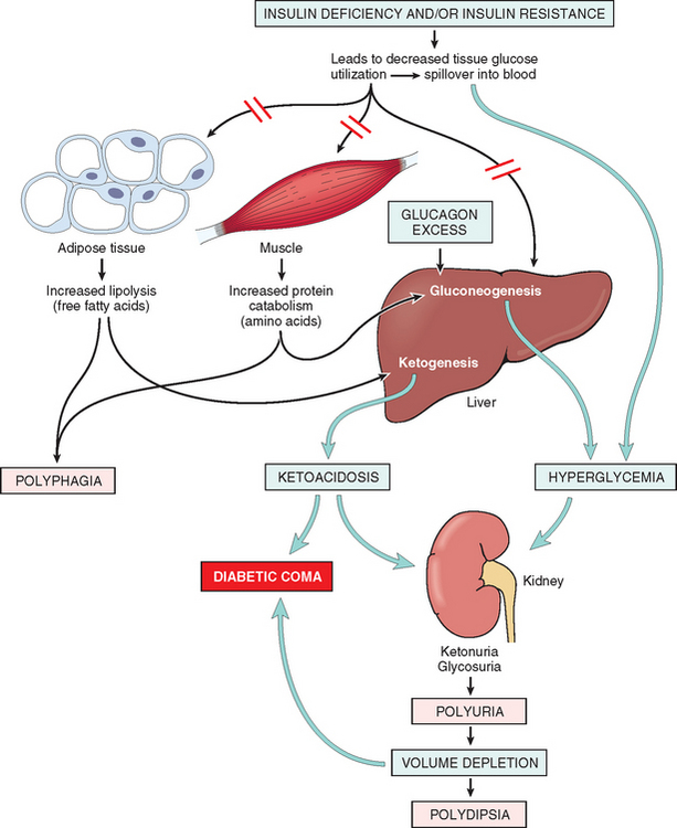
29 Pinhas-Hamiel O, Zeitler P. Acute and chronic complications of type 2 diabetes mellitus in children and adolescents. Lancet. 2007;369:1823.

30 Herman MA, Kahn BB. Glucose transport and sensing in the maintenance of glucose homeostasis and metabolic harmony. J Clin Invest. 2006;116:1767.

31 Youngren JF. Regulation of insulin receptor signaling. Cell Mol Life Sci. 2007;64:873.

32 Imagawa A, Hanafusa T, Miyagawa J, Matsuzawa Y. A novel subtype of type 1 diabetes mellitus characterized by a rapid onset and an absence of diabetes-related antibodies. Osaka IDDM Study Group. N Engl J Med. 2000;342:301.

33 Wellcome Trust Case Control Consortium. Genome-wide association study of 14,000 cases of seven common diseases and 3,000 shared controls. Nature. 2007;447:661.

34 Todd JA, Walker NM, Cooper JD, et al. Robust associations of four new chromosome regions from genome-wide analyses of type 1 diabetes. Nat Genet. 2007;39:857.

35 Jones EY, Fugger L, Strominger JL, Siebold C. MHC class II proteins and disease: a structural perspective. Nat Rev Immunol. 2006;6:271.

36 Park Y. Functional evaluation of the type 1 diabetes (T1D) susceptibility genes. Diabetes Res Clin Pract. 2007;77(Suppl 1):S110.

37 Chistiakov, et al. The crucial role of IL-2/IL-2RA-mediated immune regulation in the pathogenesis of type 1 diabetes, an evidence coming from genetic and animal model studies. Immunol Lett. 2008;118:1.

38 Hviid A, Stellfeld M, Wohlfahrt J, Melbye M. Childhood vaccination and type 1 diabetes. N Engl J Med. 2004;350:1398.

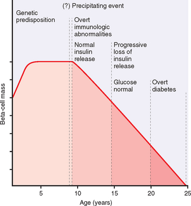
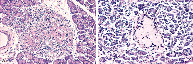
39 Eisenbarth GS. Type I diabetes mellitus. A chronic autoimmune disease. N Engl J Med. 1986;314:1360.

40 Knip M, Siljander H. Autoimmune mechanisms in type 1 diabetes. Autoimm Rev. 2008;7:550.

41 Zhang L, et al. Insulin as an autoantigen in NOD/human diabetes. Curr Opin Immunol. 2008;20:111.

42 Taplin CE, Barker JM. Autoantibodies in type 1 diabetes. Autoimmunity. 2008;41:11.

43 Frayling TM. Genome-wide association studies provide new insights into type 2 diabetes aetiology. Nat Rev Genet. 2007;8:657.

44 Zeggini E, et al. Meta-analysis of genome-wide association data and large-scale replication identifies additional susceptibility loci for type 2 diabetes. Nat Genet. 2008.

45 Michael MD, et al. Loss of insulin signaling in hepatocytes leads to severe insulin resistance and progressive hepatic dysfunction. Molecular Cell. 2000;6:87.

46 Muoio DM, Newgard CB. Mechanisms of disease: molecular and metabolic mechanisms of insulin resistance and beta-cell failure in type 2 diabetes. Nat Rev Mol Cell Biol. 2008;9:193.

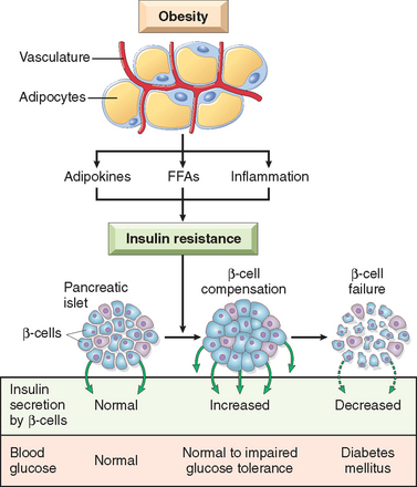
47 Kahn SE, Hull RL, Utzschneider KM. Mechanisms linking obesity to insulin resistance and type 2 diabetes. Nature. 2006;444:840.

48 Long YC, Zierath JR. AMP-activated protein kinase signaling in metabolic regulation. J Clin Invest. 2006;116:1776.

49 Lyssenko V, Lupi R, Marchetti P, et al. Mechanisms by which common variants in the TCF7L2 gene increase risk of type 2 diabetes. J Clin Invest. 2007;117:2155.

50 Babenko AP, et al. Activating mutations in the ABCC8 gene in neonatal diabetes mellitus. N Engl J Med. 2006;355:456.

51 Gloyn AL, et al. Activating mutations in the gene encoding the ATP-sensitive potassium-channel subunit Kir6.2 and permanent neonatal diabetes. N Engl J Med. 2004;350:1838.

52 Murphy R, et al. Clinical features, diagnosis and management of maternally inherited diabetes and deafness (MIDD) associated with the 3243A>G mitochondrial point mutation. Diabet Med. 2008.

53 Stoy J, et al. Insulin gene mutations as a cause of permanent neonatal diabetes. Proc Nat Acad Sci USA. 2007;104:15040.

54 Agostini M, et al. Non-DNA binding, dominant-negative, human PPARgamma mutations cause lipodystrophic insulin resistance. Cell Metab. 2006;4:303.

55 Harja E, et al. Vascular and inflammatory stresses mediate ather-osclerosis via RAGE and its ligands in apoE-/- mice. J Clin Invest. 2008;118:183.

56 Yamamoto Y, et al. Development and prevention of advanced diabetic nephropathy in RAGE-overexpressing mice. J Clin Invest. 2001;108:261.

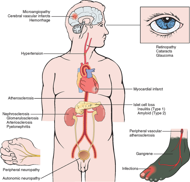
57 Clarke M, Dodson PM. PKC inhibition and diabetic microvascular complications. Best Pract Res Clin Endocrinol Metab. 2007;21:573.

58 Tomlinson DR, Gardiner NJ. Glucose neurotoxicity. Nat Rev Neurosci. 2008;9:36.

59 Daneman D. Type 1 diabetes. Lancet. 2006;367:847.

60 Stumvoll M, et al. Type 2 diabetes: principles of pathogenesis and therapy. Lancet. 2005;365:1333.

61 Boyle PJ. Diabetes mellitus and macrovascular disease: mechanisms and mediators. Am J Med. 2007;120:S12.

62 Zimmet P, et al. Global and societal implications of the diabetes epidemic. Nature. 2001;414:782.

63 Despres JP, Lemieux I. Abdominal obesity and metabolic syndrome. Nature. 2006;444:881.

64 Pi-Sunyer X, et al. Reduction in weight and cardiovascular disease risk factors in individuals with type 2 diabetes: one-year results of the look AHEAD trial. Diabetes Care. 2007;30:1374.

65 Delonlay P, et al. Neonatal hyperinsulinism: clinicopathologic correlation. Hum Pathol. 2007;38:387.

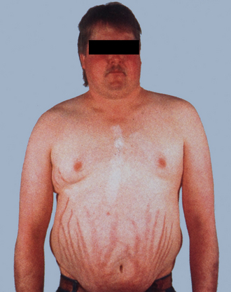
66 Newell-Price J, et al. Cushing’s syndrome. Lancet. 2006;367:1605.

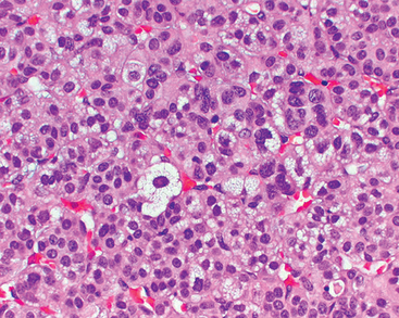
67 Cushing HW. The basophil adenomas of the pituitary body and their clinical manifestations (pituitary basophilism). Bull Johns Hopkins Hosp. 1932;50:137.

68 Mazzuco TL, et al. Aberrant GPCR expression is a sufficient genetic event to trigger adrenocortical tumorigenesis. Mol Cell Endocrinol. 2007;23:265-266.

69 Horvath A, et al. A genome-wide scan identifies mutations in the gene encoding phosphodiesterase 11A4 (PDE11A) in individuals with adrenocortical hyperplasia. Nat Genet. 2006;38:794.

70 Conn JW, Louis LH. Primary aldosteronism: a new clinical entity. Trans Assoc Am Physicians. 1955;68:215.

71 Leopold JA, et al. Aldosterone impairs vascular reactivity by decreasing glucose-6-phosphate dehydrogenase activity. Nature Medicine. 2007;13:189.

72 Goncalves J, et al. Congenital adrenal hyperplasia: focus on the molecular basis of 21-hydroxylase deficiency. Expert Rev Mol Med. 2007;9:1.

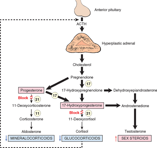
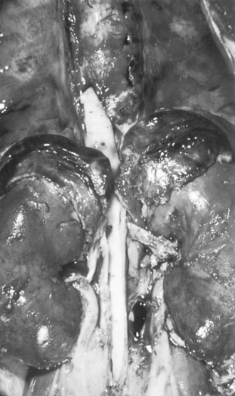
73 Merke DP, et al. Adrenomedullary dysplasia and hypofunction in patients with classic 21-hydroxylase deficiency. N Engl J Med. 2000;343:1362.

74 Waterhouse R. A case of suprarenal apoplexy. Lancet. 1911;1:577.

75 Friderichsen C. Nebennierenapoplexie bei kleinen Kindern. Jahrbuch für Kinderhilkunde. 1918;87:109.

76 Addison T. On the constitutional and local effects of disease of the supra-renal capsules. London: Samuel Highley, 1855.

77 Mathis D, Benoist C. A decade of AIRE. Nat Rev Immunol. 2007;7:645.

78 Young WFJr. Clinical practice. The incidentally discovered adrenal mass. N Engl J Med. 2007;356:601.

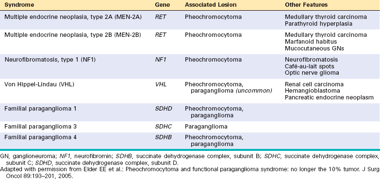
79 Elder EE, et al. Pheochromocytoma and functional paraganglioma syndrome: no longer the 10% tumor. J Surg Oncol. 2005;89:193.

80 Kaelin WG. Von hippel-lindau disease. Annu Rev Pathol. 2007;2:145.

81 Scacheri PC, et al. Genome-wide analysis of menin binding provides insights into MEN1 tumorigenesis. PLoS Genet. 2006;2:e51.

82 Agarwal SK, et al. Transcription factor JunD, deprived of menin, switches from growth suppressor to growth promoter. Proc Nat Acad Sci USA. 2003;100:10770.

83 Gujral TS, et al. Molecular mechanisms of RET receptor-mediated oncogenesis in multiple endocrine neoplasia 2B. Cancer Res. 2006;66:10741.