Pathogenesis

Many causes of pericarditis affect both the pericardium and the myocardium (myopericarditis) with varying degrees of cardiac dysfunction. Constrictive pericarditis is characterized by a fibrotic, thickened, and adherent pericardium that is compressing the heart. The heart becomes restricted in movement and function (cardiac tamponade). Diastolic filling of the heart is reduced, venous pressures are elevated, cardiac output is decreased, and eventual cardiac failure may result.

When fluid accumulates within the pericardial sac it is referred to as pericardial effusion. Blunt chest trauma or any cause of acute pericarditis can lead to pericardial effusion. Rapid distention or excessive fluid accumulation from this condition can also compress the heart and reduce ventricular filling and cardiac output.

Clinical Manifestations

The presentation and course of pericarditis are determined by the underlying etiology. For example, pericarditis may occur 2 to 5 days after infarction as a result of an inflammatory reaction to myocardial necrosis, or it may occur within the first year after radiation initiates a fibrinous and fibrotic process in the pericardium. Often there is pleuritic chest pain that is made worse by lying down and by respiratory movements and is relieved by sitting upright or leaning forward. The pain is substernal and may radiate to the neck, shoulders, upper back, upper trapezius, left supraclavicular area, or epigastrium, or down the left arm.

Other symptoms may include fever, joint pain, dyspnea, or difficulty swallowing. Auscultation of the lower left sternal border where the pericardium lies close to the chest wall will produce a pericardial friction rub, a high-pitched scratchy sound that may be heard at end expiration. This sound is produced by the friction between the pericardial surfaces that results from inflammation and occurs during heart movement. Symptoms of constrictive pericarditis develop slowly and usually include progressive dyspnea, fatigue, weakness, peripheral edema, and ascites. Constrictive disease can lead to diastolic dysfunction and eventual heart failure.

MEDICAL MANAGEMENT

DIAGNOSIS.

Clinical examination, including clinical presentation, auscultation, and client history, may be diagnostic. A classic sign of pericarditis is the pericardial friction rub heard on auscultation. Other diagnostic tools include chest x-ray (showing enlarged cardiac shadow), characteristic ECG changes (showing evidence of an underlying inflammatory process), and laboratory studies (e.g., elevated sedimentation rate or elevated white blood count [nonspecific indicators of inflammation] and elevated cardiac enzymes [post MI]). CT, MRI, and echocardiography are modalities used for imaging the pericardium and pericardial disease.

TREATMENT.

New treatments for pericardial diseases are being developed as a result of modern imaging, new understanding of molecular biology, and immunologic techniques. Comprehensive and systematic implementation of new techniques of pericardiocentesis, pericardial fluid analysis, pericardioscopy, and epicardial and pericardial biopsy, as well as the application of new techniques for pericardial fluid and biopsy analyses, have permitted early specific diagnosis, creating foundations for etiologic intervention in many cases. In cases of recurrent pericarditis resistant to conventional intervention and in the case of neoplastic pericarditis, intrapericardial application of medication has been proposed.207

Conventional treatment remains twofold, directed toward prevention of long-term complications and the underlying cause. For example, while any underlying infection is treated when possible (antibiotics for bacterial pericarditis), symptomatic treatment is provided for idiopathic, viral, or radiation pericarditis; antiinflammatory drugs are given for severe, acute pericarditis or pericarditis associated with connective tissue disorders; chemotherapy is given for neoplastic pericarditis; and dialysis is performed for uremic pericarditis. Analgesics may be prescribed for the pain and fever. Pericardiocentesis (surgical drainage with a needle catheter through a small subxiphoid incision) may be performed if cardiac compression from pericardial effusion does not resolve.

Treatment for constrictive pericarditis is both medical and surgical, including digitalis preparations, diuretics, sodium restriction, and pericardiectomy (surgical excision of the damaged pericardium).

PROGNOSIS.

The prognosis in most cases of acute viral pericarditis is excellent when there is no (or only minimal) myocardial involvement, since this is frequently a self-limited disease. Without medical intervention, shock and death can occur from decreased cardiac output with cardiac involvement. Constrictive pericarditis is a progressive disease without spontaneous reversal of symptoms. Most people become progressively disabled over time. Surgical removal of the pericardium is associated with a high mortality rate when progressive calcification in the epicardium and dense adhesions or fibrosis between the pericardial layers are present.

12-16   SPECIAL IMPLICATIONS FOR THE THERAPIST

Pericarditis

PREFERRED PRACTICE PATTERN

6D:

Impaired Aerobic Capacity/Endurance Associated with Cardiovascular Pump Dysfunction or Failure

Pericardial pain can masquerade as a musculoskeletal problem, presenting as just upper back, neck, or upper trapezius pain. In such cases, the pain may be diminished by holding the breath or aggravated by swallowing or neck or trunk movements, especially side bending or rotation.

Pain is also aggravated by respiratory movements, such as deep breathing, coughing, and laughing. The therapist must screen for medical disease by assessing aggravating and relieving factors and by asking the client about a history of fever, chills, upper respiratory tract infection (recent cold or flu), weakness, heart disease, or recent MI (heart attack).

Special precautions depend on the underlying cause of the pericarditis. Mild cases require intervention per client tolerance, and the therapist observes for any symptoms of CHF. A mild pericarditis can quickly progress to a severe condition that requires medical evaluation. The clinician is referred to each individual section in this text representing the etiology of pericarditis for precautions.

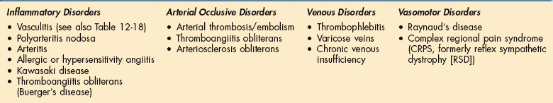
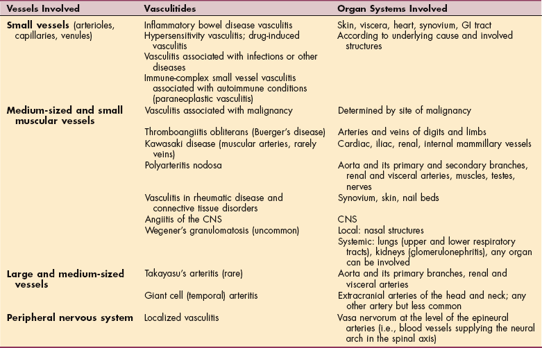
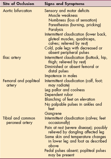
DISEASES AFFECTING THE BLOOD VESSELS

Diseases of blood vessels observed in a therapy setting can include intestinal infarction, aneurysm, PVD, vascular neoplasm, and vascular malformation; only intestinal infarction will not be discussed here.

Aneurysm

Definition and Overview

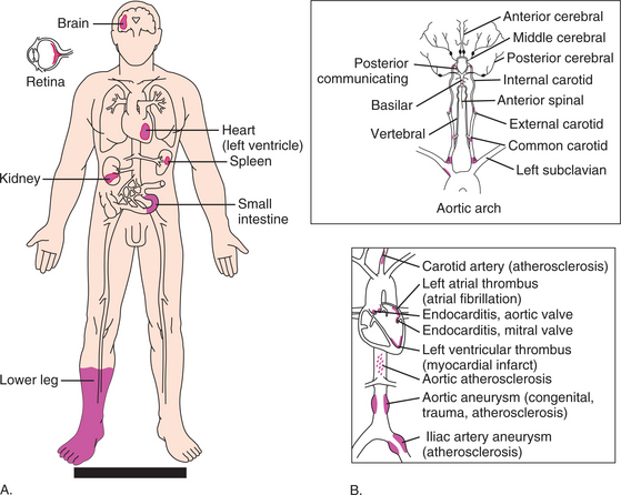
An aneurysm is an abnormal stretching (dilation) in the wall of an artery, a vein, or the heart with a diameter that is at least 50% greater than normal. When the vessel wall becomes weakened from trauma, congenital vascular disease, infection, or atherosclerosis, a permanent saclike formation develops. A false aneurysm can occur when the wall of the blood vessel is ruptured and blood escapes into surrounding tissues, forming a clot (Fig. 12-25; see also Fig. 12-27).

image

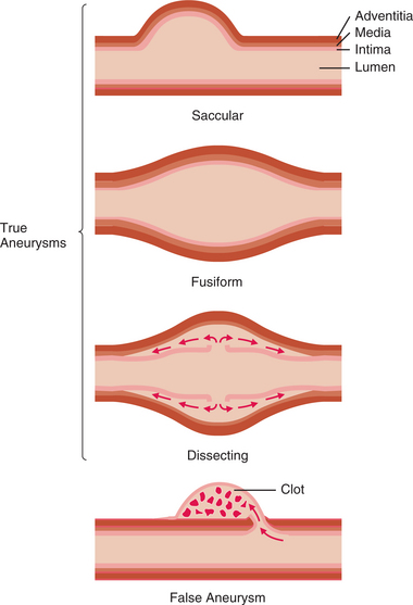
Figure 12-25 Longitudinal sections showing types of aneurysms. In a true aneurysm, layers of the vessel wall dilate in one of the following ways: saccular, a unilateral outpouching; fusiform, a diffuse dilation involving the entire circumference of the artery wall; or dissecting, a bilateral outpouching in which layers of the vessel wall separate, with creation of a cavity. In a false aneurysm, the wall ruptures, and a blood clot is retained in an outpouching of tissue.

Aneurysms are of various types (either arterial or venous) and are named according to the specific site of formation (Fig. 12-26). The most common site for an arterial aneurysm is the aorta, forming a thoracic aneurysm (which involves the ascending, transverse, or first part of the descending portion of the aorta) or an abdominal aneurysm (which generally involves the aorta between the renal arteries and iliac branches).

image

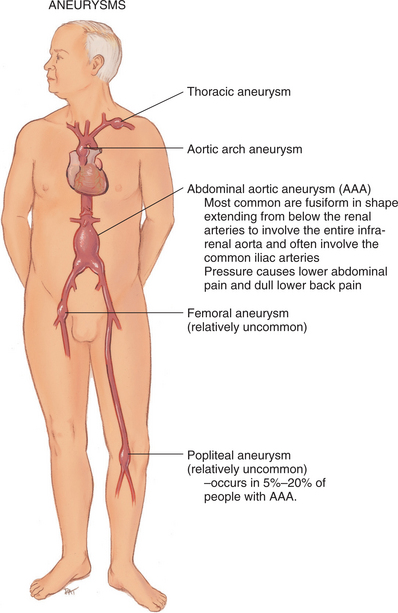
Figure 12-26 Aneurysms are named according to the specific site of formation. Abdominal aortic aneurysms are the most common type; more than 95% of abdominal aortic aneurysms are located below the renal arteries and extend to the umbilicus, causing low back pain. (From Jarvis C: Physical examination and health assessment, ed 5, Philadelphia, 2008, Saunders.)

Thoracic aortic aneurysms located above the diaphragm account for approximately 10% of all aortic aneurysms and occur most frequently in hypertensive men between the ages of 40 and 70 years. Men are more likely to have thoracic or abdominal aneurysms. Thoracic aortic aneurysms occur less often than other types but tend to be more life-threatening.

Abdominal aortic aneurysms located below the diaphragmatic border occur about four times more often than thoracic aneurysms, most likely because the aorta is not supported by skeletal muscle at this location. The incidence of abdominal aortic aneurysm is increasing, probably because of the increasing number of adults over 65 years of age.

Peripheral arterial aneurysms affect the femoral and popliteal arteries.

Incidence and Etiologic Factors

According to the Society for Vascular Surgery, approximately 200,000 people in the United States are diagnosed annually with aortic aneurysm and 15,000 of those aneu- rysms are severe enough to rupture, causing a medical emergency.312

Incidence increases with increasing age, usually beginning after age 50 years, presumably as a result of chronic inflammatory cellular changes resulting in atherosclerosis. However, someone without evidence of atherosclerosis can develop an aneurysm, especially in the presence of congenital weakness of the blood vessel walls.

Family members (parent, adult child, or sibling) of anyone with an aneurysm have a fourfold increased risk of aneurysm, and gene defects on chromosomes 11340 and 15172 have been identified with some of the connective tissue disorders associated with aneurysm. Recently, a mutation in a specific protein, transforming growth factor β receptor (TGFBR), has been identified as responsible for causing aneurysms.200 Aneurysms occur much more often in men than in women, and one half of affected persons are hypertensive.

Atherosclerosis or any injury to the middle or muscular layer of the arterial wall (tunica media) is responsible for most arterial aneurysms. Other less common causes of aneurysm include trauma (blunt or surgical), Marfan’s disease (congenital defects of the arterial wall) and other hereditary abnormalities of connective tissue, and inflammatory diseases and infectious agents (bacterial infection, syphilis, polyarteritis).

The emergence of HIV has been associated with a dramatic increase in the incidence of syphilis. Since syphilitic aortitis generally presents between 10 and 30 years after the primary infection, there may be an increased incidence of associated aneurysms in the coming years. Hypertension seems to enhance aneurysm formation.

Pathogenesis

Plaque formation erodes the vessel wall, predisposing the vessel to stretching of the inner and outer layers of the artery and formation of a sac. The stretching of the media produces infarct expansion, a weak and thin layer of necrotic muscle, and fibrous tissue that bulges with each systole. Abnormal proteolysis, the presence of elastolytic serum enzymes, and deficiencies of collagen and elastin have been implicated as factors contributing to the development of these aneurysms.184

With time, the aneurysm becomes more fibrotic, but it continues to bulge with each systole, thus acting as a reservoir for some of the stroke volume. In the case of thoracic aortic aneurysms, the shear force of elevated blood pressure causes a tear in the intima with rapid disruption and rupture of the aortic wall. Subsequent hemorrhage causes a lengthwise splitting of the arterial wall, creating a false vessel (Fig. 12-27), and a hematoma may form in either channel (i.e., the false or true lumen).

Clinical Manifestations

Aneurysms may be asymptomatic; when they do occur, manifestations depend largely on the size and position of the aneurysm and its rate of growth. Persistent but vague substernal, back, neck, or jaw pain may occur as enlargement of the aneurysm impinges adjacent structures.

Dissection over the aortic arch and into the descending aorta may be experienced as extreme, sharp pain felt at the base of the neck or along the back into the interscapular areas. When pressure from a large volume of blood is placed on the trachea, esophagus, laryngeal nerve, lung, or superior vena cava, symptoms of dysphagia; hoarseness; edema of the neck, arms, or jaw and distended neck veins; and dyspnea and/or cough may occur, respectively.

Other signs and symptoms may be present in the case of acute aortic dissection as a result of compression of branches of the aorta. These include acute MI, reversible ischemic neurologic deficits, stroke, paraplegia, renal failure, intestinal ischemia, and ischemia of the arms and legs. Acute chest pain may also result from a nondissecting hematoma of the aorta or erosion of a penetrating atherosclerotic ulcer.184

In the case of an untreated abdominal aortic aneurysm expansion and rupture can occur in one of several places, including the peritoneal cavity, the mesentery, the retroperitoneum, into the inferior vena cava, or into the duodenum or rectum. Rupture refers to a tearing of all three tunicae (tunica adventitia, tunica media, tunica intima) with bleeding into the thoracic or abdominal cavity. The most common site for an abdominal aortic aneurysm is just below the renal arteries, and it may involve the bifurcation of the aorta (see Fig. 12-26).

Most abdominal aortic aneurysms are asymptomatic, but intermittent or constant pain in the form of mild to severe mid-abdominal or lower back discomfort is present in some form in 25% to 30% of cases. Groin or flank pain may be experienced because of increasing pressure on other structures.

Early warning signs of an impending rupture may include abdominal heartbeat when lying down or a dull ache (intermittent or constant) in the mid-abdominal left flank or lower back. Rupture is most likely to occur in aneurysms that are 5 cm or larger, causing intense flank pain with referred pain to the back at the level of the rupture. Pain may radiate to the lower abdomen, groin, or genitalia. Back pain may be the only presenting symptom before rupture occurs.

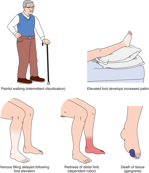
The most common site for peripheral arterial aneurysm is the popliteal space in the lower extremities. Most are caused by atherosclerosis and occur bilaterally in men. Popliteal aneurysm presents as a pulsating mass, 2 cm or more in diameter, and causes ischemic symptoms in the lower limbs (e.g., intermittent claudication, rest pain, thrombosis and embolization resulting in gangrene). Femoral aneurysm presents as a pulsating mass in the femoral area on one or both sides.

MEDICAL MANAGEMENT

DIAGNOSIS.

Detection of abdominal and peripheral aneurysms often occurs when the physician palpates a pulsating mass during routine examination or when x-rays are taken for other purposes (although not all aortic aneurysms show abnormalities on chest radiography). Radiography, ultrasonography, echocardiography with color Doppler imaging, CT and MRI, arteriography, and aortography may be used for investigation.

PREVENTION AND TREATMENT.

Annual examination to ensure early identification is recommended for family members (parent, adult child, or sibling) of anyone who has previously been diagnosed with an aortic aneurysm. Anyone with a family risk or signs of diseased arteries should take preventive measures, including smoking cessation, regular exercise, blood pressure control, and cholesterol management.

Treatment is determined based on the size of the bulge, how fast it is expanding, and the individual’s clinical presentation. For small aneurysms, watchful waiting is often advised. Preventive pharmacology (e.g., statin to lower cholesterol, β-blocker or ACE inhibitor to control blood pressure) may be prescribed depending on individual factors.

Surgical intervention before rupture provides a good prognosis; at 5 cm, the risk of rupture exceeds the risk of repair. A new, less invasive procedure known as endoluminal stent-graft may offer an alternative to open abdominal surgery. Guided by angiographic imaging, a catheter is inserted through the femoral or brachial artery to the aneurysm. A balloon within the catheter is then inflated, pushing open the stent, which attaches with tiny hooks to healthy arterial wall above and below the aneurysm. This creates a channel for blood flow that bypasses the aneurysm.

PROGNOSIS.

The standard open surgical approach to replace the diseased aorta is steadily improving but is still associated with high morbidity and substantial mortality rates. MI, respiratory failure, renal failure, and stroke are the principal causes of death and morbidity after surgical procedures performed on the thoracic aorta.

At the same time, the endoluminal stent-graft comes with its own set of complications, including fever, breakdown or migration of the device, leaks, and unknown durability. Further studies to improve treatment are ongoing. Aneurysm rupture is associated with a high mortality; frequently, aneurysms are discovered only at autopsy.

12-17   SPECIAL IMPLICATIONS FOR THE THERAPIST

Aneurysm

PREFERRED PRACTICE PATTERNS

4J:

Impaired Motor Function, Muscle Performance, Range of Motion, Gait, Locomotion, and Balance Associated with Amputation (peripheral aneurysm)

5D:

Impaired Motor Function and Sensory Integrity Associated with Nonprogressive Disorders of the Central Nervous System—Acquired in Adolescence or Adulthood

5I:

Impaired Arousal, Range of Motion, and Motor Control Associated with Coma, Near Coma, or Vegetative State

Anyone with complications associated with an aneurysm is also at risk for pulmonary complications.

Since the prevalence of all diseases of the aorta increases with age and because the population in the United States is aging, it is expected that aortic aneurysm will be encountered with increasing frequency. Knowledge of the natural history, familial history, and clinical features of this disorder may alert the therapist to the need for medical intervention.

For the person who has had a surgically repaired aneurysm, activities are restricted and are only gradually reintroduced. The therapist may be involved in bedside exercises and early mobility, which are especially important to prevent thromboembolism as a result of venous stasis during prolonged bed rest and immobility.

Because of the invasiveness of open abdominal surgery, anyone undergoing this procedure is at high risk for pulmonary complications. Incisional pain and the use of abdominal musculature in coughing discourage the person from full inspirations as well as effective forceful huffing or coughing. The acute care therapist will utilize clinical techniques to assist with cough with pillows or towel rolls at the incisional site and forceful huffing (see description in Chapter 15).150

Proper lifting techniques should be reviewed before discharge, even though the client will not be able to provide a return demonstration. Activities that require pushing, pulling, straining, or lifting more than 10 lb are restricted for 6 to 10 weeks postoperatively.

Anterior or abdominal soft tissue mobilization for persons with back pain who have postoperative abdominal scars may require indirect techniques. This precaution is especially true for the person with a previous abdominal aneurysm, the person with a known nonoperative aneurysm (less than 5 cm), or the person with a family history of aneurysm or an undiagnosed aneurysm.

The therapist must always palpate the abdomen for a pulsating mass before performing anterior or abdominal therapy. It is possible to palpate the width of the pulse beginning at the abdominal midline and progressing laterally. The pulse should be characterized by a uniform width on either side of the abdominal midline until the umbilicus is reached, at which point the aortic bifurcation results in expansion of the pulse width. Throbbing pain that increases with exertion should alert the therapist to the need to monitor vital signs and palpate pulses.

Peripheral Vascular Disease

Although PVD is usually thought to refer to diseases of the blood vessels supplying the extremities, in fact, PVD actually encompasses pathologic conditions of blood vessels supplying the extremities and the major abdominal organs, most often apparent in the intestines and kidneys.

PVD is organized based on the underlying pathologic finding (e.g., inflammatory, arterial occlusive, venous, or vasomotor disorders) (Box 12-13). Although the terms peripheral arterial disease (PAD) and peripheral vascular disease (PVD) are often used interchangeably, PVD is a broader, more encompassing grouping of disorders of both the arterial and venous blood vessels, whereas PAD only refers to arterial blood vessels. PVD typically affects the legs more often than the arms, but upper extremity involvement is not uncommon.

Box 12-13   PERIPHERAL VASCULAR DISEASES

image

Approximately 8 million Americans over age 60 years are affected by PVD, with 20% of those people over age 70 years. Like CAD and cerebrovascular disease, arterial occlusive forms of PVD are most common as a result of atherosclerosis. Intermittent claudication is the classic symptom of PAD. Like angina associated with CAD, intermittent claudication associated with PAD is predictable and nearly always develops after the same amount of exertion (e.g., walking a specific distance), generally occurs in the calves and less commonly in the thighs and buttocks,68 and usually improves rapidly with rest.

Data from the Framingham Heart Study and other population studies indicate that intermittent claudication sharply increases in late middle age and is somewhat higher among men than women. In fact the true prevalence of PAD is at least five times higher than expected based on the reported prevalence of intermittent claudication.87 Specific symptoms of the various forms of PVD depend on the underlying pathologic condition, the blood vessels involved (arteries or veins), and the location of the affected blood vessels; each form is discussed individually in the following sections.

Inflammatory Disorders

Inflammatory conditions of the blood vessels are often discussed as immunologic conditions, because inflammation and damage to large and small vessels result in end-stage organ damage. Vasculitis (e.g., arteritis, such as polyarteritis nodosa and giant cell arteritis, Kawasaki disease) is the most commonly encountered inflammatory blood vessel disease in a therapy practice.

Vasculitis is actually a group of disorders that share a common pathogenesis of inflammation of the blood vessels involving arteries, veins, or nerves, resulting in narrowing or occlusion of the lumen or formation of aneurysms that can rupture. Vascular inflammation is a central feature of many rheumatic diseases, especially rheumatoid arthritis and scleroderma. (See also the section on Rheumatoid Vasculitis in Chapter 5.)

Vasculitis.: Vasculitis can involve blood vessels of any size, type, or location and can affect any organ system, including the nervous system; classification is usually according to the size of the predominant vessels involved (Table 12-18). Vasculitis may be acute or chronic with varying degrees of involvement. The distribution of lesions may be irregular and segmental rather than continuous.

Table 12-18

Vasculitis

image

GI, Gastrointestinal; CNS, central nervous system.

Neurologic manifestations of vasculitis can occur in conjunction with any of the vasculitides listed, affecting the peripheral nervous system or the CNS. Vasculitis may occur as an isolated peripheral nerve vasculitis (localized vasculitis). Numerous vasculitic diseases have been reported in association with HIV disease. The primary target organ involvement is usually muscle and nerve, skin, testicle, kidney, and, less often, the CNS.

Immune (antibody-antigen) complexes to each disorder are deposited in the blood vessels, resulting in varying symptoms depending on the organs affected. In the case of vasculitic neuropathy, the formation of antibody-antigen complexes activates the complement cascade with generation of C3a and C5a (chemotactic agents that recruit polymorphonuclear [PMN] leukocytes to the vessel walls).

Phagocytosis of the immune complexes takes place, and release of free radicals and proteolytic enzymes disrupts cell membranes and damages blood vessel walls. The complement cascade generates the formation of complement membrane attack complex that also contributes to endothelial damage (see discussion in Chapter 6; see also Fig. 6-15). The resulting damage to endothelial cells results in thickening of the vessel wall, occlusion, and ischemia of the affected nerves with axonal degeneration and the resultant neuropathy.

Polyarteritis Nodosa:

Overview and Etiologic Factors.: Polyarteritis nodosa refers to a condition consisting of multiple sites of inflammatory and destructive lesions in the arterial system; the lesions are small masses of tissue in the form of nodes or projections (nodosum). The cause of polyarteritis nodosa is unknown, although hepatitis B is present in 50% of cases, and polyarteritis occurs more commonly among IV drug abusers and other groups who have a high prevalence of hepatitis B (see the section on Hepatitis B in Chapter 17). Any age can be affected, but it is more common among young men.

Clinical Manifestations.: Polyarteritis nodosa affects small and medium-sized blood vessels, resulting in a variety of clinical presentations depending on the specific site of the blood vessel involved. Some of the more likely symptoms include abrupt onset of fever, chills, tachycardia, arthralgia, and myositis with muscle tenderness.

Any organ of the body may be affected, but most often involved are the kidneys, heart, liver, GI tract, muscles, and testes. Abdominal pain, nausea, and vomiting are common with GI tract involvement. Pericarditis, myocarditis, arrhythmias, and MI reflect cardiac involvement. Complications may include aneurysm, hemorrhage, thrombosis, and fibrosis leading to occlusion of the lumen. Multiple asymmetric neuropathies (motor and sensory distribution) can occur when vasculitis affects the arteries of the peripheral nerves (vasa nervorum). Paresthesias, pain, weakness, and sensory loss occur, involving several or many peripheral nerves simultaneously.

MEDICAL MANAGEMENT

DIAGNOSIS, TREATMENT, AND PROGNOSIS.

Diagnosis is made by characteristic laboratory findings, biopsy of symptomatic sites (especially muscle or nerve), and, possibly, visceral angiography. When CNS vasculitis is suspected, angiography is necessary, because MRI and CT do not provide sufficient evidence to confirm the diagnosis.

Prolonged use of corticosteroids is necessary to control fever and constitutional symptoms while vascular lesions are healing. Immunosuppressants may be used in conjunction with steroids to improve survival; withdrawal from drugs is often followed by relapse. Treatment of polyarteritis nodosa associated with hepatitis B is more complicated, because cytotoxic drugs used to treat the vasculitis can exacerbate the hepatic disease. Prognosis is poor without intervention, with a 5-year survival rate of only 20%. Pharmacologic therapy with corticosteroids increases survival to 50%, and steroids combined with immunosuppressive drugs have improved 5-year survival to 90%.

Arteritis:

Overview and Incidence.: Arteritis, sometimes called giant cell arteritis (GCA) or cranial or temporal arteritis, is a vasculitis primarily involving multiple sites of temporal and cranial arteries (i.e., arteries of the head and neck and sometimes the aortic arch).

It is the most common vasculitis in the United States and affects older people; the incidence increases with age after 50 years. Postmenopausal women are affected twice as often as men, especially those individuals who have an arthritis-related disease called polymyalgia rheumatica (PMR). Other risk factors identified in women include heart murmurs and smoking.

Etiologic Factors and Pathogenesis.: Most studies have shown an association of GCA with a specific human leukocyte antigen (HLA) allele, and tumor necrosis factor appears to influence the susceptibility to both GCA and PMR. There may be an infectious origin, but additional studies are necessary to clarify the genetic influence on susceptibility to these conditions. The molecular pathogenesis of GCA involves interleukin-1, intercellular adhesion molecules, and other factors, but the exact pathogenesis remains unknown. The possible role of female sex hormones requires further investigation.

Immunologic research indicates an antigen-driven disease with local T-cell and macrophage activation in vessel walls with calcified atrophic media. The middle layer (tunica media) of the large and medium-sized arteries, particularly those blood vessels supplying blood to the head, is inflamed, causing the arteries to swell and obstruct blood flow (stenosis); ischemic complications and secondary thrombosis may occur. Healing produces fibrosis of the arterial wall and the affected blood vessel becomes cordlike, thickened, and nodular, which can be observed externally when the temporal artery is involved.

Clinical Manifestations.: The onset of arteritis is usually sudden, with severe, continuous, unilateral, throbbing headache and temporal pain as the first symptoms, with flulike symptoms or visual disturbances. The pain may radiate to the occipital area, face, or side of the neck. Visual disturbances range from blurring to diplopia to visual loss. Irreversible blindness may occur anywhere in the course of the disease from involvement of the ophthalmic artery.

Other symptoms may include enlarged, tender temporal artery; scalp sensitivity; and jaw claudication (i.e., pain in response to chewing, talking, or swallowing) when involvement of the external carotid artery causes ischemia of the masseter muscles; the pain is relieved by rest.

Approximately 40% of cases present with nonclassic symptoms of respiratory tract problems (most often dry cough), fever of unknown origin, painful paralysis of a shoulder (mononeuritis multiplex), or claudication of the arm with cold hands, arm weakness, and absent radial pulses.177 Left untreated, the condition may lead to blindness and occasionally to a stroke, heart attack, or aortic dissection.

MEDICAL MANAGEMENT

DIAGNOSIS.

Early diagnosis is important to prevent blindness caused by obstruction of the ophthalmic arteries. Diagnosis is made by recognition of the presenting symptoms, and in some cases, arteritis follows PMR, a similar condition. Although GCA and PMR may occur as separate entities, most epidemiologic surveys group these two conditions together as one disorder. These may be two forms of a common pathophysiologic process characterized by varying degrees of synovitis and arteritis. They may actually represent two points along a single disease continuum.

Biopsy of the temporal artery may be performed, but results are often negative given the focal (segmental) nature of the disease. Color ultrasonography of the temporal arteries detects characteristic signs of vasculitis with a high sensitivity and specificity even in the absence of clinical signs of vascular inflammation (helpful in diagnosing temporal arteritis in people with previously diagnosed PMR).

People with extracranial GCA present with occlusive arterial lesions that may be detected with multiple imaging modalities: arteriography, IV digital subtraction angiography (IV-DSA), CT scanning, and MRA. However, inflammation of the arterial wall cannot be detected by these means.

Standard CT imaging with contrast enhancement and certain MR sequences as well as ultrasound permit identification of the edema and inflammation of the vessel wall. This is an important marker for active disease. Laboratory findings include elevated erythrocyte sedimentation rate, reflective of the underlying inflammatory process.

TREATMENT AND PROGNOSIS.

Treatment of arteritis to prevent blindness and other vascular complications is with oral antiinflammatory drugs (usually a corticosteroid such as prednisone), providing symptomatic relief in 3 to 5 days. Visual loss can be permanent if allowed to persist for several hours without adequate intervention. With proper intervention, arteritis is a self-limiting disease, usually resolving within 6 to 12 months. About 30% of affected individuals relapse in the first year of treatment during dose tapering. Alternative therapies of combined pharmacology with corticosteroids and methotrexate may be more effective in controlling disease with fewer complications.170

Hypersensitivity Angiitis.: Hypersensitivity angiitis, a form of vasculitis, can occur at any age, but it most commonly affects children and young adults. The etiology is unknown, but the disease often follows an upper respiratory tract infection, and allergy or drug sensitivity plays a role in some cases. It is usually localized to the small vessels of the skin, first appearing on the lower extremities in a variety of possible lesions.

A classic triad of symptoms occurs in 80% of cases that includes purpura (bruising and petechiae or round purplish red spots under the skin), arthritis, and abdominal pain. Inflammation and hemorrhage may occur in the synovium and CNS. Medical management (diagnosis, treatment, prognosis) is the same as for the other forms of vasculitis already discussed.

Kawasaki Disease:

Overview and Etiologic Factors.: Kawasaki disease, also known as mucocutaneous lymph node syndrome, is an acute febrile illness associated with systemic (multiorgan) vasculitis. It can occur in any ethnic group but seems most prevalent in Asian populations (especially Japanese, with equal incidence in Japan and in the United States among Japanese or Japanese descendants).

Children under the age of 5 years comprise 80% of all cases, and 20% develop cardiac complications that can be fatal. The etiology is unknown, but because seasonal and geographic outbreaks appear to occur, an infectious cause is suspected. Current etiologic theories center on an immunologic response to an infectious, toxic, or antigenic substance.197

Pathogenesis.: Substantial evidence suggests that immune activation has a role in the pathogenesis of Kawasaki syndrome. The principal area of pathologic findings is the cardiovascular system. Kawasaki disease progresses pathologically and clinically in stages. During the acute stage of the illness (first 2 weeks) vascular inflammation and immune activation within the arterioles, venules, and capillaries occur, which later progress (stage 2, weeks 2 to 4) to include the main coronary arteries, the heart, and the larger veins.

The acute phase is also associated with the appearance of circulating antibodies that are cytotoxic against vascular endothelial cells; the presence of elevated anticardiac myosin autoantibodies may be involved in the myocardial damage that occurs in this phase. In the final stage the vessels develop scarring, intimal thickening, calcification, and formation of thrombi. If death occurs as a result of this disease (rare), it is usually the result of aneurysm, coronary thrombosis, or severe scar formation and stenosis of the main coronary artery.

Clinical Manifestations.: Clinical manifestations present in three phases: acute phase, subacute phase, and convalescent phase. In the acute phase, a sudden high fever (lasting over 5 days) that is unresponsive to antibiotics and antipyretics is followed by extreme irritability.

During the subacute phase (lasting approximately 25 days), the fever resolves, but the irritability persists along with other symptoms, such as anorexia, rash (exanthema) of the trunk and extremities with reddened palms and soles of the hands and feet, and subsequent desquamation (skin scales off) of the tips of the toes and fingers, peripheral edema of the hands and feet, cervical lymphadenopathy (usually unilateral), bilateral conjunctival infection without exudate, and changes in the oral mucous membranes (e.g., erythema, dryness and cracks or fissures of the lips, reddening or strawberry tongue).

In one third of all cases, children develop arthralgias and GI tract symptoms, typically lasting about 2 weeks. Joint involvement may persist for as long as 3 months. During this subacute phase, the person is at risk for cardiac involvement, especially the development of myocarditis, pericarditis, and arteritis that predisposes to the formation of coronary artery aneurysm in nearly 25% of cases not treated within 10 days of fever onset.

The convalescent phase occurs 6 to 8 weeks after onset of Kawasaki disease and is characterized by a resolution of all clinical signs and symptoms. However, during this phase the blood values have not returned to normal. At the end of the convalescent phase, all values return to normal and the child has usually regained his or her usual temperament, energy, and appetite.

MEDICAL MANAGEMENT

DIAGNOSIS, TREATMENT, AND PROGNOSIS.

Early recognition and prompt management of the acute syndrome are critical. Diagnosis is made on the basis of clinical manifestations and associated laboratory tests (there are no specific laboratory tests for Kawasaki disease). Echocardiograms are useful in providing a baseline and for monitoring myocardial and coronary artery status.

The introduction of high-dose IV immune globulin (IVIG) in combination with aspirin therapy to reduce fever and control inflammation and aneurysm formation has significantly reduced the prevalence of coronary artery abnormalities. The exact mechanism by which this treatment intervention reduces the vasculitis of acute Kawasaki syndrome has not been determined.

Prognosis is good for recovery with intervention, although serious cardiovascular problems (e.g., coronary thrombosis, aneurysm) may occur later in persons with cardiac sequelae. Giant aneurysms (diameter exceeding 8 mm) have the worst prognosis, since these are unlikely to regress or resolve, with death common in this subgroup population. Occasionally, severe ischemic heart disease requires cardiac transplantation.197

Thromboangiitis Obliterans (Buerger’s Disease):

Overview and Pathogenesis.: Thromboangiitis obliterans, also referred to as Buerger’s disease, is a vasculitis (inflammatory and thrombotic process) affecting the peripheral blood vessels (both arteries and veins), primarily in the extremities. The cause is not known, but it is most often found in men younger than 40 years who smoke heavily, although the incidence in women is increasing. There has been some suggestion that unrecognized cocaine use may masquerade as Buerger’s disease.210

The pathogenesis of thromboangiitis obliterans is unknown; general inflammatory concepts apply. The inflammatory lesions of the peripheral blood vessels are accompanied by thrombus formation and vasospasm occluding and eventually obliterating (destroying) small and medium-sized vessels of the feet and hands.

Recent studies have linked elevated levels of homocysteine to Buerger’s disease. Homocysteine has many potential effects: it limits the bioavailability of nitric oxide, impairs endothelium-dependent vasorelaxation, increases oxidative stress, stimulates smooth muscle cell proliferation, alters the elastic properties of vessel walls, and generates a prethrombotic state through the activation of factor V.

Clinical Manifestations.: Clinical manifestations of pain and tenderness of the affected part are caused by occlusion of the arteries, reduced blood flow, and subsequent reduced oxygenation. The symptoms are episodic and segmental, meaning that the symptoms come and go intermittently over time and appear in different asymmetric anatomic locations. The plantar, tibial, and digital vessels are most commonly affected in the lower leg and foot. Intermittent claudication centered in the arch of the foot or the palm of the hand is often the first symptom.

When the hands are affected, the digital, palmar, and ulnar arteries are most commonly involved. Pain at rest occurs, with persistent ischemia of one or more digits. Other symptoms include edema, cold sensitivity, rubor (redness of the skin from dilated capillaries under the skin), cyanosis, and thin, shiny, hairless skin (trophic changes) from chronic ischemia. Paresthesias, diminished or absent posterior tibial and dorsalis pedis pulses, painful ischemic ulceration, and eventual gangrene may develop (see Fig. 12-29). Inflammatory superficial thrombophlebitis is common.

image

Figure 12-29 Signs and symptoms of arterial insufficiency.

MEDICAL MANAGEMENT

DIAGNOSIS, TREATMENT, AND PROGNOSIS.

Arteriography may be used in the diagnosis, but definitive diagnosis of thromboangiitis obliterans is determined by histologic examination of the blood vessels (microabscesses in the vessel wall) in a leg amputated for gangrene. Given the new findings that cocaine use may present very much like Buerger’s disease, laboratory screening for drug use may be appropriate in some cases.210

Intervention should begin with cessation of smoking and avoidance of any environmental or secondhand smoke inhalation. All other treatment techniques are aimed at improving circulation to the foot or hand, including pharmacologic intervention (e.g., vasodilators, pain relief) and physical or occupational therapy (see also Atherosclerosis: Medical Management).

Regional sympathetic ganglionectomy may produce vasodilation, ulcerations require wound care, and amputation (sometimes multiple) may be required when the individual is unable to quit smoking or when conservative care fails. With the recent finding of elevated levels of plasma homocysteine associated with Buerger’s disease, screening for hyperhomocysteinemia and treatment of this condition have been recommended, especially to assess which clients may eventually require amputation.

Thromboangiitis is not life-threatening, but it can result in progressive disability from pain and loss of function secondary to amputation. Cessation of smoking is the key determinant in prognosis.

12-18   SPECIAL IMPLICATIONS FOR THE THERAPIST

Inflammatory Disorders

PREFERRED PRACTICE PATTERNS

4A:

Primary Prevention/Risk Reduction for Skeletal Demineralization (Osteoporosis)

5G:

Impaired Motor Function and Sensory Integrity Associated with Acute or Chronic Polyneuropathies (peripheral neuropathy)

7A, 7D, 7E:

(integumentary patterns) Associated with Buerger’s disease

See also Special Implications for the Therapist: Peripheral Vascular Disease. Additional practice patterns may be appropriate if there is CNS involvement.

Peripheral neuropathy is a well-known and frequently early manifestation of many vasculitic syndromes. The pattern of neuropathic involvement depends on the extent and temporal progression of the vasculitic process that produces ischemia. A severe, burning dysesthetic pain in the involved area is present in 70% to 80% of all cases.

Other symptoms may include paresthesias and sensory deficit; severe proximal muscle weakness and muscular atrophy can occur secondary to the neuropathy. In the early phase, one nerve is affected and causes symptoms in one extremity (mononeuritis multiplex), but other nerves can become involved as the disorder progresses.

The therapist should watch for anyone with neuropathy who exhibits constitutional symptoms, such as fever, arthralgia, or skin involvement. This may herald a possible vasculitic syndrome and requires medical referral for accurate diagnosis. Early recognition of vasculitis can help prevent a poor outcome. With no treatment or with a poor outcome to intervention, CNS involvement (e.g., encephalopathy, ischemic and hemorrhagic stroke, cranial nerve palsy) can occur late in the course of vasculitis.

When corticosteroids (e.g., prednisone alone or sometimes in combination with other medications) are used (e.g., in the case of vasculitic neuropathy), the therapist must be aware of the need for osteoporosis prevention and attend to the other potential side effects from the chronic use of these medications (see the section on Corticosteroids in Chapter 5).

Alternative methods of pain control may be offered in a rehabilitation setting, such as biofeedback, transcutaneous electrical nerve stimulation (TENS), and physiologic modulation (e.g., using a handheld temperature sensor to control autonomic nervous system function; see the section on Fibromyalgia in Chapter 7).

Vasculitis (Inflammatory Disease of Arteries and Veins)

The therapist’s role in management of vasculitis may be primarily for relief of painful muscular and joint symptoms when present and in the prevention of functional loss in the case of neuropathies. For the client with thromboangiitis obliterans (Buerger’s disease), exercise must be graded to avoid claudication and the client must be instructed in a home program for preventive skin care (see Box 12-14). Gangrene can occur as a result of prolonged ischemia from vessel obliteration; clients are typically treated for wound care and postoperatively after amputation. (See the section on Arteriosclerosis Obliterans below.)

Box 12-14   GUIDELINES FOR SKIN CARE AND PROTECTION

Temperature Protection

• Nicotine causes vasoconstriction of the small vessels in the hands and feet; avoid all tobacco products.

• Recognize and avoid other triggers that cause vasoconstriction (e.g., emotional distress, caffeine, cold or cough remedies that contain a decongestant).

• Wear layers of clothing made of natural fibers, such as cotton, to draw moisture away from the skin; in cold weather, wear a hat and scarf, because heat is lost through the scalp; silk is a good insulator, consider it for socks and long underwear.

• Wear thick mittens, which are warmer than gloves, and socks purchased from an outdoor clothing or ski shop designed to wick moisture away while retaining body heat.

• Avoid air conditioning; wear warmer clothes, layer light clothing, or wear a sweater or jacket in air conditioning; be careful when going into an air-conditioned environment after being out in the heat or vice versa.

• Test water temperature before bathing or showering or have a member of the family test first; use other portion of the body to test if insensitivity exists in hand or foot.

• Use a heating pad, hot water bottle, or electric blanket to warm the sheets of your bed before getting into bed, but do not apply these directly to the skin and do not sleep with any electric device left on; if necessary wear light socks and mittens or gloves to bed. Do not soak hands or feet in hot water.

• Keep household temperatures at a constant, even, and comfortable level.

• Keep protective covering available at all times, even in the summer.

• Avoid contact with extremes of temperature, such as oven, dishwasher (hot dishes), refrigerator, or freezer; wear thick oven mitts whenever reaching into the oven. Keep mittens or warm gloves by the refrigerator and freezer to prevent symptoms when reaching into them.

• Wear rubber gloves whenever cleaning, washing dishes, or rinsing or peeling vegetables under water.

• Avoid holding ice, ice-cold fruit, hot or cold drinks, or frozen foods; wear protective gloves whenever making contact with any of these items.

Skin Care

• Take care of your skin and give your hands and feet extra care and protection; examine hands and feet daily; at the first sign of bruising, skin changes (e.g., cracking, calluses, blisters, redness), swelling, infection, or ulcer, immediately contact a member of your health care team (e.g., nurse, physical therapist, physician). If vision is impaired, have a family member or health care professional inspect your hands and feet.

• Circulation problems tend to create dry skin and delay healing; keep your skin clean and well moisturized; wash with a mild, creamy, or moisturizing liquid soap or gel; clean carefully between fingers and toes; do not soak them.

• Avoid perfumed lotions, and do not put lotion on sores or between toes.

• Observe carefully for any activities that might put pressure on your fingertips, such as using a manual typewriter, playing a musical instrument (e.g., guitar, piano), and doing crafts or needlework.

• Do not go barefoot indoors or outdoors; this includes getting up at night; avoid wearing open-toed shoes, pointy-toed shoes, high heels, or sandals; always wear absorbent socks or socks that wick perspiration away from skin; avoid nylon material (including pantyhose material); avoid stockings with seams or with mends; change socks or stockings daily.

• Make sure shoes provide good support without being too tight, avoid shoes that cause excessive foot perspiration, and alternate shoes throughout the week (i.e., do not wear the same shoes every day). Do not wear shoes without socks or stockings.

• Avoid hot tubs and prolonged baths; dry carefully between toes; water temperature should be between 90° and 95° F (32.2° and 35° C).

• Use heel protectors, sheepskin, and other protective devices whenever recommended.

Other Tips

• For Raynaud’s disease or phenomenon, avoid situations that precipitate excitement, anxiety, or feelings of fear; teach yourself how to recognize early signs of these emotions and use relaxation techniques to reduce stress.

• For Raynaud’s disease or phenomenon, when you have an attack, gently rewarm fingers or toes as soon as possible; place your hands in your armpits, wiggle fingers or toes, or move or walk around to improve circulation; if possible, run warm (not hot) water over the affected body part until normal color returns.

• Do not use razor blades; use electric razors.

• Avoid medications and substances (e.g., nicotine; caffeine in chocolate, tea, coffee, and soft drinks) that can cause blood vessels to narrow; discuss all medications with your physician.

• Maintain good circulation; do not stay in one position for more than 30 minutes; use breathing and stretching exercises whenever confined to a desk, chair, car, or bed for more than 30 minutes.

• Do not wear constricting or tight clothing, especially tight socks; avoid elastic around wrists or ankles.

• Do not wear jewelry, such as watches or bracelets, to bed at night.

• Leave a night light on in dark areas; turn on lights in dark areas and hallways.

• Do not sit with legs crossed because this can cause pressure on the nerves and blood vessels.

• Avoid sunburn.

• Do not scratch insect bites; do not scratch areas of itchy skin.

• Do not do bathroom surgery on corns or calluses; do not use chemical agents for the removal of corns or calluses; see your physician.

Care of Nails

• Use clippers, not scissors; do not use razor blades; cut toenails straight across, but file fingernails in a rounded fashion to the tips of your fingers.

• Take care of your nails; use cuticle softener or moisturizing cream or lotion around cuticles; push the cuticles back very gently with a cotton swab soaked in cuticle remover; do not push cuticles back with a sharp object and do not cut the cuticles with scissors or nail clippers.

• Use lamb’s wool between overlapping toes.

Often a client with some other primary orthopedic or neurologic diagnosis has also been medically diagnosed with vasculitis (see the section on Rheumatoid Vasculitis in Chapter 5 and Special Implications for the Therapist boxes for clients with associated cardiovascular involvement, such as atherosclerosis, myocarditis, pericarditis, or aneurysm).

Arteritis

Early recognition and referral can prevent the serious complications associated with arteritis. Older adults who experience sudden or unexplained headaches, lingering flulike symptoms such as muscle aches (myalgia) and fatigue, persistent fever, unexplained weight loss, jaw pain when eating, or visual disturbances must be referred to their physicians. This is especially true for anyone with a previous diagnosis of PMR.

The use of corticosteroids can result in side effects such as osteoporosis and bone fractures, weight gain, diabetes, and high blood pressure (see complete discussion in Chapter 5). The client must be advised regarding an osteoporosis prevention program (see discussion in Chapter 24) and how to handle an increase in appetite. Remaining physically active and exercising are key components for both these issues.

Arterial Occlusive Diseases

Occlusive diseases of the blood vessels are a common cause of disability and usually occur as a result of atherosclerosis. Other causes of arterial occlusion include trauma, thrombus or embolism, vasculitis, vasomotor disorders such as Raynaud’s disease or phenomenon and reflex sympathetic dystrophy (now called complex regional pain syndrome), arterial punctures, polycythemia, and chronic mechanical irritation of the subclavian artery due to compression by a cervical rib. For each individual case, see the discussion of the underlying cause of the occlusion to understand etiologic and risk factors and pathogenesis.

Atherosclerotic occlusive disease can also affect other vessels throughout the body other than the cardiac blood vessels. For example, occlusive disease affecting the intestines results in acute intestinal ischemia or ischemic colitis (see Chapter 16), depending on the location of the occlusion.

Occlusive cerebrovascular disease (see Chapter 32) as a result of atherosclerosis accounts for many episodes of weakness, dizziness, blurred vision, or sudden cerebrovascular accident or stroke. Extracranial arterial ischemia (e.g., common carotid bifurcation, vertebral artery) accounts for over one half of these types of strokes.

Arterial Thrombosis and Embolism.: Occlusive diseases may be complicated by arterial thrombosis and embolism (Fig. 12-28). Chronic, incomplete arterial obstruction usually results in the development of collateral vessels before complete occlusion threatens circulation to the extremity. Arterial embolism is generally a complication of ischemic or rheumatic heart disease, with or without MI.

image

Figure 12-28 A, Common sites of infarction from arterial emboli. B, Sources of arterial emboli.

Signs and symptoms of pain, numbness, coldness, tingling or changes in sensation, skin changes (pallor, mottling), weakness, and muscle spasm occur in the extremity distal to the block (Fig. 12-29). Treatment may include immediate or delayed embolectomy, anticoagulation therapy (e.g., heparin), and protection of the limb.

Thromboangiitis Obliterans (Buerger’s Disease).: Thromboangiitis obliterans is discussed as a vasculitis in an earlier section (see Inflammatory Disorders) but is mentioned here as an occlusive disorder because the inflammatory lesions of the peripheral blood vessels are accompanied by thrombus formation and vasospasm, occluding blood vessels.

Arteriosclerosis Obliterans (Peripheral Arterial Disease):

Definition and Overview.: Arteriosclerosis obliterans, defined as arteriosclerosis in which proliferation of the intima has caused complete obliteration of the lumen of the artery, is also known as atherosclerotic occlusive disease, chronic occlusive arterial disease, obliterative arteriosclerosis, and peripheral arterial disease (PAD). It is the most common arterial occlusive disease and accounts for about 95% of cases. It is a progressive disease that causes ischemic ulcers of the legs and feet and is most often seen in older clients, associated with diabetes mellitus.

Etiologic and Risk Factors.: Atherosclerosis as the underlying cause of occlusive disease, with its known etiologic and associated risk factors, is discussed earlier in the chapter. PAD correlates most strongly with cigarette smoking and either diabetes or impaired glucose tolerance. Other risk factors include male gender, hypertension, low levels of HDL cholesterol, and high levels of triglycerides, apolipoprotein B, Lp(a), homocysteine, fibrinogen, and blood viscosity.

It has been reported that PAD is more prevalent in women than generally appreciated, but estimates vary greatly according to the diagnostic criteria applied. Prevalence and incidence rates do not differ significantly by gender, although incidence rates in women lag behind those in men in a pattern similar to that for CAD.229

Individuals with PAD are more likely to have CHD and cerebrovascular disease than those without PAD.87 There has been a debate as to usefulness of screening for PAD, and a conclusion has been made that targeted screening is likely to reduce heart attack, stroke, and death in patients with asymptomatic PAD.33

Pathogenesis.: See also Atherosclerosis: Pathogenesis. Since peripheral disease is one expression of atherosclerosis, understanding the pathogenesis of atherosclerosis is important. The arterial narrowing or obstruction that occurs as a result of the atherosclerotic process reduces blood flow to the limbs during exercise or at rest. Muscular reactivity is also adversely affected in PAD. Prostacyclin and nitric oxide usually activate vascular relaxation. In PAD, these relaxation factors are reduced and constrictive factors such as endothelin increase. This imbalance of vascular reactivity contributes to decreased blood flow.292

Clinical Manifestations.: In peripheral vessels, claudication symptoms appear when the diameter of the vessel narrows by 50%. PAD affecting the lower extremities is primarily one of large and medium-sized arteries and most frequently involves branch points and bifurcations. Symptoms of arterial occlusive disease usually occur distal to the narrowing or obstruction. Acute ischemia may present with some or all of the classical symptoms, such as pain, pallor, paresthesia, paralysis, and pulselessness. However, arteries can become significantly blocked without symptoms developing, a phenomenon referred to as silent ischemia.

Even though silent ischemia is not associated with symptoms, it poses the same long-term sequelae and complications as overt ischemia and must be treated. It is strongly suspected when systolic blood pressure is lower at the ankle than at the arm (see further discussion of ankle/brachial index in this section).

Occlusive disease of the distal aorta and iliac arteries usually begins just proximal to the bifurcation of the common iliac arteries, causing changes in both lower extremities (Fig. 12-30; Table 12-19). Bilateral, progres- sive, intermittent claudication (pain, ache, or cramp in the muscles causing limping) is almost always present in the calf muscles and is usually present in the gluteal and quadriceps muscles presenting as buttock, thigh, and calf pain.

Table 12-19

Arterial Occlusive Disease

image

image

Figure 12-30 Arteries in the leg. The abdominal aorta branches (aortic bifurcation) into the right and left common iliac arteries. These arteries pass through the pelvic cavity and under the inguinal ligament to become the major arteries supplying the leg, called the femoral arteries. Each femoral artery travels down the thigh until, at the lower thigh, it courses posteriorly, where it becomes the popliteal artery. Below the knee, the popliteal artery divides into the anterior tibial artery and posterior tibial artery. The anterior tibial artery travels down the front of the leg onto the dorsum of the foot, where it becomes the dorsalis pedis artery. In the back of the leg, the posterior tibial artery travels down behind the malleolus and forms the plantar arteries in the foot. (From Jarvis C: Physical examination and health assessment, ed 5, Philadelphia, 2008, Saunders.)

The distance a person can walk before the onset of pain indicates the degree of circulatory inadequacy (e.g., two blocks or more is mild; one block is moderate; one half block or less is severe). The primary symptom may only be a sense of weakness or tiredness in these same areas; both the pain and weakness or fatigue are relieved by rest.

Occlusive disease of the femoral and popliteal arteries usually occurs at the point at which the superficial femoral artery passes through the adductor magnus tendon into the popliteal space. Occlusion of these regions is also marked by intermittent claudication of the calf and foot that may radiate to the ipsilateral popliteal region and lower thigh. Although symptoms occur ipsilateral to the occlusion anywhere distal to the bifurcation of the aorta, most people have bilateral disease and therefore bilateral symptoms. There are definite changes of the affected lower leg and foot as listed in Table 12-19.

Occlusive disease of the tibial and common peroneal arteries, as well as the pedal vessels and small digital vessels, occurs slowly and progressively over months or years. The eventual outcome depends on the vessels that are occluded and the condition of the proximal and collateral vessels. Arterial ulcers may develop as a result of ischemia, usually located over a bony prominence on the toes or feet (e.g., metatarsal heads, heels, lateral malleoli). The skin is shiny and atrophic, and fissures and cracks are common.

Pain at rest indicates more severe involvement, which may mimic deep venous thrombosis (DVT), but relief from the occlusive disease can sometimes be obtained by dangling the uncovered leg over the edge of the bed. This dependent position would increase symptoms of DVT, which is usually treated by leg elevation. Exercise may cause pedal pulses to disappear in some people. Sudden occlusion of the arteries, usually at the level of one of these smaller branches, results in gangrene. The necrotic tissue may become gangrenous and infected, requiring surgical intervention.

Occlusive arterial disease for the person with diabetes is further complicated by very slow healing, and healed areas may break down easily. In the case of diabetes mellitus, diabetic neuropathy with diminished or absent sensation of the toes or feet often occurs, predisposing the person to injury or pressure ulcers that may progress because of poor blood flow and subsequent loss of sensation (Table 12-20) (see the section on Diabetes in Chapter 11). Amputation rate in people with diabetes is markedly higher than for those individuals with PAD without diabetes mellitus.

Table 12-20

Comparison of Arterial, Venous, and Neuropathic Ulcers

image

*Uliberation may also occur as a result of lymphatic disorders (see Chapter 13), skin cancer (see Chapter 9), metabolic abnormalities, and vasculitis (see Table 12-18).

MEDICAL MANAGEMENT

DIAGNOSIS.

Diagnosis is based on client history and clinical examination. Diagnostic tools may include noninvasive vascular tests (e.g., ankle/brachial index, segmental limb pressures, pulse volume recordings, duplex ultrasonography) or, if invasive tests are required, arteriography with contrast or with MRI. An in-depth discussion of the diagnosis and intervention strategies for chronic arterial insufficiency of the lower extremities is available.8

PREVENTION AND TREATMENT.

Prevention is the key to reducing the incidence of PVD caused by atherosclerosis. Risk factor reduction and lifestyle measures are the first steps, with smoking cessation (or not ever starting) as the single most effective prevention tool. A conservative approach to care that includes a program of dietary management to decrease cholesterol and fat, pain control, and daily physical training and exercise67 therapy to improve collateralization and function has been uniformly endorsed by experts in vascular disease.8 Careful attention must be given to preventive skin care (Box 12-14) to avoid even minor injuries, infections, or ulcerations.

Low-dose aspirin may be administered as an antiplatelet agent, and pentoxifylline may be used to improve capillary blood flow. Cilostazol (Pletal), with its unique combination of antiplatelet, vasodilatory, and antithrombotic effects, has been added to the pharmacologic agents for use with PAD. Although there are frequent minor adverse effects (e.g., headache, diarrhea, palpitations) the improved results with this agent (e.g., increased maximal walking distance 28% to 100%, increased pain-free walking distance 45% to 96%) may warrant its use over pentoxifylline.64

Statins, used for the prevention of cardiovascular disease because of their antithrombotic properties, have been shown effective in reducing the risk of DVT and may become useful in the primary and secondary prevention of DVT.273

Surgical intervention (revascularization procedures such as bypass graft or angioplastic treatment; Fig. 12-31) is indicated if blood flow is compromised enough to produce symptoms of ischemic pain at rest, if tissue death has occurred, or if claudication interferes with essential activities or work.68 This decision is usually made after exercise therapy combined with risk factor modification has been unsuccessful in preventing this level of impairment and subsequent disability.

image

Figure 12-31 Percutaneous transluminal angioplasty (PTA) may be used in peripheral vascular disease. A, Significant narrowing of the aortic bifurcation and both common iliac arteries. The narrowing in both iliac arteries was successfully treated by angioplasty, and bilateral stents were inserted to maintain patency. B, The client had presented with bilateral calf claudication, which was relieved by this procedure. (From Forbes CD, Jackson WF: Color atlas and text of clinical medicine, ed 3, London, 2003, Mosby.)

Cessation of smoking may be required by the physician before surgery is considered. Persons with localized occlusions of the aorta and iliac arteries less than 10 cm in length, with relatively normal vessels proximally and distally, are good candidates for angioplasty or stenting (see Figs. 12-3 and 12-5). Conversely, people with multisegmented arterial disease with more involved symptoms are at greater risk of amputation. Endovascular intervention includes a variety of catheter-based surgical techniques that are being improved by laser techniques.

PROGNOSIS.

Arterial occlusive diseases are not life-threatening, but people with symptoms such as intermittent claudication often have a decreased quality of life because of mobility limitations. Symptoms of chronic arterial insufficiency progress slowly over time, so that progressive disability from pain, ulceration, gangrene, and loss of function or limbs is more likely to occur than death as a result of peripheral occlusive diseases.

On the other hand, because people with either asymptomatic or symptomatic PAD have widespread arterial disease, they have a significantly increased risk of stroke, MI, and cardiovascular death. Twenty percent of all individuals with PVD have a heart attack or stroke, and 30% of those have a 5-year mortality rate that climbs to 62% in men and 33% in women in 10 years.14,166

12-19   SPECIAL IMPLICATIONS FOR THE THERAPIST

Arteriosclerosis Obliterans (Peripheral Arterial Disease)

See also Special Implications for the Therapist: Peripheral Vascular Disease.

Arterial Tests and Measures

Until recently, exercise testing using a 12-degree grade at a preset speed of 2.0 mph was the current practice to assess for claudication associated with PAD, but limited reproducibility and limited sensitivity to change in exercise performance were serious problems. Graded treadmill protocols have been developed to test people with PAD that give highly reproducible results and are able to evaluate change in exercise performance.

Two widely used graded protocols maintain a walking speed of 2.0 mph, one with grade increases of 3.5% every 3 minutes, the other with grade increases of 2.0% every 2 minutes. As the individual walks on the treadmill, time to pain and maximal walking time are recorded. All people limited by claudication are reproducibly brought to maximal levels of discomfort using either of these protocols.11

Venous filling time provides a reasonable assessment of the general state of perfusion but requires patent venous valves to be a valid measure. Have the client assume a recumbent position and elevate the legs to facilitate venous drainage. When the veins have collapsed below the level of the skin, quickly bring the person to a sitting position with the legs hanging in a dependent position. The time necessary for the veins to fill to skin level is the venous filling time. A filling time greater than 25 seconds implies an increased risk of ulceration, infection, and poor wound healing.

The ankle/brachial index (ABI) (Box 12-15) is another measure of arterial perfusion at the level measured available to therapists for use in documenting the need for and benefit of a prescriptive exercise program. The ABI is a simple, inexpensive, and noninvasive tool that correlates well with angiographic disease severity and functional symptoms. It is well established as an independent predictor of cardiovascular morbidity and mortality.224

Box 12-15   ANKLE/BRACHIAL INDEX*

≥1.1 Suspicious for arterial calcification; blood vessels do not compress (e.g., diabetes)
1.0 Adequate blood supply; compression acceptable
<1.0 Inadequate blood supply; impaired wound healing; requires medical evaluation; prescriptive exercise beneficial
0.5-0.9 Indicates arterial occlusion; prescriptive exercise may be beneficial; delayed wound healing; light compression acceptable
<0.5 Severe arterial occlusive disease; may require surgical revascularization procedure; wound healing unlikely; rarely compress


*Values vary slightly from institution to institution and geographically from one area of the United States to another. Consider these values a general guideline and check the standard used at your current facility/location.

Blood pressures are measured both in the arm (brachial blood pressure) and the ankle with the client in a supine position for both measures. The ankle blood pressure may be auscultated using the dorsalis pedis pulse or posterior tibialis artery with the cuff placed above the ankle or using Doppler if available. The systolic ankle pressure is divided by the brachial systolic pressure.

With increasing degrees of arterial narrowing, there is a progressive fall in systolic blood pressure distal to the sites of involvement. If both pressures are measured with the person in the supine position and the vessels are unobstructed, the ratio of ankle to brachial pressures should be 1.0.292

If flow to the lower extremity is decreased, the ratio will be less than 1.0. Based on two large population-based studies, the reference standard of ABI less than 0.90 at rest or less than 0.85 after exercise in adults older than 55 years indicates PAD.111,294 ABI measurements may be of limited value in anyone with diabetes, because calcification of the tibial and peroneal arteries may render them noncompressible.8

ABI can be measured before and after exercise to assess the dynamics of intermittent claudication. This can be accomplished by leaving the ankle pressure cuffs in place during the exercise. Once the walk is completed or pain develops, the person rapidly assumes a supine position and the ankle pressures are measured.

At modest workloads, the healthy adult can maintain ankle systolic pressures at normal levels. If the exercise is strenuous, there may be a transient fall in systolic pressure that rapidly returns to baseline levels. In people with intermittent claudication, a different response is seen, even at low workload. If the person walks to the point of claudication, ankle systolic pressure falls precipitously, often to unrecordable levels, and will not return to baseline levels for several minutes.

It is not necessary (and may be misleading) to measure arm systolic pressure after exercise since this will increase by an amount related to the workload, and the most important variable is the extent to which ankle pressure falls and the time it takes to recover (i.e., the period of postexercise ischemia). In general, if ankle pressure falls by more than 20% of the baseline value and requires more than 3 minutes to recovery, the test result is considered abnormal.8,254

Prescriptive Exercise

In prescribing an exercise program for someone with claudication secondary to occlusive disease, exercise tolerance must be determined. A training heart rate should be based on the exercise tolerance test, because persons with PVD frequently have CAD as well. Frequently, symptoms of claudication occur before training heart rate is reached, but the heart rate should be monitored and should not exceed the training heart rate, even in the absence of symptoms. Anginal chest pain is a red flag to decrease intensity.

A progressive conditioning program, including walking for fixed periods, is essential, even if the initial length of walking time is only 1 minute. Exercise can protect against atherothrombotic events by elevating t-PA365; improving peripheral circulation121; improving ambulatory function; increasing endothelial progenitor cells (cells that line the blood vessels capable of blood vessel repair),302 endothelial-dependent dilation, and calf blood flow49; and favorably altering cardiovascular risk factor profile (e.g., improved lipid profile, reduced blood pressure), an important element in the management of PAD.163

The greatest improvement occurs with intermittent exercise to near-maximal pain progressing to a long-term (6 months or more) program of structured walking for at least 30 minutes three times weekly.122 The most effective program begins with brisk treadmill walking at a pace that is comfortable for the individual until claudication begins, followed by immediate rest and continued walking when the pain subsides.

The therapist can direct the client to progress quickly to levels of exercise at maximal tolerable pain in order to obtain optimal symptomatic benefit over time. This pattern is repeated starting with intervals as short as 1 to 5 minutes, alternating with rest periods of sufficient duration to eliminate pain (usually 2 to 10 minutes). Without complicating factors, the individual is usually able to complete at least a 30-to 45-minute walk without pain or rest breaks within 6 to 8 weeks.

Claudication is influenced by the speed, incline, and surface of the walk and should be modified whenever possible to improve exercise tolerance. Impairment measures, functional measures, quality-of-life assessment, and specific walking parameters are outlined in detail elsewhere.292 Supervised exercise and social support are recommended to improve both physical function and quality of life in people with PAD and intermittent claudication.291

Altered gait pattern has been documented with PAD,120 with less time spent in the swing phase of the gait cycle and more time in double stance. This ambulatory pattern favors greater gait stability at the expense of greater walking speed and can be improved with exercise rehabilitation. People with intermittent claudication associated with PAD are also functionally limited by dorsiflexion weakness, impairing their ability to perform tasks requiring distal lower extremity strength.297

After exercise, numbness in the foot as well as pain in the calf may occur. The foot may be cold and pale, which is an indication that the circulation has been diverted to the arteriolar bed of the leg muscles. Many people with claudication are already receiving β-blockers for angina or hypertension (see also Special Implications for the Therapist: Angina Pectoris and Special Implications for the Therapist: Hypertension earlier in this chapter).

The main factor limiting success of exercise therapy is lack of client motivation. For this reason, the most successful programs combine regular, supervised outpatient sessions with home exercise programs; regularity rather than intensity should be stressed.

Comorbid diseases, such as CAD or diabetes mellitus, and severity and location of arterial occlusive disease do not preclude successful response to prescriptive exercise. Unstable cardiopulmonary conditions require more careful consideration and collaboration with the health care team.8

Precautions

When arterial thrombosis or embolism is suspected, the affected limb must be protected by proper positioning below the horizontal plane and protective skin care provided. Heat or cold application and massage are contraindicated, and family members must also be notified of these restrictions. The home health therapist must be alert to the possibility of hot water bottles, heating pads, electric blankets, and hot foot soaks being used by the client without physician approval. This precaution is especially true for people with diabetes-associated peripheral neuropathies and for people with paraplegia.

Encourage the person with vascular disease to prevent becoming chilled by keeping the thermostat at home set at 70° to 72° F (21.1° to 22.2° C) and to avoid prolonged exposure to cold outdoors (e.g., by prewarming the car, dressing properly in layers, especially protecting hands and feet).

In addition, many people with PVD and diabetes mellitus have peripheral sensory neuropathy and are at greater risk for skin breakdown on the foot from weight-bearing activities such as walking or running (see Box 12-14). These individuals should participate in alternate forms of exercise (e.g., bicycling, swimming/aquatics) even though these exercises may not improve walking ability as much as a structured walking program.230

Venous Diseases

Venous disease can be acute or chronic; acute venous disease includes thrombophlebitis, and chronic venous disease includes varicose vein formation and chronic venous insufficiency.

Venous Thrombosis and Pulmonary Embolus328:

Definition and Overview.: Venous thrombosis is a partial occlusion (mural thrombus) or complete occlusion (occlusive thrombus) of a vein by a thrombus (clot) with secondary inflammatory reaction in the wall of the vein. A venous thrombus is an intravascular collection of platelets, erythrocytes, leukocytes, and fibrin, the end result of the clotting cascade with the potential to produce significant morbidity and mortality.329

There are two types of venous thrombosis: superficial (most commonly of the saphenous vein in the lower extremity; Fig. 12-32) and deep (usually of the femoral or iliac veins of the lower extremities and pelvis). Superficial venous thrombosis of the upper extremity can occur, although it is much less common and is usually seen in people with a systemic illness in the presence of an indwelling central venous catheter (e.g., used in the treatment of cancer), malignancy, or, less often, hemodialysis.211

image

Figure 12-32 Veins in the leg. The legs have three types of veins: deep veins (femoral and popliteal) coursing alongside the deep arteries to conduct most of the venous return from the legs; superficial veins, the great and small saphenous veins; and perforators (not pictured), the connecting veins that join the two sets and route blood from the superficial into the deep veins. The great saphenous vein starts at the medial side of the dorsum of the foot and ascends in front of the medial malleolus, crossing the tibia obliquely and ascending along the medial side of the thigh. The small saphenous vein starts on the lateral side of the dorsum of the foot and ascends behind the lateral malleolus and up the back of the leg, where it joins the popliteal vein. (From Jarvis C: Physical examination and health assessment, ed 5, Philadelphia, 2008, Saunders.)

In the lower extremities, superficial venous thrombus is usually the result of varicose veins, is self-limiting, and is not a serious condition. DVT of the lower extremity may be either a calf vein or proximal thrombosis (from the trifurcation of the popliteal vein caudally). Calf vein thrombosis is usually clinically silent and benign without complications, although silent calf vein thromboses can extend into more proximal veins (approximately 30% of the time).20 Proximal DVTs are much more likely to become PEs (see the discussion in Chapter 15).329

Incidence and Etiologic and Risk Factors.: DVT is the third most common cardiovascular disease after acute coronary artery episodes and cerebrovascular accidents, affecting up to 2 million Americans annually.235

High-risk surgical candidates have a history of recent venous thromboembolism or have undergone extensive pelvic or abdominal surgery for advanced malignancy, CABG, renal transplantation, splenectomy, or major orthopedic surgery to the lower limbs (e.g., hip or knee arthroplasty, surgery for fractured hip, tibial osteotomy).

Approximately 30% to 60% of all people (women more than men) undergoing major general surgical procedures or having common pathologies such as cerebrovascular accidents develop clinical manifestations of DVT up to 4 weeks after the operation or incident7,356 (Fig. 12-33).

image

Figure 12-33 Sources and effects of venous emboli.

Fat embolism syndrome from fat thromboembolic phenomena is a well-known consequence of femoral total hip replacement arthroplasty. Intravasation of fat into the bloodstream during prosthetic implantation has been linked with postoperative confusion and cognitive decline. The risk of fat embolism syndrome is four times greater with simultaneous bilateral total knee or total hip replacements.182,188,279 Changes have been made in the arthroplasty surgical technique that may result in a reduced incidence of this complication.

Brain microemboli from cardiac surgery with subsequent neurologic dysfunction has also been reported. The major source of the microemboli is lipid droplets of the patient’s fat that drip into the blood in the surgical field. The lipid-laden blood is aspirated and returned to the patient via the cardiopulmonary bypass apparatus.318

Substantial evidence indicates that the pathologic processes of venous (and arterial) thromboembolism involve both genetic and lifestyle influences. Scientific progress over the past decade has revealed a growing number of genetic factors present in more than 1% of the population that increase the relative risk of venous thrombosis between twofold and sevenfold. Several of these factors have been demonstrated to interact adversely with lifestyle influences, such as oral contraceptives and smoking.

Thrombus formation is usually attributed to venous stasis, hypercoagulability, or injury to the venous wall, although other risk factors may be present (Box 12-16). It is commonly held that at least two of these three conditions must be present for thrombi to form. What were previously considered to be idiopathic causes of thrombosis now have been identified as abnormalities associated with thrombophilia, such as elevated levels of coagulation factors VIII and XI, a particular prothrombin mutation, elevated levels of homocysteine, and a syndrome of activated protein C resistance.106

Box 12-16   RISK FACTORS FOR DEEP VENOUS*

Immobility (Venous Stasis)

• Prolonged bed rest (e.g., burns, fracture)

• Prolonged air travel

• Neurologic disorder (e.g., spinal cord injury, stroke)

• Cardiac failure

• Absence of ankle muscle pump

Trauma (Venous Damage)

• Varicose veins

• Surgery

• Local trauma (e.g., direct injury)

• Intravenous injections

• Fracture or dislocation

• Childbirth and delivery

• Sclerosing agents

Lifestyle

• Hormonal status

• Oral contraceptive use

• Hormone replacement therapy

• Hormonal medications (e.g., tamoxifen, doxorubicin [Adriamycin])

• Pregnancy

• In vitro fertilization (?)

• Smoking

Hypercoagulation

• Hereditary thrombotic disorders

• Neoplasm (especially viscera, ovary)

• Increased levels of coagulation factors (VIII, XI)

• Prothrombin mutation

• Increased levels of homocysteine

• Activated protein C syndrome

Other

• Diabetes mellitus

• Genetic

• Obesity

• Previous deep vein thrombosis (DVT)

• Buerger’s disease

• Age >60 years

• Idiopathic


*The terms venous and vein are used interchangeably in the literature and in this text.

Acquired resistance to the anticoagulant action of activated protein C may be associated with the increased risk of venous thrombosis associated with pregnancy, hormone replacement therapy, the use of oral contraceptives, and possibly even in vitro fertilization.89

Pathogenesis.: Any trauma to the endothelium of the vein wall exposes subendothelial tissues to platelets and clotting factors in the venous blood, initiating thrombosis. Platelets adhering to the vein wall attract the deposition of fibrin, leukocytes, and erythrocytes, forming a thrombus that may remain attached to the vessel wall. There are two types of thrombus: (1) mural thrombus where the thrombus is attached to the wall of the vein but does not occlude the vessel lumen, and (2) occlusive thrombus, which begins by attachment to the vessel wall and progresses to completely occlude the vessel lumen.

Both mural and occlusive thrombi may undergo one of the following forms of evolution or resolution: (1) lysis or dissolution or recanalization, in which the thrombus is dissolved away and blood flow through the veins returns; (2) organization, with the potential for removal of thrombus and vein; (3) extension, where the thrombus enlarges either proximally or distally; and (4) release of the thrombus to form a pulmonary embolism.

As an example of resolution, mural thrombus may undergo lysis, whereby the thrombus, in the presence of the body’s own plasminogen, dissolves. These are usually clinically silent and represent the majority of DVTs, which often remain undiagnosed.

A mural thrombus may enlarge from the initial site of attachment, however. Clots can extend from the site of origin in both proximal and distal directions. The length of the vein can measure more than 1 m.

Occlusive thrombus may undergo restoration of the central venous canal and thereby recanalization may occur as a result of the healing process (this occurs in less than 10% of cases following DVT). Although blood flow is restored through the vein, the valves do not recover function, resulting in backflow of blood and other secondary functional and anatomic problems (e.g., stasis, venous stasis ulcers, risk of another DVT, pulmonary embolism from recurrent DVT).

Most occlusive thrombi heal by removal of the clot and the nonfunctional vein. Adherence of the thrombus to the vein often leads to phagocytic cell removal of both the clot and the vein followed by deposition of scar tissue (often leading to venous insufficiency under these circumstances, a condition sometimes referred to as postthrombotic syndrome). Mobilization and compression stockings following acute DVT reduce the risk of postthrombotic syndrome.256,269,319

Finally, the thrombus may break off and become free floating as an embolism. The embolus travels through the progressively enlarging venous vessels and through the right side of the heart to the progressively narrowing pulmonary artery, where it may become lodged and occlude pulmonary circulation (PE).329 If a thrombus occludes a major vein (e.g., femoral vein, vena cava, axillary vein), the venous pressure and volume rise distally. However, if a thrombus occludes a deep small vein (e.g., tibial, popliteal), collateral vessels develop and relieve the increased venous pressure and volume. This is why the majority of PEs come from proximal DVTs.

Clinical Manifestations.: In the early stages, approximately one half of the people with DVT are asymptomatic for any signs or symptoms in the affected extremity. The lower extremities are affected most often (more than 90%) but upper extremity venous thrombi can also develop, the latter usually presenting with edema of the involved extremity and pain.

When symptoms occur in the lower extremity, the client may report a dull ache, a tight feeling, or pain in the calf, often misdiagnosed as some other cause of leg pain. When symptoms are reported in the entire extremity, the condition is more extensive. In 80% of the cases with symptoms, the DVT is proximal, above the trifurcation of the popliteal vein. It is important to realize that proximal DVTs more often lead to severe consequences of DVT and at the time of diagnosis more than 50% of the affected individuals already have PEs.19

Signs are often absent; when present but taken alone, they may be variable and unreliable. Signs and symptoms include leg or calf swelling, pain or tenderness, dilation of superficial veins, and pitting edema. The skin of the leg and ankle on the affected side may be relatively warmer than on the unaffected side (check for temperature changes with the backs of the fingers or a skin thermometer). If venous obstruction is severe, the skin may be cyanotic.

Any of these symptoms can occur without DVT, possibly associated with other vascular, inflammatory, musculoskeletal, or lymphatic conditions that produce signs and symptoms similar to those of DVT.329

PEs (see Chapter 15), most often from the large, deep veins of the pelvis and legs, are the most devastating complication of DVT and can occur without apparent warning. Signs and symptoms of PE are dependent on the size and location of the PE153,278 and may include the following129:

• Possible sudden death

• Pleuritic chest pain

• Diffuse chest discomfort

• Tachypnea

• Tachycardia

• Hemoptysis

• Anxiety, restlessness, apprehension

• Dyspnea

• Persistent cough

UPPER EXTREMITY VENOUS THROMBOSIS.: In the case of upper extremity superficial venous thrombosis, dull pain and local tenderness in the region of the involved vein may be accompanied by signs of superficial induration (firm or hard cord) and redness. Upper extremity superficial venous thrombosis is self-limiting and does not cause PE, since the blood flow to deeper veins is through small perforating venous channels. Iatrogenic superficial venous thrombosis is often secondary to prolonged IV catheter use.

Upper extremity DVT is an increasing problem with prolonged use of central venous lines. A recent study showed that over an 11-year period, 586 persons were diagnosed with upper extremity DVT. PE rates were significantly lower than what is usually found with lower extremity DVT and interestingly, location of the DVT did not seem to affect PE rate or mortality (again differing from lower extremity DVT).152

MEDICAL MANAGEMENT

DIAGNOSIS.

Utilization of the Wells and colleagues clinical decision rule (CDR) in anyone with suspected DVT clusters signs, symptoms, and risk factors and classifies the person’s likelihood of having DVT as low, moderate, or high (Table 12-21). The CDR has been shown to be a reliable and valid tool for clinical assessment for predicting the risk of DVT in the lower extremity.21,22,185,350-352 The CDR has been specifically shown to be valid with orthopedic outpatients.277 A recent investigation revealed that physical therapists often underestimate the likelihood of DVT in high-risk individuals and frequently do not refer to a physician when they should.278

Table 12-21

Wells’ Clinical Decision Rule for Deep Venous Thrombosis (DVT)

Clinical Presentation Score
Active cancer (within 6 mo of diagnosis or receiving palliative care) 1
Paralysis, paresis, or recent immobilization of lower extremity 1
Bedridden for more than 3 days or major surgery in the last 4 wk 1
Localized tenderness in the center of the posterior calf, the popliteal space, or along the femoral vein in the anterior thigh/groin 1
Entire lower extremity swelling 1
Unilateral calf swelling (more than 3 mm larger than uninvolved side) 1
Unilateral pitting edema 1
Collateral superficial veins (nonvaricose) 1
An alternative diagnosis is as likely (or more likely) than DVT (e.g., cellulitis, postoperative swelling, calf strain) −2
Total points  

Key:  
−2 to 0 Low probability of DVT (3%)
1 to 2 Moderate probability of DVT (17%)
3 or more High probability of DVT (75%)

Medical consultation is advised in the presence of low probability; medical referral is required with moderate or high score.

From Wells PS, Anderson DR, Bormanis J, et al: Value of assessment of pretest probability of deep-vein thrombosis in clinical management, Lancet 350:1795-1798, 1997. Used with permission.

Low-risk clients as assessed by history and clinical examination with the CDR receive a D-dimer blood test (checking for fibrin breakdown products released from a thrombus). Moderate-to high-risk individuals receive Doppler duplex ultrasonography as a rapid screening procedure to detect thrombosis. Venous duplex ultrasonographic scanning has replaced contrast venography as the primary diagnostic test for DVT because it allows noninvasive visualization of the vein while simultaneously providing information on venous flow.

It is recognized that often other calf muscle strain or contusion may be difficult to differentiate from venous thrombosis; further diagnostic testing may be required to determine the correct diagnosis. Occasionally, a ruptured Baker cyst may produce unilateral pain and swelling in the calf. A history of arthritis in the knee of the same leg and the disappearance of the popliteal cyst at the time symptoms develop are clues the physician can use to make the differentiation. Although Homans’ sign was once used for differential diagnosis of acute DVT, it is no longer considered a sensitive or specific test for ruling in or out DVT.

PREVENTION.

Primary prevention of DVT is important through the use of early mobilization for low-risk individuals and prophylactic use of anticoagulants (see Table 12-5) in people considered at moderate to high risk for DVT. While such interventions reduce the risk of DVT, it must be understood that even people receiving anticoagulant therapy may still develop DVTs. The highest incidence of DVT occurs with abdominal, thoracic, pelvic, hip, or knee surgical procedures; neurologic or other conditions leading to paresis or paralysis; and prolonged immobilization, cancer, and CHF.

Routine use of knee elastic stockings in all postoperative clients has been adopted in most hospitals, and many facilities use pneumatic pressure devices with on/off cycles applied for the first few hours after major surgery to mimic the calf pump. Once the person is able, ankle pumping is added, since this has been shown effective in increasing average peak venous velocity (flow) from the lower extremity with dorsiflexion of the ankle by over 200%, thereby reducing DVT while the person is immobilized.357

TREATMENT.

The goals of DVT management are to prevent progression to pulmonary embolism, limit extension of the thrombus, limit damage to the vein, and prevent another clot from forming. Current therapy is to administer low-molecular-weight heparin (LMWH) followed by long-term oral anticoagulation (warfarin). Anticoagulation therapy for acute DVT prevents enlargement of the thrombus and allows for further attachment of the thrombus to the vessel wall, thereby reducing the likelihood of PE.329

Management of DVT changed dramatically with the introduction of LMWH used as a bridge to warfarin. This medication is more effective than the previously used unfractionated heparin, has fewer major bleeding complications, and does not require laboratory monitoring of coagulation test results to adjust medications, allowing for earlier hospital discharge or treatment at home. However, anticoagulation therapy does not effectively address the need to restore venous function in the thrombosed veins.

Formerly, symptomatic intervention included bed rest for 3 to 5 days to prevent emboli and pressure fluctuations in the venous system that occur with walking; elevation of the leg with the knee flexed until the edema and tenderness subsided; and continuous local application of heat to relieve venospasm, produce analgesia, and promote resolution of inflammation. Ambulation (while wearing elastic stockings) was permitted if local tenderness and swelling had resolved (usually after 7 days for calf thrombosis and 10 to 14 days for thigh or pelvic thrombosis).

Today, routine practice is to authorize ambulation in all cases of DVT after adequate anticoagulation by LMWH or unfractionated heparin has been administered if local symptoms and general condition permit. The concern in an acute care or rehabilitation setting is the increased risk of PE in clients who are aggressively mobilized too soon after a diagnosis of a DVT and before adequate anticoagulation has been administered. Bed rest (up to 24 hours) may be advised before returning a person with acute DVT to ambulation and physical therapy.

A large cohort prospective study to assess the risk of PE with early mobilization has been recommended.18 Several randomized controlled trials and large registry trials have shown no increased risk of PE in the mobilized (ambulation with compression) group as compared to the immobilized (strictly bed rest) group.40,171,255-257,334

Elastic stockings must be worn whenever the person is ambulating or in the upright position. The standard of care for DVT is moving toward the following protocol: inject with LMWH, discharge to home with additional doses the client can use to inject at home over the next week while taking warfarin. Return for follow-up in 1 week with evaluation of INR. Clients are advised to remain active but avoid any straining maneuvers.

For cases of massive DVT, thrombolysis, thrombectomy, and embolectomy (often performed in an interventional angiography laboratory) are being used with increasing skill and improved outcomes. New research is leading to the next generation of antithrombotic compounds, such as direct coagulation factor inhibitors; tissue factor pathway inhibitors; the use of statins273; and gene therapy.

Gene therapy for antithrombotic strategies can involve a number of different approaches, such as inhibition of coagulation factors, overexpression of anticoagulant factors, or modulation of endothelial biology to make thrombus formation or propagation unfavorable.

Other investigators are looking at the systemic administration of recombinant tissue factor pathway inhibitor to decrease intimal hyperplasia after vascular injury and to suppress systemic mechanisms of blood coagulation and thrombosis.369

PROGNOSIS.

DVTs that are not diagnosed can lead to life-threatening consequences, such as PE. With appropriate intervention and in the absence of complications, a return to normal health and activity can be expected within 1 to 3 weeks for the person with a calf DVT and within 6 weeks for the person with thigh or pelvic DVT.

Prognosis depends on the size of the vessel involved, the presence of collateral circulation, and the underlying cause of the thrombosis (e.g., spinal cord injury, stroke, or neoplasm may prevent return to former health). Recurrence occurs in 5% of DVT cases and 1% of PE cases and may be related to risk factors listed in Box 12-16 or too short a time on anticoagulants.

It remains unknown whether anticoagulant therapy should be extended for longer periods (more than 3 months), if lower intensity should be recommended during this extension (i.e., with a target INR of less than 2.0), or if the benefits of extended anticoagulant therapy outweigh the risk of bleeding complications.

A potential long-term complication of DVT is venous stasis or insufficiency (postthrombotic syndrome) when permanent damage to the vein has occurred; see the section on Chronic Venous Insufficiency in this chapter. Between 25% and 30% of people who had DVT treated with anticoagulants will develop some form of postthrombotic syndrome in the first 10 years following DVT.

The National Institutes of Health are investigating the combined use of anticoagulants and thrombolytics to preserve the patency of the veins, thereby reducing the frequency of postthrombotic syndromes compared to anticoagulation therapy alone. Previously, the prohibitive cost, need for hospitalization, and procedural complications associated with thrombolytic therapy have prevented their use in proximal DVT.

12-20   SPECIAL IMPLICATIONS FOR THE THERAPIST

Venous Thrombosis and Pulmonary Embolism

PREFERRED PRACTICE PATTERNS

6A:

Primary Prevention/Risk Reduction for Cardiovascular/Pulmonary Disorders

7A:

Primary Prevention/Risk Reduction for Integumentary Disorders

Risk Assessment

Populations at risk (see Box 12-16), especially postoperative, postpartum, and immobilized clients, should be identified by the medical staff and observed carefully. Risk factor assessment scales (e.g., Autar DVT scale) for use in a therapy practice are available.28,29,329 Using seven risk categories (i.e., increasing age, BMI, immobility, special DVT risk, trauma, surgery, high-risk disease), the therapist can accurately predict and categorize each person’s risk for venous thromboembolic disease as no risk (less than 10%), low risk (10%), moderate risk (11% to 40%), or high risk (more than 41%).329

The person at risk for DVT secondary to fracture and subsequent immobility involving a lower extremity cast should be carefully evaluated when the cast is removed. Normally, calf muscle atrophy is easily observed when the cast is removed. Normal calf size (less than 1-cm difference between left and right) without atrophy on cast removal may signal swelling associated with DVT.

For the client with diagnosed thrombophlebitis, the therapist should monitor and report any signs of PE, such as chest pain, hemoptysis, cough, diaphoresis, dyspnea, and apprehension. Clients with a history of DVT may develop chronic venous insufficiency even years later and therefore must be monitored periodically for life.

Anyone receiving anticoagulant therapy (e.g., warfarin [Coumadin]) must be monitored for manifestations of bleeding, as evidenced by blood in the urine, in the stool, or along the gums or teeth; subcutaneous bruising; or back, pelvic, or flank pain. The presence of any of these signs or symptoms must be reported to the physician immediately.

The risk for bleeding is increased with alcohol use, especially if there is concomitant liver disease, since alcohol also can potentiate warfarin. Many herbs have natural anticoagulant effects that can potentiate the effect of warfarin, and others can counteract its effect. Ginkgo biloba, garlic, dong quai, dan shen, and ginseng (herbs commonly ingested in supplemental form) should not be used or taken at the same time as warfarin. Anyone using these products should be encouraged to discuss medication dosage with the prescribing physician. Eating large quantities of vitamin K–rich foods can also interfere with the drug’s anticoagulant effects, requiring careful monitoring of food intake while on warfarin.

Bleeding under the skin and easy bruising in response to the slightest trauma can occur when platelet production is altered. This condition necessitates extreme care in the therapy setting, especially any intervention requiring soft tissue mobilization, manual therapy, or the use of any equipment, including any modalities and weight-training devices. (See the section on Platelets in Chapter 40; see also Tables 40-8, 40-9, and 40-12.) Rarely, skin necrosis associated with the use of warfarin occurs, presenting as large hemorrhagic blisters on the breasts, buttocks, thighs, and penis requiring wound management.

Prevention and Intervention

Prevention is the key to treatment of thrombophlebitis, both preventing thrombus formation and preventing thrombi from becoming emboli. Preventive therapy can be tailored to the individual’s level of risk and may include active and passive range-of-motion exercise, early ambulation for brief but regular periods whenever possible, coughing and deep-breathing exercises, and proper positioning.

The person at risk must be taught the importance of avoiding one position for prolonged periods and avoiding pillows under the legs postoperatively to facilitate venous return. At the same time, elevation of the legs just above the level of the heart aids blood flow by gravitational force and prevents venous stasis as a contributing factor to the formation of new thrombi.

Placing the foot of the bed in Trendelenburg’s position (6-inch elevation with slight knee bend to prevent popliteal pressure) decreases venous pressure and helps relieve pain and edema. Prolonged sitting in a chair in the early postoperative period should be avoided.

After thrombosis of a deep calf vein, elastic support hose should be worn for at least 6 to 8 weeks or longer if risk assessment is moderate or high. Helping the client find easier ways to put the hose on and explaining the purpose may increase compliance in using the hose consistently and correctly.

Support pantyhose may be an acceptable alternative for some people who have trouble putting on the compressive stockings or who live in very hot climates. The hypothesis for the use of compressive or elastic stockings is that the compressive force applied by the stocking causes the vessel wall to become applied to the thrombus, thereby keeping the thrombus in its location and preventing movement inside the blood vessel. Without the external compressive force of the stocking, once the person stands, increased hydrostatic pressure causes venous distention and permits the thrombus to become free floating inside the vessel.329

Varicose Veins:

Definition and Incidence.: Varicose veins are an abnormal dilation of veins, usually the saphenous veins of the lower extremities, leading to tortuosity (twisting and turning) of the vessel, incompetence of the valves, and a propensity to thrombosis. Women are affected with leg varicosities more often than men (secondary to pregnancy) until age 70 years, when the gender difference disappears. Forty-one percent of women ages 40 to 50 years and 72% of women ages 60 to 70 years have varicose veins.142

This condition most often develops between the ages of 30 and 50 years for all persons. A separate but similar condition called spider veins or telangiectasia (broken capillaries) results in fine-lined networks of red, blue, or purple veins, usually on the thighs, calves, and ankles. The veins may form patterns resembling a sunburst, a spider web (see Fig. 10-3), or a tree with branches but can also appear as short, unconnected, or parallel lines.

Etiologic and Risk Factors.: Varicose veins may be an inherited trait, but it is unclear whether the valvular incompetence is secondary to defective valves in the saphenous veins or to a fundamental weakness of the walls of the vein leading to dilation of the vessel.

Periods of high venous pressure associated with heavy lifting or prolonged sitting or standing are risk factors. Hormonal changes (e.g., pregnancy, menopause, hormonal therapy) often contribute to the development of this condition by relaxing the vein walls.

Other risk factors include pressure associated with pregnancy or obesity, heart failure, hemorrhoids, constipation, esophageal varices, and hepatic cirrhosis. Risk factors for spider veins are similar (age, hormones, familial predisposition) but also include local injury (past or present).

Pathogenesis.: Blood returning to the heart from the legs must flow upward through the veins, against the pull of gravity. This blood is milked upward, principally by the massaging action of the muscles against the veins. To prevent the blood from flowing backward, the veins contain one-way valves located at intervals, which operate in pairs by closing to stop the reverse movement of the blood.

The vessels most commonly affected by varicosities are located just beneath the skin superficial to the deep fascia and function without the kind of support deep veins of the legs receive from surrounding muscles. As the one-way valves become incompetent or the veins become more elastic, the veins engorge with stagnant blood and become pooled.

Any condition accompanied by pressure changes places a strain on these veins, and the lack of pumping action of the lower leg muscles causes blood to pool. Other sites involved include the hemorrhoidal plexus of the rectum and anal canal (either inside or outside the anal sphincter), submucosal veins of the distal esophagus, and the scrotum (varicocele).

The weight of the blood continually pressing downward against the closed venous valves causes the veins to distend and eventually lose their elasticity. When several valves lose their ability to function properly, the blood collects in the veins, causing the veins to become swollen and distended. During pregnancy, the uterus may press against the veins coming from the lower extremities and prevent the free flow of returning blood. More force is required to push the blood through the veins, and the increased back-pressure can result in varicose veins.

Clinical Manifestations.: The clinical picture is not directly correlated with the severity of the varicosities; extensive varicose veins may be asymptomatic, but minimal varicosities may result in multiple symptoms. The development of varicose veins is usually gradual; the most common symptom reported is a dull, aching heaviness, tension, or feeling of fatigue brought on by periods of standing. Cramps of the lower legs may occur, especially at night, and elevation of the legs often provides relief. Itching from an associated dermatitis may also occur above the ankle.

The most visible sign of varicosities is the dilated, tortuous, elongated veins beneath the skin, which are usually readily visible when the person is standing (Fig. 12-34). Varicosities of long duration may be accompanied by secondary tissue changes, such as a brownish pigmentation of the skin and a thinning of the skin above the ankle. Swelling may also occur around the ankles.

image

Figure 12-34 A, Diagrams of normal (top) and varicose (bottom) veins. B, Person with varicose veins. (A, from O’Toole M, ed: Miller-Keane encyclopedia and dictionary of medicine, nursing, and allied health, ed 6, Philadelphia, 1997, Saunders, p 1702. B, from Forbes CD, Jackson WF: Color atlas and text of clinical medicine, ed 3, London, 2003, Mosby.)

Untreated, the veins become thick and hard to the touch; impaired circulation and skin changes may lead to ulcers of the lower legs, especially around the ankles (see Table 12-20). (See also the section on Esophageal Varices in Chapter 16.) One of the most important distinctions between varicose veins and spider veins is that, in some cases, varicose veins can result in thromboses (blood clots) and phlebitis (inflammation of the vein) or venous insufficiency ulcers. Spider veins are merely a cosmetic issue with no adverse effects.