The cause of psoriasis is unknown, but it appears to be hereditary; that is, the tendency to develop psoriasis is genetically determined. Researchers have discovered a significantly higher than normal incidence of certain human leukocyte antigens (HLAs) in families with psoriasis, suggesting a possible immune disorder.
In the search for a psoriasis gene, researchers have discovered that, although there may be a primary “gatekeeper” gene, multiple genes seem to be involved. If one parent has psoriasis, a child has a 10% to 15% chance of developing the disease; if both parents have psoriasis, the chances increase to 50%.
Although psoriasis is believed to be genetically linked, it may be triggered by mechanical, UV, and chemical injury; various infections (especially by β-hemolytic streptococci); prescription drug use; psychologic stress; smoking; and pregnancy and other endocrine changes.113
Cold weather and severe anxiety or emotional stress tend to aggravate psoriasis. Flare-ups are often related to specific systemic and environmental factors but may be unpredictable. New epidemiologic studies present evidence that both smoking and drinking have an influence on psoriasis, suggesting that simple modifications in lifestyle may reduce both the prevalence and the severity of psoriasis.66
The underlying abnormality in psoriasis has not been definitively identified. It is a disorder of the keratinocytes, which form in the lower epidermis, flatten with age, and move toward the surface as new cells are generated below.
Normally, the life cycle of a skin cell is 26 to 28 days: 14 days to move from the basal layer to the stratum corneum and 14 days of normal wear and tear before the cell is sloughed off. In contrast, the turnover time of psoriatic skin is 3 to 4 days. This shortened cycle does not allow time for the cell to mature; thus cells stick and build up on the skin, resulting in a thick and flaky stratum corneum, which in turn produces the cardinal manifestations of psoriasis.
A second component in the pathogenesis of psoriasis is the immune system reaction, since T cells appear at the sites of heightened keratinocyte activity, much as they would at the site of an infection or tumor. The accelerated activity also triggers capillary growth supplying blood and nutrients to the tissue at that site.
It remains uncertain whether the accelerating keratinocyte turnover initiates the immune system reaction. One theory is that psoriasis is an autoimmune condition in which T-cell attack is provoked by a protein present in the skin, yet other animal studies suggest the pathogenesis begins in the T lymphocytes and initiates the disease process.107
Psoriasis appears as erythematous papules and plaques covered with silvery scales. The lesions in ordinary cases have a predilection for the scalp, chest, nails, elbows, knees, groin, skin folds, lower back, and buttocks. The occurrence may vary from a solitary lesion to countless patches covering large areas of the body in a symmetric pattern. Two clearly distinguishing features are the tendency for this condition to recur and to persist.
Lesions that develop at the site of a previous injury are known as the Koebner phenomenon. Flare-ups are more common in the winter as a result of dry skin and lack of sunlight, and, as is true for many skin ailments, the severity of psoriasis varies over time, and its exacerbations and remissions often correlate with stress levels and mental outlook.
The most common subjective complaint is itching and, occasionally, pain from dry, cracked, encrusted lesions. In approximately 30% of cases, psoriasis spreads to the fingernails, producing small indentations and yellow or brown discoloration. In severe cases, the accumulation of thick, crumbly debris under the nail causes it to separate from the nail bed (nail dystrophy).
Approximately 10% of people with psoriasis (usually moderate to severe) develop arthritic symptoms referred to as psoriatic arthritis (see Chapter 27). Psoriatic arthritis usually affects one or more joints of the fingers or toes, or sometimes the sacroiliac joints, and may progress to spondylitis. These clients report morning stiffness that lasts more than 30 minutes.
Joint symptoms show no consistent linkage to the course of the cutaneous manifestations of psoriasis but rather demonstrate remissions and exacerbations similar to those of rheumatoid arthritis. No other systemic effects of psoriasis have been reported, but hyperuricemia (gout) is fairly common in clients, precipitated by treatment with methotrexate and as a result of nucleic acid turnover caused by cellular breakdown in lesions of psoriasis.163
Diagnosis depends on the previous history, clinical presentation, and, if needed, skin biopsy to identify psoriatic changes in skin or rule out other causes for the lesions. Typically, the serum uric acid level is elevated because of accelerated nucleic acid degradation but without the corresponding gout usually associated with increased uric acid levels. Psoriasis must be distinguished from eczema, seborrheic dermatitis, and lichenlike papules.
In the absence of a cure, the goal of treatment is to maximize remission and lessen outbreaks. Psoriasis therapy is highly individualized and often determined by trial and error because different people respond to different treatments. Psoriasis does not spread, and early treatment does not prevent the condition from progressing.
New options exist that adequately suppress the disease process and help provide better control of the psoriasis through the use of a combination of therapies. Various forms of local or systemic treatment routinely offered fall into five general categories: (1) topical preparations, (2) phototherapy, (3) antimetabolites, (4) oral retinoid therapy, and (5) immunosuppressants. New biologic systemic drugs for moderate to severe psoriasis are now available.80
Identifying individuals who would be good candidates for systemic therapy is under investigation; use of a formal diagnostic tool called the Koo-Menter Psoriasis Instrument (KMPI) has been advocated in making this treatment decision.41
Topical treatment of psoriasis is usually the first line of therapy, and therapeutic agents include corticosteroids; synthetic vitamin D3; vitamin A analogues (retinoids); occlusive ointments (e.g., petroleum jelly, salicylic acid preparations, urea-containing topical ointments); oatmeal baths and emollients to relieve pruritus; and occasionally tar preparations.
Corticosteroids are the most commonly prescribed therapy for psoriasis but should be used sparingly because of the incidence of side effects, which have increased with the use of the superpotent fluorinated preparations. Only weak preparations, such as 0.5% or 1.0% hydrocortisone, should be used on the face, perineum, or other sensitive areas (e.g., the flexor surfaces of the arms, abdomen).
The major concerns with all corticosteroid preparations are dermal atrophy, skin fragility, fast relapse times, and, in rare cases, adrenal suppression resulting from systemic absorption. See also the section on Corticosteroids in Chapter 5.
Crude coal tar, one of the oldest remedies for psoriasis, is assumed to work by an antimitotic effect (helps retard rapid cell production). This treatment consists of the daily application of 2% to 5% crude coal tar combined with a tar bath and UV light. The disadvantages of this treatment are the extended time commitments required by the client and the associated mess. More recently, the use of liquid carbonis detergens (LCD) has replaced the use of crude coal tar.
Exposure to UV light (phototherapy), such as UVB or natural sunlight, also helps retard rapid cell production. Widespread involvement may improve with whole-body irradiation with UV light. PUVA refers to the combination of an orally administered photosensitizing drug plus exposure to 1 to 1½ hours of UVA radiation. It is more effective for the thick plaque type of psoriasis, pustular psoriasis, and generalized erythroderma.
PUVA also has its disadvantages as a treatment option, including premature skin aging, increased risk of nonmelanoma skin and other cancers, and premature cataract formation. This type of therapy is contraindicated in pregnancy and for anyone with abnormal moles or otherwise at risk for skin cancer.
Methotrexate, originally an anticancer drug, affects DNA synthesis and inhibits reproduction in rapidly growing cells, such as the prolific keratinocytes in psoriasis. Methotrexate also has an immunosuppressant effect, tempering the inflammatory response. Cyclosporine (Neoral), an immunosuppressant most often used to prevent organ transplant rejection, has also been approved as a psoriasis treatment. These pharmaceuticals have potentially serious side effects and must be monitored closely.
A treatment strategy called sequential therapy involving a deliberate sequence to optimize therapeutic outcome is being explored for those who require systemic therapy without methotrexate. The rationale for this strategy in psoriasis is that it is a chronic disease requiring long-term maintenance therapy as well as quick relief of symptoms and that some medical interventions are better suited for rapid clearance whereas others are more appropriate for long-term care. In sequential therapy, an acute exacerbation of psoriasis is brought under control promptly with the use of cyclosporine followed by phototherapy and then acitretin is administered in a maintenance phase.78,79
To minimize side effects and maximize efficacy of rapidly clearing lesions and maintaining remission, topical sequential therapy is widely used. A class I corticosteroid and calcipotriene are applied in three different phases—the clearance, transition, and maintenance phases.81
Psoriasis usually recurs at intervals and lasts for increasingly longer periods, but treatment advances bring relief during flare-ups in approximately 85% to 90% of cases. Spontaneous cure is uncommon, and the risk of infection is high because of the greater than normal amounts of staphylococci present on psoriatic plaques.
People with psoriasis who are HIV positive are at high risk of infection from self-inoculation. As many as 20% of clients who develop psoriatic arthritis may sustain early and severe joint damage with accompanying deformity and disability.
Finally, psoriasis treatment involving PUVA has been shown to contribute to an increased risk of skin cancer decades after the treatment has stopped.144 New research shows that it may be possible to reduce the risk of skin cancer from PUVA with meditation and stress reduction techniques that reduced healing time to half, thus reducing UV exposure.73
Lupus erythematosus is a chronic inflammatory disorder of the connective tissues. It appears in several forms, including cutaneous lupus erythematosus primarily affecting the skin and systemic lupus erythematosus (SLE), which affects multiple organ systems (including the skin) with considerably more morbidity and associated mortality (see complete discussion in Chapter 7). Lupus is the Latin word for “wolf,” referring to the belief in the 1800s that the skin erosion of this disease was caused by a wolf bite. The characteristic rash of lupus is red, hence the term erythematosus.
Overview and Incidence.: The subsets of lupus erythematosus (LE) involving the skin include chronic cutaneous LE, acute cutaneous LE, and subacute cutaneous LE. Only the skin-related components of LE are discussed in this chapter. See also the section on Systemic Lupus Erythematosus in Chapter 7.
Chronic cutaneous LE, formerly known as discoid lupus, is marked by chronic skin eruptions on sun-exposed skin that can lead to scarring and permanent disfigurement if left untreated. Usually a systemic disorder does not develop, but in 5% to 10% of cases SLE does develop later; conversely, discoid lesions occur in 20% of people with SLE.134 It is estimated that approximately 60% of persons with chronic cutaneous LE are women in their late twenties or older. The disease is rare in children.
Acute cutaneous LE occurs in 30% to 50% of clients who have SLE and includes malar erythema, widespread erythema, and bullous lesions. Association with systemic disease is highest in acute cutaneous LE, with virtually all clients meeting the American College of Rheumatology criteria for SLE (see Chapter 7).
Etiologic and Risk Factors and Pathogenesis.: The exact cause of cutaneous LE is unknown, but evidence suggests an autoimmune defect. There appear to be interrelated immunologic, environmental, hormonal, and genetic factors involved. Smoking is considered a risk factor for the development of the discoid lesions associated with chronic cutaneous LE and for resistance to treatment with antimalarial agents in this subgroup.46,71
Just how the sun causes skin rash flare-ups remains unknown. One theory is that the DNA of people with lupus, when exposed to sunlight, becomes more antigenic (able to induce a specific immune response). This antigenicity causes accelerated antigen-antibody reactions and thus more deposition of immune complexes in the skin at the dermal-epidermal junction. The photosensitivity is most commonly associated with LE and not other rheumatologic diseases.
Clinical Manifestations.: Discoid lesions (chronic cutaneous LE) can develop from the rash typically seen in lupus and become raised, red, smooth plaques with follicular plugging and central atrophy. The raised edges and sunken centers give them a coinlike appearance (Fig. 10-20). Although these lesions can appear anywhere on the body, they usually erupt on the face, scalp, ears, neck, and arms or any part of the body that is exposed to sunlight. Lesions more typical of systemic lupus discussed in Chapter 7 are shown in Fig. 10-21.

Figure 10-20 Discoid lupus erythematosus. Skin changes associated with discoid lupus erythematosus can present in a variety of ways. A, Hypertrophic discoid lupus erythematosus with prominent adherent scale. B, Round or oval cutaneous lesions can occur on the face or other parts of the body. C, Round or oval cutaneous lesions as they appear on a dark-skinned individual. (From Callen JP, Greer K, Paller A, Swinyer L: Color atlas of dermatology, ed 2, Philadelphia, 2000, Saunders.)

Figure 10-21 The lesions here are typical of systemic lupus found on the lower extremities. They are ulcerated, punched-out wounds with necrotic bases. Discoid lupus lesions (Fig. 10-20) are usually found on the face and scalp, and are raised, flat, coin-shaped wounds. (Courtesy Harriett B. Loehne, PT, DPT, CWS, FCCWS, Archbold Center for Wound Management, Thomasville, GA, 2006. Used with permission.)
Hair tends to become brittle, and scalp lesions can cause localized alopecia (bald patches). Facial plaques sometimes assume the classic butterfly pattern with lesions appearing on the cheeks and the bridge of the nose. The rash may vary in severity from a sunburned appearance to discoid (plaquelike) lesions. These lesions can occur in the absence of other lupus-related symptoms and tend to leave hypopigmented and hyperpigmented scars that can become a cosmetic concern.
The most recognized skin manifestation of SLE (acute cutaneous LE) is the classic butterfly rash over the nose, cheeks, and forehead (Fig. 10-22) commonly precipitated by exposure to sunlight (UV rays). This classic rash over the nose and cheeks occurs in a large percentage of affected people, but rash can occur on the scalp, neck, upper chest, shoulders, extensor surface of the arms, and dorsum of the hands. These rashes begin abruptly and last from hours to days. They may be precipitated by sun exposure and often coincide with a flare of systemic disease.134
Other skin manifestations may point to the presence of vasculitis (inflammation of cutaneous blood vessels) leading to infarctive lesions in the digits (see Fig. 27-14), necrotic leg ulcers, or digital gangrene.
Acute cutaneous LE is usually accompanied by other symptoms of SLE, commonly including malaise, overwhelming fatigue, arthralgia, fever, arthritis, anemia, hair loss, Raynaud’s phenomenon, and urologic symptoms associated with kidney involvement.
The client history and appearance of the rash itself are diagnostic. Skin biopsy of the discoid lesions may be performed. The client must report any changes in the lesions to the attending physician. Drug treatment consists of topical, intralesional, or systemic medication.
Potential side effects of systemic therapy (antimalarial agents) for chronic cutaneous LE include diarrhea, nausea, myopathy, cardiomyopathy, and anemia. The lesions resolve spontaneously in 20% to 40% of affected individuals or may cause hypopigmentation or hyperpigmentation, atrophy, and scarring. Discoid lesions are not life-threatening (unless accompanied by complications of SLE) but are associated with psychologic distress and altered quality of life.
Skin lesions require topical treatment, maintaining an optimal wound environment (moist enough to allow tissue healing but not swamplike) while preventing further deterioration or infection. Most often, topical corticosteroid creams are used. The disease process can cause loss of skin integrity and subsequent loss of function.
Clients with any form of cutaneous lupus should avoid prolonged exposure to the sun, fluorescent lighting, or reflected sunlight. They are encouraged to wear protective clothing, use sun-screening agents, and avoid engaging in outdoor activities during periods of intense sunlight (see Box 10-4).
The survival rate has improved dramatically in recent years, although death can occur from renal failure when there is kidney involvement causing progressive changes in the glomeruli; cardiac involvement with deposition of immune complexes in the coronary vessels, myocardium, and pericardium; or cerebral infarct.
Systemic sclerosis (SSc, progressive systemic sclerosis [PSS], scleroderma) is a diffuse connective tissue disease that causes fibrosis of the skin, joints, blood vessels, and internal organs. SSc is a chronic disease, lasting for months, years, or a lifetime, and is classified according to the degree and extent of skin thickening.
The presence of a distinctive, widespread vascular lesion characterized by endothelial abnormalities as well as by proliferative reaction of the vascular intima was a significant factor in changing terminology from scleroderma to systemic sclerosis. General clinical vernacular still refers to this condition as scleroderma, although that term simply refers to thickening or hardening of the skin.
There are two distinct subtypes: systemic scleroderma and localized scleroderma (Box 10-12). Systemic scleroderma can take one of three forms: limited (lSSc), diffuse (dSSc), and an overlap form with either diffuse or limited skin thickening.
Limited cutaneous SSc was previously known as the CREST syndrome from its manifestations (calcinosis, Raynaud’s phenomenon, esophageal dysmotility, sclerodactyly, telangiectasia). Persons with this form of SSc have a much lower incidence of serious internal organ involvement, although pulmonary hypertension and esophageal disease are not uncommon. Skin tightness is limited to the hands and face (excluding the trunk).
Although the diffuse form is less common than the limited form, it is by far the more debilitating because of the more frequent renal and pulmonary involvement. Some measurable degree of heart, lung, or kidney involvement, or any combination of these, can be found in the majority of people with SSc.
Diffuse scleroderma is characterized by involvement of all body parts, including the skin. In most people, this involvement tends to progress slowly, if at all, but if involvement is to become severe, it tends to do so early, within the first 5 years. The severity of the disease depends on the number of organs affected and the extent of the effect.
Localized scleroderma affects primarily the skin in one or many different areas without visceral organ involvement and is therefore a benign form of this disease. Localized scleroderma should not be confused with limited cutaneous scleroderma. The latter is a form of systemic rather than localized disease.
There is further differentiation of localized scleroderma: morphea is characterized by hard, oval-shaped patches on the skin, generally on the trunk. These patches are usually white with a purple ring around them. Linear refers to the bandlike lesions that occur in the areas of the arms, legs, and forehead. The bones and muscles beneath these areas may also be affected. Ultimately, ROM and a child’s growth are greatly affected. Linear scleroderma often occurs in childhood.
Other forms include chemically induced localized scleroderma, eosinophilic myalgia syndrome (previously associated with ingestion of L-tryptophan), toxic oil syndrome (associated with ingestion of contaminated rapeseed oil), and graft-versus-host disease.
The annual incidence of SSc based on epidemiologic studies of hospital records and death certificates is 10 to 20 cases per 1 million, affecting approximately 400,000 Americans. Until the 1980s, SSc was considered a rare (1 per 2000 individuals) disease, but studies at that time reported a prevalence as much as five times greater than the highest prevalence rate previously reported. Some evidence suggests continued rising incidence rates among adults as well as children, but whether this reflects worldwide incidence or merely regional differences remains unknown.14,40
SSc affects women two to three times more often than men, with the female/male ratio peaking at 15: 1 during the childbearing years. Preliminary studies show that fetal cells persist in maternal blood for as long as 27 years postpartum even if the pregnancy does not go to full term. This phenomenon, referred to as fetomaternal cell trafficking, may provide an explanation for the increased prevalence of autoimmune disorders such as SSc in adult women following childbearing.12
The cause of scleroderma is reportedly unknown. However, several groups suggest that scientific evidence accumulated over the last 50 years strongly points to SSc as an acquired disease triggered by bacteria (mycoplasma).22,86 It has also been suggested that an autoimmune mechanism is the underlying cause, because specific autoantibodies occur in the sera of these clients. Other possible triggers suggested include cytomegalovirus (CMV; increased levels of anti-CMV antibodies present in scleroderma) or immune reactions to viral or environmental factors.74
The potential role of placental transfer of fetal cells in the pathogenesis of autoimmune diseases has been mentioned (see Incidence). In childbearing women with scleroderma, fetal cell–derived DNA is detected more frequently in the peripheral blood than in controls. This finding of a limited number of fetal cells in maternal tissues leading to microchimerism (see explanation of chimerism in Chapter 21) has been proposed to have a role in the induction of scleroderma.148
The occasional onset after trauma suggests the possibility of trophoneurosis (a trophic disorder consequent to disease or injury to nerves). The onset of scleroderma immediately following a severe emotional shock is in some cases a manifestation and result of a psychosomatic disturbance that causes vascular spasm.
Chemicals, especially from occupational exposure to silica, vinyl chloride, or various organic solvents (whether through direct contact or by inhalation), may also induce scleroderma-like changes. Exposure to organic solvents such as benzene and trichloroethane, chemicals common in paint thinners, stains, epoxy resins, and degreasers, associated with recreational hobby or occupation causes the body to produce an antibody called Scl-70 associated with scleroderma-like illnesses.105,106 The toxic oil syndrome and eosinophilia-myalgia syndrome are best-known examples of chemically induced localized forms of scleroderma.33
Widespread small-vessel vasculopathy and fibrosis set SSc apart from other connective tissue diseases. The relentless deposition of extracellular matrix (collagen) in the intima of blood vessels, the pericapillary space, and the interstitium of the skin is distinctive for SSc and distinguishes it from other autoimmune disorders.
Endothelial injury, obliterative microvascular lesions, and increased vascular wall thickness preferentially affecting small arteries, arterioles, and capillaries are present in all involved organs. Autonomic nerve dysfunction (parasympathetic impairment and marked sympathetic overactivity) seems to be linked to the development of microvascular, cardiac, and GI alterations.50
The vascular pathologic condition is characterized by altered vascular function with increased vasospasm, reduced vasodilatory capacity, and increased adhesiveness of the blood vessels to platelets and lymphocytes. The connection between the vascular pathologic condition and development of tissue fibrosis remains unknown, but it is hypothesized that SSc modifies the activity of both the endothelium and the peripheral nervous system, eventually leading to the clinical manifestations of this condition.51 The extent of injury and dysfunction is reflected by changes in the circulating levels of vascular markers.74
The three stages in the clinical development of scleroderma are the edematous stage, the sclerotic stage, and the atrophic stage. In the edematous stage, bilateral nonpitting edema is present in the fingers and hands and, rarely, in the feet. The edema can progress to the forearms, arms, upper chest, abdomen, back, and face. After a few weeks to several months, edema is replaced by thick, hard skin.
The replacement of edema takes place in the sclerotic stage, when the skin becomes tight, smooth, and waxy and seems bound down to underlying structures. Accompanying changes include a loss of normal skin folds, decreased flexibility, and skin hyperpigmentation and hypopigmentation.
The skin changes may stabilize for periods (years) and may then either progress to the third stage or soften and return to normal. Actual atrophy of skin may occur, particularly over joints at sites of flexion contractures, such as the proximal interphalangeal joints and the elbows. Such thinning of the skin contributes to the development of ulcerations at these sites. Softening and return to normal of the skin may occur to some extent. Improvement typically begins centrally, so that the last areas to become classically involved are the first to show regression.
Not all people pass through all the stages. Subcutaneous calcification (calcinosis) is a late-developing complication that is considerably more frequent in lSSc. Sites of trauma are often affected, such as the fingers, forearms, elbows, and knees. These calcifications vary in size from tiny deposits to large masses ulcerating the overlying skin.
Raynaud’s Phenomenon.: Scleroderma affects everyone in a different fashion. Each previously mentioned form affects the body in different ways (Box 10-13). Raynaud’s phenomenon is very often the first manifestation of SSc, preceding the onset of all the other signs and symptoms of the disease by months or years.51 It appears almost universally in lSSc and in approximately 75% of cases of dSSc.
Raynaud’s phenomenon is characterized by sudden blanching, cyanosis, and erythema of the fingers and toes as the walls of the blood vessels that supply the hands and feet become narrowed, making it difficult for the blood to pass through. Closure of the muscular digital arteries, precapillary arterioles, and arteriovenous shunts of the skin causes the hands or feet to become white and numb and then bluish in color as blood flow remains blocked.
As the spasm eases and blood flow returns (approximately 10 to 15 minutes after the triggering stimulus has ended), rewarming occurs and the fingers or toes become red and painful. This cycle is initiated in response to stress or exposure to cold. Progressive phalangeal resorption may shorten the fingers, and compromised circulation resulting from abnormal thickening of the arterial intima may cause slowly healing ulcerations on the tips of the fingers or toes that may lead to gangrene.158
Skin.: Other symptoms include pain, stiffness, and swelling of the fingers and joints. Skin thickening produces taut, shiny skin over the entire hand and forearm. As tightening progresses, contractures may develop. Flexion contractures are especially severe in people with dSSc (Fig. 10-23).

Figure 10-23 The progression of skin thickening in systemic sclerosis is greatest in the early period (first 5 years), especially in those with diffuse disease. (Modified from Clements PJ: Systemic sclerosis: natural history and management strategies, J Musculoskel Med 11[11]:43-50, 1994.)
Facial skin may also become tight and inelastic, and the face takes on a stretched and masklike appearance, with thin lips and a pinched nose. Peripheral nervous system involvement affects nerve terminals, reducing sensory fibers in SSc skin. Neuropeptides released by sensory nerve endings are reduced, resulting in vasoconstriction in the skin.
Neuromusculoskeletal System.: Most persons with dSSc have disuse atrophy of muscle because of limited joint motion secondary to skin, joint, or tendon involvement. A small percentage of people may have overlap syndromes and demonstrate marked weakness and inflammatory myopathy indistinguishable from polymyositis or dermatomyositis.
Some individuals develop myositis or erosive arthropathy that complicates the joint retraction induced by skin fibrosis. SSc also targets the peripheral nervous system with distal mononeuropathy of the median nerve as a frequent and early feature.50 Neuropathy from carpal tunnel syndrome is also common.
Polyarthralgias affect both small and large joints and are especially frequent early in dSSc; polyarthritis is unusual. Tenosynovial involvement with inflammation and fibrosis of the tendon sheath or adjacent tissues is characterized by the presence of carpal tunnel syndrome and by coarse, leathery friction rubs palpated during motion over the extensor and flexor tendons of the fingers, distal forearms, knees, ankles, and other sites. These friction rubs are found almost exclusively in persons with dSSc, and their presence signifies a poorer overall clinical outcome.158
Viscera.: GI motility dysfunction affects the esophagus and anorectal regions, causing frequent reflux, heartburn, dysphagia, and bloating after meals. Other effects include abdominal distention, diarrhea, constipation, and malodorous, floating stools. In advanced disease, cardiac and pulmonary fibrosis develops.
Cardiac involvement can be manifested as myocardial disease, pericardial disease, conduction system disease, or arrhythmias. Pulmonary involvement is characterized by impaired diffusing capacity for carbon monoxide. Kidney involvement and scleroderma renal crisis are now considered rare because of the introduction of angiotensin-converting enzyme (ACE) inhibitors.50
Early diagnosis and accurate staging of visceral involvement are fundamental for appropriate management and therapeutic approach to this disease. However, diagnosis can be delayed because there is no single laboratory test diagnostic for SSc. A thorough physical examination and history are the first steps to a definitive diagnosis.
Laboratory tests (skin biopsy, urinalysis, blood studies, including erythrocyte sedimentation rate [ESR], presence of rheumatoid factor [results found to be positive in 30% of SSc cases], presence of antinuclear antibodies) are used to determine the extent of involvement and rule out other disease processes. Distinctive serum autoantibodies are found in more than 90% of cases.96 Other tests may include chest films and pulmonary function studies, GI series, and electroencephalogram (EEG).
Presently there is no cure for SSc. A global vision of SSc is necessary for this multisystem disease, and each treatment program is individualized to manage the specific disease process. Treatment ranges from merely symptomatic for a person with only limited skin involvement after 5 years to aggressive treatment for a person with early, diffuse skin involvement.
When organ involvement occurs, it most often develops early in the disease course, and in the acute phase it requires aggressive management. The program may include medications (e.g., immunosuppressants, penicillamine, antiinflammatory drugs), exercises, joint protection techniques, skin protection techniques, and stress management. See also the section on Raynaud’s Phenomenon in Chapter 12.
Penicillamine, a disease-modifying drug, is a penicillin-derivative immunomodulating agent that has been shown to improve the skin by interfering with cross linking of collagen and to prolong survival in people with early, rapidly progressive SSc. Oral tetracyclines have been found to be the most effective and have the fewest side effects, with minocycline or doxycycline being the antibiotic of choice. Brand-name drugs (Minocin, Vibramycin) are preferred because some generics are ineffective.153
Treatment of the pulmonary complications (pulmonary hypertension, interstitial lung disease) remains difficult. Home blood pressure monitoring can screen for acute hypertension signaling a renal crisis; treatment with ACE inhibitors (see Chapter 12) may be lifesaving.
Research remains ongoing to investigate various treatment regimens for scleroderma, including the use of recombinant human relaxin to reduce skin thickening, improve mobility, and improve function in people with moderate to severe diffuse scleroderma.137 The use of antibiotics such as minocycline without the use of any disease-modifying drugs or steroids has shown improvement in the disease, reduction of pain and severity of condition, and better quality of life.25,84
The prognosis in SSc principally depends on early diagnosis; the intensity and rapidity of involvement of the lungs, heart, gut, and kidneys; and appropriate medical management. A model to predict mortality based on a combination of three factors (proteinuria, elevated ESR, low carbon monoxide diffusing capacity) has been reported to have an accuracy of more than 80% in predicting mortality. The absence of these three factors is associated with 93% survival.19
Spontaneous recovery is common in children, but approximately 30% of clients with SSc die within 5 years of onset. Persons with dSSc who have lived beyond the 5-year mark with no significant visceral involvement are unlikely to experience such organ involvement. Those in whom significant visceral disease developed early can expect a slowing in its progression or at least a stabilization of its course. This 5-year mark is also a time when skin softening begins and musculoskeletal aches and pains begin to ease.
Treatment with ACE inhibitors, started early, now prevents previously fatal complications (acute hypertension, renal failure). Aggressive treatment of early interstitial lung disease may further survival.56 Localized scleroderma may reach an end point beyond which the disease does not progress.
Polymyositis and dermatomyositis are the two most common idiopathic inflammatory diseases of muscle (Box 10-14). Other types of inflammatory muscle disease have been distinguished, but no satisfactory classification of the idiopathic inflammatory myopathies exists; however, histologic analysis allows differentiation among the types of dermatomyositis.17 They are diffuse, inflammatory myopathies that produce symmetric weakness of striated muscle, primarily the proximal muscles of the shoulder and pelvic girdles, neck, and pharynx. These related illnesses belong to the family of rheumatic diseases. These diseases often progress slowly, with frequent exacerbations and remissions.
Polymyositis and dermatomyositis are not very common in the United States, affecting approximately 5 to 10 persons per 1 million; the incidence appears to be increasing. Myositis can affect people of any age, but mostly adults between 45 and 65 years and children between 5 and 15 years are affected. Twice as many women as men are affected with the exception of dermatomyositis associated with malignancy, which is most common in men over age 40 years.
The cause of these conditions remains unknown, although there appears to be some autoimmune mechanism whereby the T cells inappropriately recognize muscle fiber antigens as foreign and attack muscle tissue. Autoantibodies are present in most cases. Polymyositis and dermatomyositis may be drug induced; possibly triggered by a virus; or associated with other disorders as listed in Box 10-14. The association of dermatomyositis with malignancy, particularly ovarian, gastric, and colonic malignancies, may suggest that the neoplasm may stimulate the dermatomyositis.17
If these conditions are caused by an autoimmune reaction, diffuse or focal muscle fiber degeneration is followed by regeneration of new muscle cells, producing remission. Muscle biopsy reveals focal or diffuse inflammatory infiltrates consisting primarily of lymphocytes and macrophages surrounding muscle fibers and small blood vessels. Muscle cells show evidence of degeneration and regeneration, and fiber atrophy is often most severe at the periphery of the muscle bundle. Extensive interstitial fibrosis and fatty replacement are common in longstanding cases.
Symmetric proximal muscle weakness is the dominant feature of these diseases, although it is variable in its onset, progression, and severity. In some people, symptoms appear suddenly, progress rapidly, and quickly result in a bedridden state, sometimes requiring ventilatory assistance and tube feeding.
More typically, malaise and weight loss develop insidiously over months or even years, with some people either unable to identify the onset of the disease or unaware of the gradual disability developing. Fatigue, rather than weakness, is a commonly reported symptom, but close questioning usually reveals functional losses indicating weakness as well. Pain is not a key feature of these diseases in the adult population, although aching muscles are not uncommon. Muscle wasting is observed in longstanding or severe cases.
Cardiac involvement is not uncommon and contributes significantly to mortality. Nearly half of all people with polymyositis or dermatomyositis have arrhythmias, congestive heart failure, conduction defects, ventricular hypertrophy, or pericarditis.
Pulmonary disease (progressive pulmonary fibrosis) can result from weakness of the respiratory muscles, intrinsic lung pathologic conditions, or aspiration. Swallowing difficulties, nasal regurgitation, and esophageal dysphagia and reflux are common, especially in severe cases.
Polymyositis.: Polymyositis begins acutely or insidiously with muscle weakness, tenderness, and discomfort. The proximal muscles of the shoulder and pelvic girdle are affected more often than the distal muscles, usually in a symmetric pattern, but asymmetry is common.
The legs are affected more often than the arms, and the anterior thigh is more frequently involved than the posterior thigh. Initially, the muscles may be slightly swollen, but as the disease progresses muscular atrophy and induration become more noticeable, reflecting the deposition of fibrous tissue. Some persons have a mild peripheral neuropathy with loss of deep tendon reflexes.
Early signs of muscle weakness may include impaired functional status, such as difficulty climbing stairs, getting up from a chair, reaching into an overhead cupboard, combing the hair, or lifting the head from a pillow; difficulty with balance; or a tendency to fall, often resulting in a fracture.
Other muscular effects may include decreased deep tendon reflexes, contractures, arthralgias, arthritis, an inability to move against resistance (e.g., pushing open a heavy door, opening a car door), proximal dysphagia (difficulty swallowing), and dysphonia (difficulty speaking).
Dermatomyositis.: When a rash is associated with polymyositis, it is referred to as dermatomyositis. A characteristic purplish rash appears on the eyelids (heliotrope erythema), accompanied by periorbital edema (puffy eyelids). The rash may progress to the anterior neck, upper chest and back, shoulders, and arms and may appear around the nail beds. Gottron’s papules (red or violet, smooth or scaly patches) may appear on the knuckles, elbows, knees, or medial malleoli (Fig. 10-24).

Figure 10-24 Gottron’s papules or Gottron’s sign. Typical lesions over bony prominences on the extensor surfaces of the hand. (From Bolognia JL, Jorizzo JL, Rapini RP: Dermatology, 2 volume set, St Louis, 2003, Mosby.)
Although the disease usually begins with erythema and swelling of the face and eyelids, cutaneous manifestations can develop concomitantly with muscle involvement or even afterward. The cutaneous lesions of dermatomyositis are nearly always present by the time proximal muscle weakness manifests itself. In some persons, muscle involvement is minimal, whereas in others it may progress to wasting and contractures associated with extreme disability.163
The diagnosis of myositis is often difficult because it resembles closely several other diseases and the pathologic manifestation can be localized, sometimes resulting in nondiagnostic biopsies. The physician must rule out internal malignancy first, requiring appropriate medical testing.
Laboratory studies to evaluate muscle enzymes, biopsy to assess muscle fibers, and electromyography (EMG) to measure the electrical activity of the muscles are all necessary to properly diagnose myositis.
Most people with these diseases have an elevated creatine kinase (CK) level at presentation. The CK represents striated muscle involvement, although in people with chronic disease CK may be of the cardiac MB isotype (see Table 40-15). MRI can reveal muscle inflammation and may help to select the site on which to do a biopsy in difficult cases.
The treatment must be individualized; the components include medication, exercise, and rest. High- dose daily oral systemic corticosteroid therapy is the usual initial pharmacologic treatment for polymyositis or dermatomyositis. Steroids reduce the inflammation, shorten the time to normalization of muscle enzymes, and reduce morbidity. Persons who do not respond well to steroids or who are unable to tolerate the high dosages required may be treated with immunosuppressive drugs.
The adult prognosis varies depending on age and progression of the disease process, but overall prognosis has improved with the introduction of systemic glucocorticoid therapy. At present, 85% of people with dermatomyositis can be expected to survive. Approximately 50% are left with residual weakness and have persistently elevated serum CK levels or experience a relapse when corticosteroids are reduced, and 20% are substantially disabled.
Generally, the prognosis is worse with visceral organ involvement, and death occurs from associated malignancy, respiratory disease, or heart failure. Side effects of therapy (corticosteroids, immunosuppressants) contribute to long-term morbidity. The prognosis for children is guarded if the disease is left untreated; it progresses rapidly to disabling contractures and muscular atrophy.
Cold injuries result from overexposure to cold air or water and occur in two major forms: localized injuries (e.g., frostbite) and systemic injuries (e.g., hypothermia). Untreated or improperly treated frostbite can lead to gangrene and may necessitate amputation requiring therapy and rehabilitation. Hypothermia is a medical emergency and is not discussed in detail here.
Cold injuries, once almost exclusively a military problem, are becoming more prevalent among the general population, especially in athletes using localized cryotherapy or participating in outdoor sports. Frostbite results from prolonged exposure to dry temperatures far below freezing.
The risk of serious cold injuries is increased by lack of insulating body fat, old age, homelessness, drug or alcohol use, cardiac disease, psychiatric illness, motor vehicle problems, or smoking when combined with unplanned circumstances leading to cold exposure without adequate protective clothing.146
Research is ongoing to reduce the risk of hypothermia and frostbite through the use of nuclear, biologic, and chemical (NBC) protective clothing combined with the Extreme Cold Weather Clothing System (ECWCS) for those individuals engaging in cold weather outdoor activities. It is undetermined yet whether wearing protective clothing may increase the risk of hypothermia during periods of strenuous activity followed by subsequent periods of inactivity accompanied by sweat accumulation in clothing, which may compromise insulation.165
Cold-induced injuries can be local or systemic. Severe cold affects all organ systems and especially the central nervous and cardiovascular systems. Many biologic reactions and pathways become distorted or slowed at low body core temperatures. Low body shell temperature can interfere with athletic ability by weakening and slowing muscle contractions, by delaying nerve conduction time, and by facilitating injury.135
Typically, an initial vasoconstriction in the skin will protect body parts from a drop in core temperature, but when tissue temperature drops to 35.6° F (2° C), ice crystals form in the tissues and expand extracellular spaces, resulting in localized cold injuries. With compression of cells, cell membranes rupture, interrupting enzymatic and metabolic activities.
Additional injury occurs with thawing when increased capillary permeability accompanies the release of histamine, resulting in aggregation of red blood cells and microvascular occlusion. Research into the pathophysiology of cold injuries has revealed marked similarities in inflammatory processes to those seen in thermal burns and ischemia/reperfusion injury.102
Frostbite may be deep or superficial. Superficial frostbite affects the skin and subcutaneous tissue, especially of the face, ears, extremities, and other exposed body areas. Although it may go unnoticed at first, after return to a warm place, frostbite produces burning, tingling, numbness, swelling, and a mottled, blue-grey skin color.
When the affected area begins to rewarm, the person will feel pain and numbness followed by hypoesthesia. Deep frostbite extends beyond subcutaneous tissue and usually affects the hands or feet. The skin becomes white until it has thawed and then turns purplish blue. Deep frostbite produces pain, blisters, tissue necrosis, and gangrene (Fig. 10-25).
Diagnosis is usually made based on the history and presenting symptoms; measures to prevent and treat general hypothermia are taken before managing the local frostbite injuries. Evidence of the role of thromboxanes and prostaglandins in cold injuries has resulted in more active approaches in the medical treatment of frostbite wounds, including the use of vasodilators, thrombolysis, and hyperbaric oxygen.102
Triple-phase bone scans can be used to distinguish between tissue that is irreversibly destined for necrosis and tissue that is at risk for necrosis but potentially salvageable. These improvements in radiologic assessment have led to earlier surgical intervention to provide at-risk tissue with a new blood supply and preserve both function and length in an extremity.146
In a localized cold injury, treatment consists of rewarming the injured part without rubbing or massaging the area to avoid further tissue damage, and supportive measures (e.g., analgesics for pain [200 mg of ibuprofen every 6 hours]122 and proper positioning to avoid weight bearing with gauze between the toes to prevent maceration). More severe and deeper injuries should not be thawed until medical treatment can be given in a hospital.
The management of blisters is still somewhat controversial, but current practice indicates that clear blisters (shallow injury) should be aspirated; hemorrhagic blisters (deep injury) should not be debrided to avoid desiccation and infection of underlying deep tissue28 (see the section on Skin Lesions: Special Implications for the Therapist).
All frostbitten areas should be treated with topical aloe vera cream. Foam dressings may be applied to maintain a moist wound bed, absorb drainage, and provide protection. A bed cradle may be needed to keep the weight of bedcovers off the affected part or parts.
In the case of a developing compartment syndrome a fasciotomy may be performed to increase circulation by lowering edematous tissue pressure. If gangrene occurs, amputation may be necessary. Smoking causes vasoconstriction and slows healing; the client should be advised to quit smoking, at least during the recovery period.
The prognosis depends on the extent of localized cold injury and development of any complications, such as compartment syndrome, necrosis, or gangrene. Rapid triage and treatment of frostbite can lead to dramatic improvements in outcome and prognosis.122 Long-term effects may include increased sensitivity to cold, burning and tingling on reexposure to cold, and increased sweating of the affected area.
Future cold injuries may be prevented through the use of wind-proof, water-resistant, many-layered clothing; moisture-wicking socks; a head covering; mittens instead of gloves; and heat-generating devices (except for those with peripheral neuropathy) in pockets or batteryoperated socks.
Injuries that result from direct contact with or exposure to any thermal, chemical, electrical, or radiation source are termed burns. Burn injuries occur when energy from a heat source is transferred to the tissues of the body. The depth of injury is a function of temperature or source of energy (e.g., radiation) and duration of exposure.
The severity of burn injury is assessed with respect to the risk of infection, mortality, and cosmetic or functional disability.117 Factors that influence injury severity include burn depth, burn size (percentage of total body surface area [TBSA]), burn location, age, general health, and mechanism of injury. Burn depth can be divided into categories based on the elements of the skin that are damaged (Fig. 10-26). Most burn wounds that require medical intervention are a combination of partial-and full-thickness burns.

Figure 10-26 Burn injury classification according to depth of injury. This information is important to review because it will help determine the practice pattern to use when making a physical therapy diagnosis. A partial-thickness burn involves loss of epidermis and/or a portion of the dermis. Because part of the dermis is intact and that is where the regenerating elements are, a partial-thickness wound has the ability to heal via epithelialization. A full-thickness burn involves total destruction of the epidermis and dermis and cannot heal independently without granulation and contraction, sometimes requiring a flap or skin graft procedure.38
Burn size is determined by one of two techniques: the rule of nines (Fig. 10-27) and the Lund-Browder method (Figs. 10-28 and 10-29). The rule of nines is based on the division of the body into anatomic sections, each of which represents 9% or a multiple of 9% of the TBSA. This is an easy method to quickly assess the percentage of TBSA injured and is most commonly used in emergency departments where the initial evaluation takes place.

Figure 10-28 A sample chart for recording the extent and depth of a burn injury using the Lund-Browder formula.

Figure 10-29 A, Pediatric scald burn. B, Corresponding Lund-Browder chart. (Courtesy Katherine S. Biggs, PT, Yale New Haven Hospital, New Haven, Conn.)
The Lund-Browder method modifies the percentages for body segments and provides a more accurate estimate of burn size according to age. For the most accurate estimate of burn size, the burn diagram should be confirmed following the initial wound debridement.90
In the United States, approximately 1.4 to 2 million burn injuries occur each year; 70,000 people are hospitalized with severe injuries; and 7500 are fatalities. Extensive autografts are required in over 1500 third-degree burns every year and 40,000 second-degree burns.
Burn injuries are the third leading cause of accidental death in all age groups. Males tend to be injured more frequently than females, except for the older population (older than 70 years).116 The incidence of burn injuries is expected to increase as an aging society characterized by a striving for independence becomes more apparent.94
Burn injuries are categorized according to their mechanism of injury: thermal, chemical, electrical, or radiation. Thermal burns are caused by exposure to or contact with sources such as flames, hot liquids, steam, semisolids (tar), or hot objects.
Chemical burns are caused by tissue contact with or ingestion, inhalation, or injection of strong acids, alkalis, or organic compounds. Chemical burns can result from contact with certain household cleaning agents and various chemicals used in industry, agriculture, and the military.
Electrical burns are caused by heat that is generated by the electrical energy as it passes through the body. Electrical burns can result from contact with exposed or faulty electrical wiring, high-voltage power lines, or lightning.
Radiation burns are the least common burn injury and are caused by exposure to a radioactive source. These types of injuries have been associated with the use of ionizing radiation in industry or with therapeutic radiation sources in medicine. A sunburn from prolonged exposure to UV rays is also considered a type of radiation burn.
Data collected from the National Burn Information Exchange indicate that 75% of all burn injuries result from the actions of the injured person, occurring most often in the home. Children under 3 years and adults over 70 years are at the highest risk for burn injury.
Risk factors include inadequate adult supervision (in the case of children), psychomotor disorders (e.g., impaired judgment, impaired mobility, drug or alcohol use), rural location, mobile home residence, occupation, lack of smoke detectors, fireworks, and misuse of cigarettes.27,94,95
New data demonstrate a change in the epidemiology of burns from previous studies. These data point out the relationship between epileptic seizures and domestic scald injuries. Scald injuries are now the major cause of burns in people with epilepsy and account for approximately 2% of all burn admissions.72
Safety recommendations for prevention of burn injuries while showering have been made, including nonlever water handles, limited-temperature devices on water heaters, and curtains rather than cubicles for easy escape.154
Cutaneous Burns.: The pathophysiologic changes that occur immediately following a cutaneous burn injury depend on the extent or size of the burn. For smaller burns, the body’s response to injury is localized to the injured area. With more extensive burns (25% or more of the TBSA), the response is systemic, potentially affecting all major systems of the body. The systems more obviously affected include the cardiovascular, renal, GI, immune, and respiratory systems.
Cardiovascular changes occur immediately following a burn injury as vasoactive substances (catecholamines, histamine, serotonin, leukotrienes, prostaglandins) are released from the injured tissue, causing an increase in capillary permeability.
Extensive burns result in generalized body edema in both burned and nonburned tissues and a decrease in circulating intravascular blood volume. Heart rate increases in response to catecholamine release and to the hypovolemia, but overall cardiac output falls. If the intravascular space is not replenished with IV fluids, hypovolemic (burn) shock and death may result.
Within 18 to 36 hours after the burn, capillary permeability decreases and continues to return to normal for several weeks following the injury. Cardiac output returns to normal and then increases approximately 24 hours after the injury to meet the hypermetabolic needs of the body. The body begins to reabsorb the edema fluid and excretes the excess fluid over the ensuing days and weeks. See also the section on Common Causes of Fluid and Electrolyte Imbalances in Chapter 5.
The renal and GI systems are affected as the body responds initially by shunting blood from the kidneys and intestines, leading to oliguria (decreased urine output) and intestinal dysfunction, respectively, in clients with burns of greater than 25% of TBSA. Immune system function is depressed, increasing the risk of infection and life-threatening sepsis. The respiratory system may respond with pulmonary artery hypertension and decreased lung compliance, even when there has been no inhalation injury.
Smoke Inhalation.: Smoke inhalation may result in injury secondary to inhalation of carbon monoxide, smoke poisoning from the inhalation of by-products of combustion, or direct thermal burns to the pulmonary airways. See the section on Noxious Gases, Fumes, and Smoke Inhalation in Chapter 15.
Electrical and Chemical Burns.: In electrical burns, heat is generated as the electricity travels through the body, resulting in internal tissue damage. However, entrance and exit wounds may be significant, distracting medical personnel from internal injuries.
Cutaneous burn injuries associated with electrical burns may be negligible, but soft tissue and muscle damage may be extensive, particularly in high-voltage electrical injuries. However, it is possible for electrical sources to ignite the person’s clothes, causing thermal burns as well.
The voltage, type of current (direct or alternating), contact site, and duration of contact are important factors in the amount and type of damage sustained. Alternating current is more dangerous than direct and is often associated with cardiopulmonary arrest, ventricular fibrillation, and tetanic muscle contractions.
Other significant injuries, such as long-bone or vertebral compression fractures, spinal cord injury, or traumatic brain injury, can occur if the victim falls on electrical contact. Chemical burns are associated with systemic toxicity from cutaneous absorption. See also Chapter 4.
Appearance, sensation, and course of injury of superficial, partial-thickness, and full-thickness burns are outlined in Fig. 10-26. Burn location influences injury severity in that burns of certain areas of the body are commonly associated with specific complications. For example, burns of the head, neck, and chest frequently have associated pulmonary complications.
Burns involving the face may have associated corneal abrasions. Burns of the hands (Fig. 10-30) and joints can result in permanent physical and vocational disability requiring extensive therapy and rehabilitation. Circumferential burns of extremities may produce a tourniquet-like effect and lead to total occlusion of circulation (Fig. 10-31).

Figure 10-30 Pediatric burn of the hand. This 2-year-old grabbed a hot iron, sustaining a partial-thickness burn to her hand. An intact blister remains on her middle finger. (Courtesy Harriett B. Loehne, PT, DPT, CWS, FCCWS, Archbold Center for Wound Management, Thomasville, GA, 2006. Used with permission.)

Figure 10-31 Circumferential burns of extremities may produce a tourniquet-like effect and lead to total occlusion of circulation. This 3-year-old child reached into the microwave oven and spilled macaroni in boiling water, resulting in a partial-thickness scald burn. (Courtesy Harriett B. Loehne, PT, DPT, CWS, FCCWS, Archbold Center for Wound Management, Thomasville, GA, 2006. Used with permission.)
Theoretically, with a full-thickness burn the nerve endings have been destroyed and no pain should be associated with this type of injury. However, most full-thickness burns occur with superficial and partial-thickness burns in which nerve endings are intact and exposed. Excised eschar (dead tissue) and donor sites expose nerve fibers as well. As peripheral nerves regenerate, painful sensation returns. Consequently, people with burn injuries often experience severe pain that is related to the size and depth of the burn.
The clinical course of the (major) burn client can be divided into three phases: the emergent and resuscitation phase, the acute phase, and the rehabilitation phase. The emergent period begins at the time of injury and concludes with the restoration of capillary permeability, usually 48 to 72 hours following injury.
The resuscitation period begins with initiation of fluid resuscitation measures and ends when capillary integrity returns to near-normal levels and the large fluid shifts have decreased. The acute phase of recovery begins when the person is hemodynamically stable, capillary permeability is restored, and diuresis has begun, usually 48 to 72 hours after the initial injury occurred. The acute phase continues until wound closure is achieved.
The rehabilitation phase represents the final phase of burn care, often overlaps the acute care phase, and lasts well beyond the period of hospitalization. This phase focuses on gaining independence through achievement of maximal functional recovery.
Infection is the most common and life-threatening complication of burn injuries. Burn wound infections can be classified on the basis of the causative organism, the depth of invasion, and the tissue response.
Individuals with extensive burns and in whom wound closure is difficult to achieve are at greatest risk for infection and other complications. Inhalation injury with major burns and added staphylococcal septicemia are often fatal.47 The multiple organ system response that occurs following a burn injury may result in the multiple organ dysfunction syndrome and death (see Chapter 5).
Hypertrophic scarring is a second complication that although not life-threatening is associated with considerable morbidity and potential lifelong disfigurement. Children and African Americans are at greatest risk for hypertrophic scarring, presumably because of the abundance of collagen in these groups. Aging Caucasian adults with wrinkled, loose skin have little to no hypertrophic scarring because of the absence of collagen.
The therapist may be involved in wound care for minor burns consisting of cleansing; removal of any damaging agents (e.g., chemicals, tar); debridement of loose, nonviable tissue; and application of topical antimicrobial creams or ointment and a sterile dressing. Blister management usually includes debridement of the blister (see the section on Skin Lesions: Special Implications for the Therapist 10-1). Although the blister fluid is theoretically sterile, most blisters break, and the fluid is an ideal medium for bacteria.130
Instructions for home care include observation for clinical manifestations of infection and active ROM exercises to maintain normal joint function, decrease edema formation, and decrease possible scar formation.
Treatment of major burns includes lifesaving measures (ABCs: airway, breathing, circulation) immediately after the injury followed by restorative care (e.g., infection control, wound care, skin grafts, pain management) during the acute phase until wound closure is achieved. Therapists are closely involved early in the acute phase of recovery to maximize functional recovery and cosmetic outcome.
Therapeutic interventions include wound management—irrigation, debridement, advanced wound dressings, positioning and immobilization following skin grafting to prevent unwanted movement and shearing of grafts, scar and contracture prevention and management, exercise, ambulation, and ADLs. Elasticized garments help reduce scar hypertrophy and may be worn for months to 2 years after hospitalization.
Bioengineered temporary biologic dressings may be used to minimize fluid and protein loss from the burn surface, prevent infection, and reduce pain. Types of temporary grafts include allografts (homografts), which are usually cadaver skin; xenografts (heterografts), which are typically pigskin; and biosynthetic grafts, which are a combination of collagen and synthetics. To treat a full-thickness burn, an autograft (the person’s own skin) may be required.
The transplanted skin graft will be used intact over areas where appearance or joint movement is important, but the graft may be meshed (fenestrated) to cover up to three times its original size. Several new permanent skin substitutes are being utilized to aid in replacing dermal thickness and to assist in coverage of large surface area injuries.115 Cultured skin is usually used in conjunction with allograft dermis. See the section on Skin Transplantation in Chapter 21.
Burn care has improved in recent decades, resulting in a lower mortality rate for victims of burn injuries. Current techniques of burn wound management, such as effective topical antimicrobials and early burn wound excision, have significantly reduced the overall occurrence of invasive burn wound infections.117
The client’s age affects the severity and outcome of the burn. Mortality rates are higher for children less than 4 years of age and for clients over 65 years, although survival rates after burns have improved significantly for children. At present most children, even children with large burns, should survive.138 Survival rate for older clients is 70%, with at least 60% of those individuals becoming fully functional 6 months after hospital discharge.94
Factors such as obesity, alcoholism, and cardiac disorders affecting general health, especially disorders that impair peripheral circulation, such as peripheral vascular disease, increase the complication and mortality rates for adults with burns.
Delay in amputation results in prolonged hospital stay, delayed rehabilitation, and a higher mortality rate. Early amputation is associated with a 14% mortality rate compared with a 50% mortality rate for cases of delayed amputation. Earlier identification of nonsalvageable limbs may decrease infectious complications and improve chances of survival.166
Integumentary ulcers can be caused by a variety of underlying disorders, including diabetes, arterial insufficiency, radiation damage, SSc, vasculitis, and prolonged pressure. In keeping with the focus of this text of recognizing the underlying pathology for various conditions, integumentary ulcers are discussed in individual sections according to the pathogenesis (e.g., diabetic ulcers, see the section on Diabetes Mellitus in Chapter 11; arterial insufficiency ulcers, see the section on Peripheral Vascular Disease in Chapter 12).
A pressure ulcer (formerly called bed sore, decubitus ulcer) is a lesion caused by unrelieved pressure resulting in damage to underlying tissue. Pressure ulcers usually occur over bony prominences, such as the heels, sacrum, ischial tuberosities, greater trochanters, elbows, and scapula, and are staged to classify the degree of tissue damage observed (Fig. 10-33; Box 10-15).

Figure 10-33 Staging pressure ulcers is based on the depth and type of tissue damage. This system was developed by the National Pressure Ulcer Advisory Panel (NPUAP).
In 1975 a landmark paper was published describing a method of classifying pressure ulcers, defined by the anatomic depth of soft tissue damage137a (see Fig. 10-33). Since that time the original staging system has been modified and developed into the current staging system adopted by the Agency for Health Care Policy and Research (AHCPR) Pressure Ulcer Guideline Panels and published in both sets of Pressure Ulcer Clinical Practice Guidelines.9,65 The revised stage I definition adopted by the National Pressure Ulcer Advisory Panel (NPUAP) in 1998 that is more inclusive of the range of skin pigmentation is included in Box 10-15.
Wounds cannot be backstaged. Once a pressure ulcer is designated as stage II, III, or IV, it will always remain classified the same for documentation. As the lesion fills with granulation tissue and closes with epithelial tissue, grafts, or flaps, it should be documented as healing stage II, III, or IV (still using the original deepest level noted). Some agencies require backstaging for reimbursement purposes; hopefully this will be changed in the future so that no backstaging will exist in any form.
It should be noted that this staging classification is only for pressure ulcers. Other types of ulcers, such as vascular (arterial, venous) are designated partial or full thickness. Neuropathic ulcers (see Table 12-20) are staged using Wagner’s classifications (Table 10-9). The term neuropathic ulcer is used interchangeably with diabetic ulcer, but a diabetic ulcer is really a neuropathic ulcer in someone with diabetes. Neuropathic ulcers can occur in anyone with loss of sensation (e.g., alcoholic neuropathy, peripheral neuropathy).
Table 10-9
Wagner’s Ulcer Grade Classification
| Grade | Characteristics |
| 0 | Preulcerative lesions; healed ulcers; presence of bony deformity |
| 1 | Superficial ulcer without subcutaneous tissue involvement |
| 2 | Penetration through the subcutaneous tissue; may expose bone, tendon, ligament, or joint capsule |
| 3 | Osteitis, abscess, or osteomyelitis |
| 4 | Gangrene of digit |
| 5 | Gangrene of foot requiring disarticulation |
This classification scheme for ulceration is used for neuropathic ulcers and does not represent pressure ulcers. It is included here for comparison with the stages of pressure ulcers (see Box 10-15).
From Wagner REW: The dysvascular foot: a system for diagnosis and treatment, Foot Ankle:64-122, 1981.
Consistent data concerning incidence of pressure ulcers in the United States are difficult to find. Studies differ in a number of significant ways, and methodologies vary considerably.10,11 Given these limitations it is estimated that 1.8 million34 people develop pressure ulcers each year, including 500,000 people in nursing homes and another 400,000 people with diabetic foot ulcers.
Pressure ulcers are viewed as high-volume, high-risk problems in most health care settings. In long-term care facilities, regulatory agencies have designated the development of pressure ulcers as an indicator of quality of care provided to clients.44
Healthy People 2010 has set as an objective to reduce the proportion of nursing home residents with current diagnosis of pressure ulcers from 16 per 1000 residents to no more than 9 per 1000 residents.
The target-setting method used by Healthy People 2010 in conjunction with the NPUAP is based on a 50% reduction in prevalence and improvement over the baseline. Incidence refers to the rate at which new cases occur in a population over a given period of time, whereas prevalence refers to the number of both new and old cases at any one time in the population.
In 2005 the Centers for Medicare and Medicaid Services (CMS) issued new guidelines for nursing homes, giving surveyors specific criteria for inspections. Included is guidance for F Tag 314 for the prevention, assessment, and treatment of pressure ulcers in nursing homes.26 It is anticipated that hospitals will be held to the same standards in the near future.
Pressure ulcers are caused by unrelieved pressure that results in damaged skin, muscle, and underlying tissue, usually over bony prominences. The primary causative factors for the development of pressure ulcers are (1) interface pressure (externally); (2) friction (rubbing of the skin against another surface); (3) shearing forces (two layers sliding against each other in opposite directions causing damage to the underlying tissues); (4) maceration (softening caused by excessive moisture); (5) decreased skin resilience (e.g., dehydration); (6) malnutrition; and (7) decreased circulation.
Pressure contributes to other types of ulcers (e.g., arterial, venous, neuropathic), and likewise, the underlying cause of the other types of ulcers can contribute to the development of pressure ulcers (see Table 12-20). However, pressure ulcers are a separate entity from these other types of ulcers. A systemic risk assessment evaluating both sensation and physiologic risk of pressure ulcers can be made using a validated risk assessment tool, such as the Braden Scale (Table 10-10) or the Norton Scale (Table 10-11).
Table 10-10
The Braden Scale for Predicting Ulcer Risk


Key: A score of 15 to 16 (15 to 18 if >75 yr) indicates minimum risk; 13 to 14, moderate risk; ≤12, high risk. IV, Intravenous; NPO, nothing by mouth; TPN, total parenteral nutrition.
Copyright Barbara Braden and Nancy Bergstrom, 1988. Used with permission of Braden B, Frantz R: Selecting a tool to measure skin integrity. In Stromberg M, ed: Instruments for clinical nursing research, ed 2, Norwalk, CT, 1997, Appleton and Lange.
Table 10-11

Key: The Norton Scale is a summated rating scale made up of five subscales scored from 1-4 (1 for low level of functioning and 4 for highest level functioning), for total scores that range from 5-20. The subscales measure functional capabilities of the person that contribute to their risk of developing pressure ulcers. A lower Norton Scale score indicates lower level of functioning and, therefore, higher level of risk for pressure ulcer development. A score of 5 to 14 rates the client “at risk.”
From Norton D, McLaren R, Exton-Smith AN: An investigation of geriatric nursing problems in the hospital, London, 1962, National Corporation for the Care of Old People (now the Centre for Policy on Ageing).
Intrinsic factors most commonly associated with pressure ulcer development include decreased sensation, impaired mobility or activity levels, incontinence, diaphoresis, impaired nutritional status, and altered levels of consciousness. Extrinsic factors include pressure, shear, friction, and moisture.
Bed-and chair-bound clients and those with impaired ability to reposition themselves should be assessed for additional factors that increase the risk of developing pressure ulcers. These factors include decreased mobility or immobility; hip or femoral fractures; contractures; increased muscle tone; loss of sensation; incontinence; obesity; nutritional factors; chronic disease accompanied by anemia, edema, renal failure, or sepsis; and altered level of consciousness.
Nutritional factors may include malnutrition or inadequate nutrition leading to weight loss and subsequent reduction of subcutaneous tissue and muscle bulk. The Agency for Health Care Policy and Research guidelines9 for clinically significant malnutrition impairing wound healing include serum albumin less than 3.5 mg/dl and total lymphocyte count less than 1800/mm3 (see Tables 40-5 and 10-11).
Prealbumin, which determines protein over the previous 48 hours, rather than over the previous 3 weeks as with albumin, is a better indicator of the current nutritional status of the client. Prealbumin should be more than 20 for optimal wound healing. It is considered the gold standard for monitoring nutritional progress, allowing for documentation and appropriate interventions.31
Pressure is the external factor causing ischemia and tissue necrosis. Continuous pressure on soft tissues between bony prominences and hard or unyielding surfaces compresses capillaries and occludes blood flow. Normal capillary blood pressure at the arterial end of the vascular bed averages 32 mm Hg.
When tissues are externally compressed, that pressure may be exceeded, reducing blood supply to, and lymphatic drainage of, the affected area.64,65 Shearing (when the skin layers move in opposite directions) is the intrinsic factor that contributes to ripping or tearing of blood vessels, further damaging the integument.
If the pressure is relieved, a brief period of rebound capillary dilation (called reactive hyperemia) occurs and no tissue damage develops. If the pressure is not relieved, the endothelial cells lining the capillaries become disrupted by platelet aggregation, forming microthrombi that occlude blood flow and cause anoxic necrosis of surrounding tissues. Necrotic tissue predisposes to bacterial invasion and subsequent infection, preventing healthy granulation. Muscle and tendon tissue can tolerate less pressure loading than skin before incurring ischemic damage (Fig. 10-35).91

Figure 10-35 Evolving stages of a stage IV pressure ulcer. This diagram represents a trochanter partially surrounded by tendon, with subcutaneous, dermis, and epidermis layers (vertical lines left to right). As pressure occurs from the outside (arrow), the tendon becomes ischemic first, and then the subcutaneous layer is affected because it is less vascular than the dermis. The last tissue to become ischemic is the epidermis. The observer would initially identify the epidermal tissue change as a stage I pressure ulcer. Initially, the ischemic inner tissue layers would not be known. Only as the necrosis moves superficially will the full impact of tissue damage become observable, identifying the area as a stage IV pressure ulcer with extensive damage to the bone. (Courtesy Karen Kendall, PT, CWS, Medical Center for Continuing Education, Gulf Breeze, FL, 2000.)
In the case of neuropathic ulcers associated with diabetes, the primary pathogenesis is the absence of protective sensation combined with high pressure. The absence of protective sensation indicates a high risk for pressure ulcers on the feet; diabetic ulcers are typically present on the soles of the feet (see the section on Diabetes Mellitus: Ulceration in Chapter 11).
Pressure ulcers usually occur over bony prominences and often in a circular pattern shaped like an inverted volcano with the greatest tissue ischemia at the apex next to the bone, or they may assume the shape of objects causing the pressure, such as tubing or clamps. Irregular patterns indicate additional shearing forces or other contributing factors.
Sacral ulcers are often large, undermined, and deep to the bone since the tissue mass over the sacrum is thin and erodes easily to the deep tissues. Pressure ulcers are manifested at the surface as the deeper tissues die, so that a stage I ulcer can become a stage III or IV quickly without further injury.
The wounds (Fig. 10-36) can be described, measured, and categorized with respect to surface area, exudates, and type of wound tissue. Therapists may want to utilize the PUSH Tool to assess and document pressure ulcers. This tool is available from the National Pressore Ulcer Advisory Panel (NPUAP): www.npuap.org. When present, infection can be localized and self-limiting or can progress to sepsis. Proteolytic enzymes from bacteria and macrophages dissolve necrotic tissues and cause a foul-smelling discharge that appears like, but is not, pus.

Figure 10-36 Unstageable (due to eschar) trochanteric pressure ulcer. (Courtesy Harriett B. Loehne, PT, DPT, CWS, FCCWS, Archbold Center for Wound Management, Thomasville, GA, 2006. Used with permission.)
Necrosis associated with pressure ulcers is not painful, but the surrounding tissue is often painful in individuals who do not have loss of sensation from spinal cord trauma or neuropathy. Trauma to the tissues produces an acute inflammatory response with hyperemia, fever, and increased white blood cell count.
Many individuals never initiate a significant acute inflammatory response because of the heavy bioburden from large amounts of necrotic tissue, but develop an unresolved chronic inflammation. Individuals who are immunosuppressed or who have diabetes mellitus are often unable to mount a sufficient inflammatory response to start the healing cascade and thus are at greater risk for infection.
Prevention is the key to this condition (Box 10-16), starting with assessment of people at high risk for the development of pressure ulcers. In fact, risk prediction should be an ongoing assessment carried out by all health care professionals. In addition to the Braden Scale (see Table 10-10) or Norton Scale (see Table 10-11), laboratory data on hemoglobin, hematocrit, prealbumin, total protein, and lymphocytes should be assessed by all health care professionals involved.
The diagnosis is reached by looking at the location of the wound and the type of tissue response. The pressure ulcer is then staged (see Box 10-15 and Table 10-10). If there is evidence of infection, the wound is cleaned with isotonic saline and debrided if necrosis is present, and then viable tissue is cultured (not a swab specimen of the exudates or necrotic tissue).
The definition of infection is invasion into viable tissue. Cultures of the organisms that have invaded the tissue causing the infection must be determined following these procedures. Clinical practice of wound cultures must be careful to avoid culturing wound exudate contaminants, of which there are usually a minimum of three per wound.76
Sensitivity testing to identify infecting organisms and to help determine appropriate topical or systemic antibiotics may be needed. The AHCPR (No. 15) recommends blood cultures for ulcer-related sepsis to determine appropriate systemic antibiotics.
Prevention and removing the causative factor are the first step in the treatment intervention for pressure ulcers. Preventing shear and friction forces requires education of the client and primary caretakers. The pressure ulcer is cleansed thoroughly. Healing will occur optimally when the ulcer is kept moist.
Topical antibiotics (e.g., Polysporin, Neosporin, bacitracin, Bactroban, MetroGel) can be effective on local infections without systemic involvement to control bacterial concentration, being mindful of allergic reactions, especially to neomycin and bacitracin. Antiseptics are not recommended because these are cytotoxic.
Some physicians continue to advocate the initial use of wet-to-dry dressing for debridement (application of open wet dressing, allowing it to dry on the ulcer, and mechanically debriding exudate by removal of the dressing). Because there is a risk of removing viable tissue, damaging new granulation tissue, as well as bleeding with this procedure, it is not acceptable for debridement if any viable tissue is evident and should be used only rarely. Wet-dry is not permissible as a dressing change order—only for debridement.8,75
The use of antiseptics such as hydrogen peroxide or povidone iodine (cadexomer iodine is an excellent antimicrobial dressing choice) is not recommended because these are cytotoxic and can be damaging to granulation tissue. Hyperbaric oxygen therapy (HOT) has not been approved for pressure ulcers except when osteomyelitis is present that has failed systemic antibiotic treatment or there are complications from a flap or graft.
Successful healing requires continued adequate redistribution of pressure (e.g., turning, positioning, support surfaces) and absence of infection. The presence of necrotic tissue in a wound may provide an optimal environment for bacteria to grow, hence the importance of removing necrotic material from a wound as rapidly as possible.
Therapeutic intervention may include hydrotherapy (e.g., PLWS; see Fig. 10-32), electrical stimulation, ultrasound, debridement (autolytic, enzymatic, mechanical, sharp), or any combination of these. An appropriate wound dressing is then applied to provide an optimal wound environment.
Large deep pressure ulcers may require sharp or surgical debridement of necrotic tissue and opening of deep pockets for drainage. A slower method of debridement is the use of proteolytic enzymes. A variety of skin-grafting techniques may be used if the wound requires surgical closure.
In stage III ulcers, undamaged tissue near the wound is rotated to cover the ulcer. In stage IV ulcers, musculoskeletal flaps (a single unit of skin with its underlying muscle and vasculature), as well as a variety of other skin-grafting techniques, may be used effectively to close the wound.
Bioactive human dermal tissue capable of interacting with the wound bed is now available commercially for use in pressure and neuropathic ulcer wound management.
These skin substitutes derived from living human tissue (human fibroblasts) represent an important advance in the treatment of burns and skin ulcers, including neuropathic foot ulcers, venous ulcers, and pressure ulcers. See the extensive section on Skin Transplantation in Chapter 21.
Most clients have multiple complicating medical factors that contribute to poor wound closure. Each client responds differently to a course of therapy. Provided there is no infection, there is a good blood supply, the pressure has been eliminated or redistributed, and the client is not malnourished and has no medical complications, the wound should heal successfully. The presence of any of these factors alters the prognosis negatively.
Skin color or pigmentation is determined by the deposition of melanin, a dark polymer found in the skin, as well as in the hair, ciliary body, choroid of the eye, pigment layer of the retina, and certain nerve cells.
Melanin is formed in the melanocytes in the basal layer of the epidermis and is regulated (dispersion and aggregation) through the release of melatonin, a pineal hormone.
Hyperpigmentation is the abnormally increased pigmentation resulting from increased melanin production. Hypopigmentation is the abnormally decreased pigmentation resulting from decreased melanin production.
Pigmentary disorders (either hyperpigmentation or hypopigmentation) may be primary or secondary. Secondary pigmentary changes occur as a result of damage to the skin, such as irritation, allergy, infection, excoriation, burns, or dermatologic therapy, such as curettage, dermabrasion, chemical peels, or freezing with liquid nitrogen.
The formation and deposition of melanin can be affected by external influences such as exposure to heat, trauma, solar or ionizing radiation, heavy metals, and changes in oxygen potential. These influences can result in hyperpigmentation, hypopigmentation, or both. Local trauma may destroy melanocytes temporarily or permanently, causing hypopigmentation, sometimes with hyperpigmentation in surrounding skin.
Other pigmentary disorders may occur from exposure to exogenous pigments, such as carotene, certain metals, and tattooing inks. Carotenemia occurs as a result of excessive carotene in the blood, usually from ingesting certain foods (e.g., carrots, yellow fruit, egg yolk). It may also occur in diabetes mellitus and in hypothyroidism. Exposure to metals such as silver can cause argyria, a poisoning marked by a permanent ashen grey discoloration of the skin, conjunctivae, and internal organs. Gold, when given long term for rheumatoid arthritis, can also cause pigmentary changes.
Hyperpigmentation.: Primary disorders in the hyperpigmentation category include pigmented nevi, mongolian spots, juvenile freckles (ephelides), lentigines (also called liver spots) from sun exposure, café au lait spots associated with neurofibromatosis, and hypermelanosis caused by increased melanocyte-stimulating hormone (e.g., Addison’s disease).
Secondary hyperpigmentation most commonly occurs after another dermatologic condition, such as acne (e.g., postinflammatory hyperpigmentation seen in dark-skinned people). Melasma, a patterned hyperpigmentation of the face, can occur as a result of steroid hormones, estrogens, and progesterones, such as occurs during pregnancy and in 30% to 50% of women taking oral contraceptives. Secondary hyperpigmentation may also develop as a phototoxic reaction to medications, oils in perfumes, and chemicals in the rinds of limes, other citrus fruits, and celery.
Hypopigmentation and Depigmentation.: The disorder most commonly seen by a therapist in the hypopigmentation/depigmentation category is vitiligo. In vitiligo, pigment cells (melanocytes) are destroyed resulting in small or large circumscribed areas of depigmentation often having hyperpigmented borders and enlarging slowly (Fig. 10-39). This condition may be associated with hyperthyroidism, hypothyroidism, pernicious anemia, diabetes mellitus, Addison’s disease, and carcinoma of the stomach.

Figure 10-39 Vitiligo is a term derived from the Greek word for “calf” used to describe patches of light skin caused by loss of epidermal melanocytes. Note the patchy loss of pigment on the face, trunk, and axilla. This condition can affect any part of the face, hands, or body and can be very disfiguring, especially in dark-skinned individuals. (From Swartz MH: Textbook of physical diagnosis, ed 5, Philadelphia, 2006, Saunders.)
Hypopigmentation can also occur on African American skin from the use of liquid nitrogen. Intraarticular injections of high concentrations of corticosteroids may also cause localized temporary hypopigmentation.
Definition, Incidence, and Etiologic Factors
On occasion, blistering diseases may be seen in a therapy practice when severe enough to warrant localized treatment intervention (wound management). Blisters occur on skin and mucous membranes in a condition called pemphigus, which is an uncommon intraepidermal blistering disease in which the epidermal cells separate from one another. This disease occurs almost exclusively in middle-aged or older adults of all races and ethnic groups.
The exact cause of blistering diseases is unknown, but they may occur as a secondary event associated with viral or bacterial infections of the skin (e.g., herpes simplex, impetigo) or local injury of the skin (e.g., burns, ischemia, dermatitis), or they may be drug induced (e.g., penicillamine, captopril). In other diseases, blistering of the skin occurs as a primary autoimmune event characterized by the presence of autoantibodies directed against specific adhesion molecules of the skin and mucous membranes.37
Paraneoplastic pemphigus, an autoantibody-mediated mucocutaneous disease associated with underlying neoplasm, is a syndrome that has a distinct clinical and histologic presentation. This form of pemphigus has a poor prognosis because of the underlying malignancy.
Blistering diseases are characterized by the formation of flaccid bullae, or blisters. These bullae appear spontane- ously, often on the oral mucous membranes or scalp, and are relatively asymptomatic. Erosions and crusts may develop over the blisters, causing toxemia and a mousy odor. The lesions become extensive, and the complications of the disease, especially infection, can lead to great toxicity and debility. Disturbances of electrolyte balance are also common because of fluid losses through the involved skin in severe cases. See the section on Fluid and Electrolyte Balance in Chapter 5.
Medical management may include hospitalization (bed rest, IV antibiotics and feedings) when the disease is severe. For others, treatment may be with corticosteroids (e.g., prednisone) and local measures. The course of this disorder tends to be chronic in most people, and high-dose corticosteroids can mask the signs and symptoms of infection. If untreated, this condition is usually fatal within 2 months to 5 years as a result of infection. In the case of paraneoplastic pemphigus, early diagnosis and treatment of the underlying neoplasm are imperative.
Sarcoidosis is a multisystemic disorder characterized by the formation of granulomas, inflammatory lesions containing mononuclear phagocytes usually surrounded by a rim of lymphocytes. These granulomas may develop in the lungs, liver, bones, or eyes (see Box 15-10) and may be accompanied by skin lesions (see Fig. 15-21).
Subcutaneous nodules around the knee and elbow joints may occur in association with pulmonary or cardiac involvement and resolve in response to systemic corticosteroids. In the United States, sarcoidosis occurs predominantly among African Americans, affecting twice as many women as men. Acute sarcoidosis usually resolves within 2 years. Chronic, progressive sarcoidosis, which is uncommon, is associated with pulmonary fibrosis and progressive pulmonary disability. See Chapter 15 for a complete discussion of this condition.
1. Aboulafia, DM. The epidemiologic, pathologic, and clinical features of AIDS-associated pulmonary Kaposi’s sarcoma. Chest. 2000;117(4):1128–1145.
2. Akula, SM, Ford, PW, Whitman, AG, et al. B-Raf-dependent expression of vascular endothelial growth factor-A in Kaposi Sarcoma associated herpesvirus-infected human B cells. Blood. 2005;105(11):4516–4522.
3. Arasteh, K, Hannah, A. The role of vascular endothelial growth factor (VEGF) in AIDS-related Kaposi’s sarcoma. Oncologist. 2000;5(suppl 1):28–31.
4. Atkinson, W, Wolfe, C, Humiston, S, et al. Epidemiology and prevention of vaccine-preventable diseases. Bethesda, MD: Centers for Disease Control and Prevention, Department of Health and Human Services, 2000.
5. Baddour, LM. Breast cellulitis complicating breast conservation therapy. J Intern Med. 1999;245(1):5–9.
6. Bagel, J. Establishing a practical and effective psoriasis treatment center. Dermatol Clin. 2000;18(2):349–357.
7. Ballard, T, Lagorio, S, De Angelis, G, et al. Cancer incidence and mortality among flight personnel: a meta-analysis. Aviat Space Environ Med. 2000;71(3):216–224.
8. Belsito, DV. The diagnostic evaluation, treatment, and prevention of allergic contact dermatitis in the new millennium. J Allergy Clin Immunol. 2000;105(3):409–420.
9. Bergstrom, N. Treatment of pressure ulcers. Bethesda, MD: Department of Health and Human Services, Public Health Service, Agency for Health Care Policy and Research, 1994. [Clinical practice guideline no. 15].
10. Bergstrom, N, Braden, B, Kemp, M, et al. Multi-site study of incidence of pressure ulcers and the relationship between risk level, demographic characteristics, diagnoses, and prescription of preventive interventions. J Am Geriatr Soc. 1996;44(1):22–30.
11. Berlowitz, DR, Anderson, JJ, Ash, AS, et al. Reducing random variation in reported rates of pressure ulcer development. Med Care. 1998;36(6):818–825.
12. Bianchi, DW. Fetomaternal cell trafficking: a new cause of disease? Am J Med Genet. 2000;91(1):22–28.
13. Blettner, M, Zeeb, H, Auvinen, A, et al. Mortality from cancer and other causes among male airline cockpit crew in Europe. Int J Cancer. 2003;106(6):946–952.
14. Bodemer, C, Belon, M, Hamel-Teillac, D, et al. Scleroderma in children: a retrospective study of 70 cases. Ann Dermatol Venereol. 1999;126(10):691–694.
15. Bouffard, D, Wong, TY, Hernandez, M, Mihm, MO. Suspicion of early desmoplastic melanoma. Melanoma Lett. 1993;11(3):1.
16. Brinckerhoff, LH, Thompson, LW, Slingluff, CL. Melanoma vaccines. Curr Opin Oncol. 2000;12(2):163–173.
17. Brown, CD, Zitelli, JA. The prognosis and treatment of true local cutaneous recurrent malignant melanoma. Dermatol Surg. 1995;21(4):285–290.
18. Brown, CW, Marschall, SF. Connective tissue update: focus on dermatomyositis. Consultant. 1999;39(10):2867–2875.
19. Bryan, C, Knight, C, Black, CM. Prediction of five-year survival following presentation with scleroderma: development of a simple model using three disease factors at first visit. Arthritis Rheum. 1999;42(12):2660–2665.
20. Cafiero, F, Peressini, A, Percivale, PL, et al. Selective lymph node dissection in patients with intermediate thickness melanoma: our experience. Anticancer Res. 2000;20(1B):497–500.
21. Callaghan, S. Skin considerations with silicone-type interfaces. Adv Phys Ther. 1996;7:22–23.
22. Cantwell, A, Infection with bacteria as the cause of scleroderma. Physician’s Page. 2008;5(1):5–8. Available on-line at www.roadback.org Accessed April 26, 2008.
23. Carrougher, GJ. Burn care and therapy. St Louis: Mosby, 1998.
24. Cathomas, G. Human herpesvirus 8: a new virus discloses its face. Virchows Arch. 2000;436(3):195–206.
25. Cella, G Road Back Foundation. Antibiotic Therapy for Rheumatic Diseases. Patient survey by Harris Interactive, Inc., 2006. Available on-line at www.roadback.org Accessed August 14, 2006.
26. Centers for Medicare and Medicaid Services. Principles of documentation, November 12, 2005. Available on-line at www.cms.hhs.gov/manuals/downloads/som107_exhibit_007a.pdf Accessed August 14, 2006.
27. Cerovac, S, Roberts, AH. Burns sustained by hot bath and shower water. Burns. 2000;26(3):251–259.
28. Chang D, Hackshaw D, Dattarck, et al: Frostbite, March 2006. Available on-line at www.emedicine.com Accessed April 26, 2008.
29. Chosidow, O. Scabies and pediculosis. Lancet. 2000;355(9206):819–826.
30. Clements, PJ, Furst, DE, Wong, WK, et al. High-dose versus low-dose D-penicillamine in early diffuse systemic sclerosis: analysis of a two-year, double-blind, randomized, controlled clinical trial. Arthritis Rheum. 1999;42(6):1194–1203.
31. Collins, N. The difference between albumin and prealbumin. Adv Skin Wound Care. 14(5), 2001. [235-235].
32. Davis M: Top ten contact dermatitis allergens identified in Mayo Clinic Study. Available on-line at www.mayoclinic.org Accessed March 3, 2006.
33. D’Cruz, D. Autoimmune diseases associated with drugs, chemicals and environmental factors. Toxicol Lett. 2000;112-113:421–432.
34. Deep tissue injury. Washington, D.C., 2002, National Pressure Ulcer Advisory Panel. Available on-line at www.npuap.org/DOCS/DTI.doc Accessed April 26, 2008.
35. Descamps, V. HIV-1 infected patients with toxic epidermal necrolysis: an occupational risk for healthcare workers. Lancet. 1999;353(9167):1855–1856.
36. deVries, TJ, Fourkour, A, Punt, CJ, et al. Melanoma-inhibiting activity (MIA) mRNA is not exclusively transcribed in melanoma cells: low levels of MIA mRNA are present in various cell types and in peripheral blood. Br J Cancer. 1999;81(6):1066–1170.
37. Diaz, LA, Giudice, GJ. End of the century overview of skin blisters. Arch Dermatol. 2000;136(1):106–112.
38. Driscoll, J. Integumentary management of the patient with multiple traumatic injuries. Acute Care Perspect. 1999;7(3):1. [3-4, 16-18].
39. Elgart, ML. Current treatments for scabies and pediculosis. Skin Therapy Lett. 2000;5(1):1–3.
40. Englert, H, Small-McMahon, J, Davis, K, et al. Systemic sclerosis prevalence and mortality in Sydney 1974-1988. Aust N Z J Med. 1999;29(1):42–50.
41. Feldman, SR, Koo, JY, Menter, A, et al. Decision points for the initiation of systemic treatment for psoriasis. J Am Acad Dermatol. 2005;53(1):101–107.
42. Fink, CA, Bates, MN. Melanoma and ionizing radiation: is there a causal relationship? Radiat Res. 2005;164(5):701–710.
43. Frances, C. Smoker’s wrinkles: epidemiological and pathologic considerations. Clin Dermatol. 1998;16(5):565–570.
44. Frantz, RA. Measuring prevalence and incidence of pressure ulcers. Adv Wound Care. 1997;10(1):21–24.
45. Fuller, J. Cover up and clean up to prevent deadly infections. Nursing2005. 2005;35(1):31.
46. Gallego, H, Crutchfield, CE, Lewis, EJ, et al. Report of an association between discoid lupus erythematosus and smoking. Cutis. 1999;63(4):231–234.
47. Gang, RK, Sanyal, SC, Bang, RL, et al. Staphylococcal septicaemia in burns. Burns. 2000;26(4):359–366.
48. Gascon, P, Schwartz, RA. Kaposi’s sarcoma: new treatment modalities. Dermatol Clin. 2000;18(1):169–175.
49. Geisse, J. Imiquimod 5% cream for the treatment of superficial basal cell carcinoma: results from two phase III, randomized, vehicle-controlled studies. J Am Acad Dermatol. 2004;50(5):722–733.
50. Generini, S, Fiori, G, Moggi, PA, et al. Systemic sclerosis: a clinical overview. Adv Exp Med Biol. 1999;455:73–83.
51. Generini, S, Matucci, CM. Raynaud’s phenomenon and vascular disease in systemic sclerosis. Adv Exp Med Biol. 1999;455:93–100.
52. Ghossein, RA, Carusone, L, Bhattacharya, S. Review: polymerase chain reaction detection of micrometastases and circulating tumor cells: application to melanoma, prostate, and thyroid carcinomas. Diagn Mol Pathol. 1999;8(4):165–175.
53. Gnann, JW, Pellett, PE, Jaffe, HW. Human herpesvirus 8 and Kaposi’s sarcoma in persons infected with human immunodeficiency virus. Clin Infect Dis. 2000;30(suppl 1):S72–S76.
54. Green, A. Daily sunscreen application and beta-carotene supplementation in prevention of basal-cell and squamous-cell carcinomas of the skin: a randomized controlled trial. Lancet. 1999;354(9180):723–729.
55. Griffiths, CEM. Aging of the skin. In: Tallis RC, Fillit HM, Brocklehurst JC, eds. Brocklehurst’s textbook of geriatric medicine and gerontology. ed 5. New York: Churchill Livingstone; 1998:1293–1298.
56. Gulin, J, Korn, JH. Systemic sclerosis: challenges in diagnosis and management. J Musculoskel Med. 1999;16:288–300.
57. Habif, TP. Skin disease: diagnosis and treatment. St Louis: Mosby, 2000.
58. Haldorsen, R, Reitan, JB, Tveten, U. Cancer incidence among Norwegian airline pilots. Scand J Work Environ Health. 2000;26(2):106–111.
59. Halpern A: P.T.s can help patients by recognizing signs of skin disorders. Presented at the Hospital of the University of Pennsylvania (HUP), 1993.
60. Harrison, RA, Haque, AU, Roseman, JM, et al. Socioeconomic characteristics and melanoma incidence. Ann Epidemiol. 1998;8(5):327–333.
61. Healy, E, Flannagan, N, Ray, A, et al. Melanocortin-1-receptor gene and sun sensitivity in individuals without red hair. Lancet. 2000;355(9209):1072–1073.
62. Henry, KD. Effect of physical therapy on a patient with dermatomyositis. Phys Ther Case Reports. 1999;2(4):157–161.
63. Hess, CT, Salcido, R. Wound care: clinical guide. Springhouse, PA: Springhouse Publishing, 1999.
64. HHS (U.S. Department of Health and Human Services). Pressure ulcer treatment. Rockville, MD: The Department, 1995. [AHCPR publication no. 95-0652].
65. HHS (U.S. Department of Health and Human Services). Pressure ulcers in adults: predilection and prevention. Rockville, MD: The Department, 1992. [AHCPR publication no. 92-0047].
66. Higgins, E. Alcohol, smoking, and psoriasis. Clin Exp Dermatol. 2000;25(2):107–110.
67. Hill, LL, Ouhtit, A, Loughlin, SM, et al. Fas ligand: a sensor for DNA damage critical in skin cancer etiology. Science. 1999;285(5429):898–900.
68. Hofer, MF. Atopic dermatitis: the first allergic step in children. Rev Med Suisse Romande. 2000;120(3):263–267.
69. Iscovich, J, Boffetta, P, Franceschi, S, et al. Classic Kaposi sarcoma: epidemiology and risk factors. Cancer. 2000;88(3):500–517.
70. Jemal, A, Siegel, MPH, Ward, E, et al. Cancer statistics, 2006. CA Cancer J Clin. 2006;56(2):106–130.
71. Jewell, ML, McCauliffe, DP. Patients with cutaneous lupus erythematosus who smoke are less responsive to antimalarial treatment. J Am Acad Dermatol. 2000;42(6):983–987.
72. Josty, IC, Narayanan, V, Dickson, WA. Burns in patients with epilepsy: changes in epidemiology and implications for burn treatment and prevention. Epilepsia. 2000;41(4):453–456.
73. Kabat-Zinn, J, Wheeler, E, Light, T, et al. Influence of a mindfulness mediation based stress reduction intervention on rates of skin clearing in patients with moderate to severe psoriasis. Psychosom Med. 1998;60(5):625–632.
74. Kahaleh, MB, LeRoy, EC. Autoimmunity and vascular involvement in systemic sclerosis (SSc). Autoimmunity. 1999;31(3):195–214.
75. Kalka, K, Merk, H, Mukhtar, H. Photodynamic therapy in dermatology. J Am Acad Dermatol. 2000;42(3):389–413.
76. Kendall, K: Personal communication, Gulf Breeze, FL, 2000, Medical Center for Continuing Education.
77. Kohen, R. Skin antioxidants: their role in aging and in oxidative stress new approaches for their evaluation. Biomed Pharmacother. 1999;53(4):181–192.
78. Koo, J. Systemic sequential therapy of psoriasis: a new paradigm for improved therapeutic results. J Am Acad Dermatol. 1999;41(3, pt 2):S25–S28.
79. Koo, J, Liao, W. Update on psoriasis therapy: a perspective from the USA. Keio J Med. 2000;49(1):20–25.
80. Koo, JY, Bagel, J, Sweetser, MT, et al. Alefacept in combination with ultraviolet B phototherapy for the treatment of chronic plaque psoriasis: results from an open-label, multicenter study. J Drugs Dermatol. 2006;5(7):623–628.
81. Koo, JYM. New developments in topical sequential therapy for psoriasis. Skin Therapy Lett. 2005;10(9):1–4.
82. Krogstad, AL, Lonnroth, P, Larson, G, et al. Capsaicin treatment induces histamine release and perfusion changes in psoriatic skin. Br J Dermatol. 1999;141(1):87–93.
83. Langner, I, Blettner, M, Gundestrup, M, et al. Cosmic radiation and cancer mortality among airline pilots: results from a European cohort study (ESCAPE). Radiat Environ Biophys. 2004;42(4):247–256.
84. Le, C, Morales, A, Trentham, DE. Minocycline in early diffuse scleroderma. Lancet. 1998;352(9142):1755–1756.
85. Leung, DY. Atopic dermatitis: new insights and opportunities for therapeutic intervention. J Allergy Clin Immunol. 2000;105(5):860–876.
86. Livingston Wuerthele-Caspe, V, Brodkin, E, Mermod, C. Etiology of scleroderma: preliminary clinical report. J Med Soc N J. 1947;44:256–259.
87. Loehne, H. Pulsatile lavage with suction. In Sussman C, Bates-Jensen BM, eds.: Wound care notebook: a collaborative practice manual for physical therapists and nurses, ed 3, Ambler, PA: Lippincott Williams & Wilkins, 2006.
88. Loehne H, Streed SA, Gaither B, et al: Aerosolization of microorganisms during pulsatile lavage with suction, 1999.
89. Luedtke-Hoffmann, KA, Schafer, DS. Pulsed lavage in wound cleansing. Phys Ther. 2000;80(3):292–300.
90. Lund, CC, Browder, NC. The estimation of areas of burn. Surg Gynecol Obstet. 1994;79:352–358.
91. Maklebust, J, Sieggreen, M. Pressure ulcers: guidelines for prevention and nursing management, ed 2. Springhouse, PA: Springhouse Publishing, 1996.
92. Maragakis, LL, Cosgrove, SE, Song, X, et al. An outbreak of multidrug-resistant Acinetobacter baumannii associated with pulsatile lavage wound treatment. JAMA. 2004;292(24):3006–3011.
93. McCulloch JM, Kloth L, eds. Wound healing: alternatives in management (contemporary perspectives in rehabilitation). Philadelphia: FA Davis, 1994.
94. McGill, V, Kowal-Vern, A, Gamelli, RL. Outcome for older burn patients. Arch Surg. 2000;135(3):320–325.
95. McGwin, G, Chapman, V, Rousculp, M. The epidemiology of fire-related deaths in Alabama, 1992-1997. J Burn Care Rehabil. 2000;21(1, pt 1):75–83.
96. Medsger, TA. Systemic sclerosis (scleroderma). In: Stein JH, ed. Internal medicine. ed 4. St Louis: Mosby; 1994:2443–2449.
97. Mitsuyasu, RT. Update on the pathogenesis and treatment of Kaposi’s sarcoma. Curr Opin Oncol. 2000;12(2):174–180.
98. Morgan J: Kaposi’s sarcoma. DermNet NZ: The dermatology resource, New Zealand, 2005. Available on-line at www.dermnetnz.org Accessed August 10, 2006.
99. Morton, D. Therapeutic vaccine for disseminated melanoma prolongs survival. J Clin Oncol. 2002;20:4549–4554.
100. Morton, DL, Ollila, MD, Eddy, C, et al. Cytoreductive surgery and adjuvant immunotherapy: a new management paradigm for metastatic melanoma. CA Cancer J Clin. 1999;49:101–116.
101. Muller, MG, Borgstein, PJ, Pijpers, R, et al. Reliability of the sentinel node procedure in melanoma patients: analysis of failures after long-term follow-up. Ann Surg Oncol. 2000;7(6):461–468.
102. Murphy, JC, Banwell, PE, Roberts, AH, et al. Frostbite: pathogenesis and treatment. J Trauma. 2000;48(1):171–178.
103. Navid, F, Furman, WL, Fleming, M, et al. The feasibility of adjuvant interferon alpha-2b in children with high-risk melanoma. Cancer. 2005;103(4):780–787.
104. Nicol, NH. Managing atopic dermatitis in children and adults. Nurse Pract. 2000;25(4):58–70.
105. Nietert, PJ, Sutherland, SE, Silver, RM, et al. Is occupational organic solvent exposure a risk factor for scleroderma? Arthritis Rheum. 1998;41(6):1111–1118.
106. Nietert, PJ, Sutherland, SE, Silver, RM, et al. Solvent oriented hobbies and the risk of systemic sclerosis. J Rheumatol. 1999;26(11):2369–2372.
107. Ortonne, JP. Recent developments in the understanding of the pathogenesis of psoriasis. Br J Dermatol. 1999;140(suppl 54):1–7.
108. O’Sullivan, SB, Schmitz, TJ. Physical rehabilitation: assessment and treatment, ed 5. Philadelphia: FA Davis, 2006.
109. Otley, CC, Zitelli, JA. Review of sentinel lymph node biopsy and systemic interferon for melanoma: promising but investigational modalities. Dermatol Surg. 2000;26(3):177–180.
110. Owen-Schaub, L, Chan, H, Cusack, JC, et al. Fas and fas ligand interactions in malignant disease. Int J Oncol. 2000;17(1):5–12.
111. Oxman, MN. A vaccine to prevent herpes zoster and postherpetic neuralgia. N Engl J Med. 2005;352(22):2271–2284.
112. Pacifico, MD, Grover, R, Richman, PI, et al. Development of a tissue array for primary melanoma with long-term follow-up: discovering melanoma cell adhesion molecule as an important prognostic marker. Plast Reconstr Surg. 2005;115(2):367–375.
113. Peters, BP, Weissman, FG, Gill, MA. Pathophysiology and treatment of psoriasis. Am J Health Syst Pharm. 2000;57(7):645–659.
114. Ponten, J. How do skin cancers get their start? Skin Cancer Found J. 1999;17:34–35. [94].
115. Pruitt, BA. The evolutionary development of biologic dressings and skin substitutes. J Burn Care Rehabil. 1997;18:S2–S5.
116. Pruitt, BA, Mason, AD, Goodwin, CW. Epidemiology of burn injury and demography of burn care facilities. Probl Gen Surg. 1990;7(2):235–251.
117. Pruitt, BA, McManus, AT, Kim, SH, et al. Burn wound infections: current status. World J Surg. 1998;22(2):135–145.
118. Pukkala, E, Aspholm, R, Auvinen, A, et al. Cancer incidence among 10,211 airline pilots: a Nordic study. Aviat Space Environ Med. 2003;74(7):699–706.
119. Rafnsson, V, Hrafnkelsson, J, Tulinius, H. Incidence of cancer among commercial airline pilots. Occup Environ Med. 2000;57(3):175–179.
120. Ranieri, JM, Wagner, JD, Wenck, S, et al. The prognostic importance of sentinel lymph node biopsy in thin melanoma. Ann Surg Oncol. 2006;13(7):927–932.
121. Ranieri, JM, Wagner, JD, Wiebke, EA, et al. Lack of prognostic importance of reverse-transcriptase polymerase chain reaction detection of circulating messenger RNA in patients with melanoma. Plast Reconstr Surg. 2005;115(4):1058–1063.
122. Reamy, BV. Frostbite: review and current concepts. J Am Board Fam Pract. 1998;11(1):34–40.
123. Rees, JL, Birch-Machin, M, Flanagan, N, et al. Genetic studies of the human melanocortin-1 receptor. Ann N Y Acad Sci. 1999;885:134–142.
124. Reimann, S, Luger, R, Metze, D. Topical administration of capsaicin in dermatology for treatment of itching and pain. Hautarzt. 2000;51(3):164–172.
125. Richard R, Staley M, eds. Burn care and rehabilitation: principles and practice. Philadelphia: FA Davis, 1994.
126. Richard, R, Ward, RS. Splinting strategies and controversies. J Burn Care Rehabil. 2005;26(5):392–396.
127. Rigel, DS. Recent advances in diagnostic techniques. Skin Cancer Found J. 1998;16:36–37.
128. Rigel, DS, Rigel, EG, Rigel, AC. Effects of altitude and latitude on ambient UVB radiation. J Am Acad Dermatol. 1999;40(1):114–116.
129. Surgery. Worldwide Melanoma Update. 1997;1:11. [J Rivers, ed.].
130. Rockwell, WB, Ehrlich, HP. Fibrinolysis inhibition in human burn blister fluid. J Burn Care Rehabil. 1990;11:1–6.
131. Rolewski, SL. Clinical review: topical retinoids. Dermatol Nurs. 2003;15(5):447–450. [459-465].
132. Romagnolo, SC, Benedetto, AV. Rosacea in a new light. SKINmed. 2005;4(1):47–48.
133. Rupp, JF, Kaplan, DL. Pruritus: causes cures, part 1. Consultant. 1999;39(11):3157–3160.
134. Sabir, AM, Werth, VP. Cutaneous manifestations of lupus erythematosus. J Musculoskel Med. 1999;16:482–491.
135. Sallis, R, Chassay, CM. Recognizing and treating common cold-induced injury in outdoor sports. Med Sci Sports Exerc. 1999;31(10):1367–1373.
136. Schindl, A, Schindl, M, Pernerstorfer-Schon, H, et al. Low intensity laser irradiation in the treatment of recalcitrant radiation ulcers in patients with breast cancer. Photodermatol Photoimmunol Photomed. 2000;16(1):34–37.
137. Seibold, JR, Korn, JH, Simms, R, et al. Recombinant human relaxin in the treatment of scleroderma: a randomized, double-blind, placebo-controlled trial. Ann Intern Med. 2000;132(11):871–879.
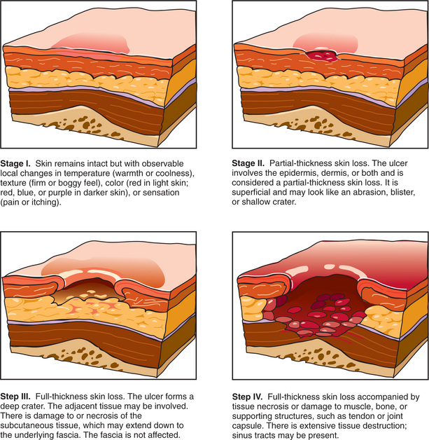
137a. Shea, JD, Pressure sores: classification and management. Clin Orthop Relat Res. 1975(112):89–100.
138. Sheridan, RL, Remensnyder, JP, Schnitzer, JJ, et al. Current expectations for survival in pediatric burns. Arch Pediatr Adolesc Med. 2000;154(3):245–249.
139. Sicherer, SH, Leung, DY. Advances in allergic skin disease, anaphylaxis, and hypersensitivity reactions to foods, drugs, and insects. J Allergy Clin Immunol. 2005;116(1):153–163.
140. Sidbury, R, Hanifin, JM. Old, new, and emerging therapies for atopic dermatitis. Dermatol Clin. 2000;18(1):1–11.
141. Skin Cancer Physicians, Skin cancer net. Am Acad Dermatol 2006. Available on-line at www.skincarephysicians.com Accessed August 10, 2006.
142. Soltani, MH, Pichardo, R, Song, Z, et al. Microtubule-associated protein 2, a marker of neuronal differentiation, induces mitotic defects, inhibits growth of melanoma cells, and predicts metastatic potential of cutaneous melanoma. Am J Pathol. 2005;166(6):1841–1850.
143. Stallone, G, Schena, A, Infante, B, et al. Sirolimus for Kaposi’s sarcoma in renal-transplant recipients. N Engl J Med. 2005;352(13):1317–1323.
144. Stern, RS, Liebman, EJ, Vakeva, L. Oral psoralen and ultraviolet-A light (PUVA) treatment of psoriasis and persistent risk of nonmelanoma skin cancer: PUVA follow-up study. J Natl Cancer Inst. 1998;90(17):1278–1284.
145. Stratton, SP, Dorr, RT, Alberts, DS. The state-of-the-art in chemoprevention of skin cancer. Eur J Cancer. 2000;36(10):1292–1297.
146. Su, CW, Lohman, R, Gottlieb, LJ. Frostbite of the upper extremity. Hand Clin. 2000;16(2):235–247.
147. Sussman C, Bates-Jensen BM, eds. Wound care: a collaborative practice manual for physical therapists and nurses, ed 2, Gaithersburg, MD: Aspen Publishers, 2001.
148. Tanaka, A, Lindor, K, Ansari, A, et al. Fetal microchimerisms in the mother: immunologic implications. Liver Transpl. 2000;6(2):138–143.
149. Thiboutot, DM. Acne and rosacea. Dermatol Clin. 2000;18(1):63–71.
150. Thissen, MR, Schroeter, CA, Neumann, HA. Photodynamic therapy with delta-aminolaevulinic acid for nodular basal cell carcinomas using a prior bulking technique. Br J Dermatol. 2000;142(2):338–339.
151. Thomas, JM, Patocskai, EJ. The argument against sentinel node biopsy for malignant melanoma: its use should be confined to patients in clinical trials. BMJ. 2000;321(7252):3–4.
152. Trefzer, U, Weingart, G, Chen, Y, et al. Hybrid cell vaccination for cancer immune therapy: first clinical trial with metastatic melanoma. Int J Cancer. 2000;85(5):618–626.
153. Trentham, DE, Minocycline in early diffuse scleroderma SSc the next step. Physician’s Page. 2008;3(2):3. Available on-line at www.roadback.org Accessed April 26, 2008.
154. Unglaub, F, Woodruff, S, Demir, E, et al. Patients with epilepsy: a high-risk population prone to severe burns as a consequence of seizures while showering. J Burn Care Rehabil. 2005;26(6):526–528.
155. Valenta, R, Seiberler, S, Natter, S, et al. Autoallergy: a pathogenetic factor in atopic dermatitis? J Allergy Clin Immunol. 2000;105(3):432–437.
156. Wagner, JD, Corbett, L, Park, HM, et al. Sentinel lymph node biopsy for melanoma: experience with 234 consecutive procedures. Plast Reconstr Surg. 2000;105(6):1956–1966.
157. Weisinger, GF, Quittan, M, Nuhr, M, et al. Aerobic capacity in adult dermatomyositis/polymyositis patients and healthy controls. Arch Phys Med Rehabil. 2000;81(1):1–5.
158. Wigley, FM. Systemic sclerosis and related syndromes. B. Clinical features. In: Klippel JH, ed. Primer on the rheumatic diseases. ed 11. Atlanta: Arthritis Foundation; 1997:267–272.
159. Williams, H, Flohr, C. How epidemiology has challenged three prevailing concepts about atopic dermatitis. J Allergy Clin Immunol. 2006;118(1):209–213.
160. Winchester, R. Psoriatic arthritis and the spectrum of syndromes related to the SAPHO (synovitis, acne, pustulosis, hyperostosis, and osteitis) syndrome. Curr Opin Rheumatol. 1999;11(4):251–256.
161. Wolf, SE, Herndon, DN. Burn care. Austin, TX: Landes Bioscience Publishing, 1999.
162. Wong, SL. The role of sentinel lymph node biopsy in the management of thin melanoma. Am J Surg. 2005;190(2):196–199.
163. Wooldridge, WE. Psoriasis, joint swelling, and draining plaques. J Musculoskel Med. 1996;13(7):61–62.
164. Yarchoan R: Bevacizumab in treating patients with Kaposi’s sarcoma. National Cancer Institute, Clinical Trial NCT00058136, June 2006.
165. Young, AJ, O’Brien, C, Sawka, MN, et al. Physiological problems associated with wearing NBC protective clothing during cold weather. Aviat Space Environ Med. 2000;71(2):184–189.
166. Yowler, CJ, Mozingo, DW, Ryan, JB, et al. Factors contributing to delayed extremity amputation in burn patients. J Trauma. 1998;45(3):522–526.
167. Zandi, S, Shamsadini, S, Zahedi, MJ, et al. Helicobacter pylori and rosacea. East Mediterr Health J. 2003;9(1-2):167–171.
168. Zitelli, JA, Brown, CD, Hanusa, BH. Surgical margins for excision of primary cutaneous melanoma. J Am Acad Dermatol. 1997;37(3, pt 1):422–429.
*For further information contact the National Rosacea Society, Barrington, Illinois, (847) 382-8971 or www.rosacea.com.