DYSREGULATION OF CANCER-ASSOCIATED GENES

The genetic damage that activates oncogenes or inactivates tumor suppressor genes may be subtle (e.g., point mutations) or may involve segments of chromosomes large enough to be detected in a routine karyotype. Activation of oncogenes and loss of function of tumor suppressor genes by mutations were discussed earlier in this chapter. Here we discuss chromosomal abnormalities. We end this section by discussing the epigenetic changes that contribute to carcinogenesis.

Chromosomal Changes

In certain neoplasms, karyotypic abnormalities are nonrandom and common. Specific chromosomal abnormalities have been identified in most leukemias and lymphomas, many sarcomas, and an increasing number of carcinomas. In addition, whole chromosomes may be gained or lost. Although changes in chromosome number (aneuploidy) and structure are generally considered to be late phenomena in cancer progression, it has been suggested that aneuploidy and chromosomal instability may be the initiating events in tumor growth.

The study of chromosomal changes in tumor cells is important on two accounts. First, molecular cloning of genes in the vicinity of chromosomal breakpoints or deletions has been extremely useful in identification of oncogenes (e.g., BCL2, ABL) and tumor suppressor genes (e.g., APC, RB). Second, certain karyotypic abnormalities are specific enough to be of diagnostic value, and in some cases they are predictive of clinical course. The translocations associated with the ABL oncogene in CML and with c-MYC in Burkitt lymphoma have been mentioned earlier, in the context of molecular defects in cancer cells (see Fig. 7-27). Several other karyotype alterations in cancer cells are presented in the discussion of specific tumors in later chapters.

Two types of chromosomal rearrangements can activate proto-oncogenes—translocations and inversions. Chromosomal translocations are much more common (Table 7-9) and are discussed here. Translocations can activate proto-oncogenes in two ways:

In lymphoid tumors specific translocations result in overexpression of proto-oncogenes by swapping their regulatory elements with those of another gene.
In many hematopoietic tumors, sarcomas, and certain carcinomas, the translocations allow normally unrelated sequences from two different chromosomes to recombine and form hybrid fusion genes that encode chimeric proteins that variously promote growth and survival, or enhance self-renewal and block differentiation.

TABLE 7-9 Selected Examples of Oncogenes Activated by Translocation

Malignancy Translocation Affected Genes*
Chronic myeloid leukemia (9;22)(q34;q11) image
Acute leukemias (AML and ALL) (8;21)(q22;q22) AML 8q22
(15;17)(q22;q21) image
Burkitt lymphoma (8;14)(q24;q32) image
Mantle cell lymphoma (11;14)(q13;q32) image
Follicular lymphoma (14;18)(q32;q21) image
T-cell ALL (10;14)(q24;q11) image
Ewing sarcoma (11;22)(q24;q12) image
Prostatic adenocarcinoma (21;21)(q22;q22) TMPRSS2 (21q22.3)
(7:21)(p22;q22) ERG (21q22.2)
(17:21)(p21;q22) ETV1 (7p21.2)
ETV4 (17q21)

AML, acute myeloid leukemia; ALL, acute lymphoblastic leukemia.

* Genes in boldface are involved in multiple translocations.

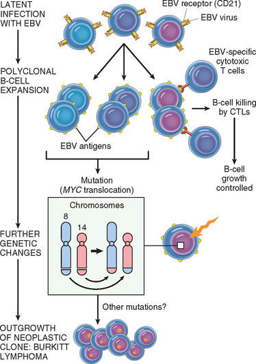
Overexpression of a proto-oncogene caused by translocation is best exemplified by Burkitt lymphoma. All such tumors carry one of three translocations, each involving chromosome 8q24, where the MYC gene has been mapped, as well as one of the three immunoglobulin gene–carrying chromosomes. At its normal locus, MYC is tightly controlled, and is most highly expressed in actively dividing cells. In Burkitt lymphoma the most common form of translocation results in the movement of the MYC-containing segment of chromosome 8 to chromosome 14q32 (see Fig. 7-27), placing it close to the IGH gene. The genetic notation for the translocation is t(8:14)(q24;q32). The molecular mechanisms of the translocation-associated activation of MYC are variable, as are the precise breakpoints within the gene. In most cases the translocation causes mutation or loss of the regulatory sequences of the MYC gene, replacing them with the control regions of the IGH locus, which is highly expressed in B-cell precursors. As the coding sequences remain intact, the gene is constitutively expressed at high levels. The invariable presence of the translocated MYC gene in Burkitt lymphomas attests to the importance of MYC overactivity in the pathogenesis of this tumor.

There are other examples of oncogenes translocated to antigen receptor loci in lymphoid tumors. As mentioned earlier, in mantle cell lymphoma the cyclin D1 gene (CCND1) on chromosome 11q13 is overexpressed by juxtaposition to the IGH locus on 14q32. In follicular lymphomas, a t(14;18)(q32;q21) translocation, the most common translocation in lymphoid malignancies, causes activation of the BCL2 gene. Not unexpectedly, all these tumors in which the immunoglobulin gene is involved are of B-cell origin. In an analogous situation, overexpression of several proto-oncogenes in T-cell tumors results from translocations of oncogenes into the T-cell antigen receptor locus. The affected oncogenes are diverse, but in most cases, as with MYC, they encode nuclear transcription factors.

The Philadelphia chromosome, characteristic of CML and a subset of acute lymphoblastic leukemias, provides the prototypic example of an oncogene formed by fusion of two separate genes. In these cases, a reciprocal translocation between chromosomes 9 and 22 relocates a truncated portion of the proto-oncogene c-ABL (from chromosome 9) to the BCR (breakpoint cluster region) on chromosome 22 (see Fig. 7-27). The hybrid fusion gene BCR-ABL encodes a chimeric protein that has constitutive tyrosine kinase activity. As mentioned, BCR-ABL tyrosine kinase has served as a target for leukemia therapy, with remarkable success so far. Although the translocations are cytogenetically identical in CML and acute lymphoblastic leukemias, they usually differ at the molecular level. In most cases of CML the chimeric protein has a molecular weight of 210 kD, whereas in the more aggressive acute leukemias a 190-kD BCR-ABL fusion protein is typically formed.48,49

Transcription factors are often the partners in gene fusions occurring in cancer cells. For instance, the MLL (myeloid, lymphoid leukemia) gene on 11q23, which itself is a component of a chromatin-remodeling complex, is known to be involved in 50 different translocations with several different partner genes, some of which encode transcription factors (see Table 7-9). Ewing sarcoma/primitive neuroectodermal tumor (PNET) is defined by translocation of the Ewing sarcoma (EWSR1) gene at 22q12, which is involved in numerous translocations, and all of its partner genes analyzed so far also encode a transcription factor. In Ewing sarcoma/PNET, for example, the EWSR1 gene fuses with the FLI1 gene, also a member of the ETS transcription factor family; the resultant chimeric EWS-FLI1 protein has transforming ability. One might ask, why are particular translocations so strongly associated with specific tumors? This is incompletely understood, but one recurrent theme is that at least one of the affected genes often encodes a transcription factor that is required for the development and differentiation of normal cells of the same lineage as the tumor. For example, in acute leukemias many genes involved by recurrent translocations (such as MLL) play essential roles in regulating the self-renewal of hematopoietic stem cells and the normal differentiation of lymphoid and myeloid cells. The fusion proteins resulting from translocations most often inhibit, but occasionally increase, transcriptional function. Until recently, most known translocations were discovered in leukemias/lymphomas and sarcomas; few common translocations had been identified in carcinomas, even though carcinomas are more common. The complex karyotypes of most carcinomas have made identifying translocations difficult. Recently, however, a translocation involving an androgen-regulated gene, TMPRSS2 (21q22), and one of three ETS family transcription factors (ERG [21q22], ETV1 [7p22.2], or ETV4 [17q21]) was found to be present in 50% or more prostate adenocarcinomas.137,138 Development of this translocation seems to occur early in carcinogenesis, in that it is also present in high-grade prostatic intraepithelial neoplasia, a precursor lesion. Although the mechanism by which this translocation causes cancer is not completely understood, it removes the ETS family gene from its normal control region and fuses it to the androgen-regulated TMPRSS2. Thus, the ETS family transcription factor is inappropriately expressed in prostate cells, and as noted above with Ewing sarcoma, when ETS proteins are inappropriately expressed they have transforming ability. There is significant interest in identifying additional fusion genes in other carcinomas. Many fusion genes are thought to be initiators in carcinogenesis, and it is postulated that many cancers may be “addicted” to their properties, similar to the oncogene addiction seen in CML with the BCR-ABL fusion. Thus, inhibition of these genes may provide an avenue for targeted therapy.

Deletions.

Chromosomal deletions are the second most prevalent structural abnormality in tumor cells. Compared with translocations, deletions are more common in nonhematopoietic solid tumors. Deletion of specific regions of chromosomes is associated with the loss of particular tumor suppressor genes. As discussed, deletions involving chromosome 13q14, the site of the RB gene, are associated with retinoblastoma. Deletions of 17p, 5q, and 18q have all been noted in colorectal cancers; these regions harbor three tumor suppressor genes. Deletion of 3p, noted in several tumors, is extremely common in small-cell lung carcinomas, and the hunt is on for one or more cancer suppressor genes at this locale.

Gene Amplification

Activation of proto-oncogenes associated with overexpression of their products may result from reduplication and amplification of their DNA sequences.139 Such amplification may produce several hundred copies of the proto-oncogene in the tumor cell. The amplified genes can be readily detected by molecular hybridization with appropriate DNA probes. In some cases the amplified genes produce chromosomal changes that can be identified microscopically. Two mutually exclusive patterns are seen: multiple small, centric structures called double minutes and homogeneous staining regions. The latter derive from the insertion of the amplified genes into new chromosomal locations, which may be distant from the normal location of the involved genes; because regions containing amplified genes lack a normal banding pattern, they appear homogeneous in a G-banded karyotype (see Fig. 7-28). The most interesting cases of amplification involve N-MYC in neuroblastoma and ERBB2 in breast cancers. N-MYC is amplified in 25% to 30% of neuroblastomas, and the amplification is associated with poor prognosis. In neuroblastomas with N-MYC amplification, the gene is present both in double minutes and homogeneous staining regions. ERBB2 amplification occurs in about 20% of breast cancers, and antibody therapy directed against this receptor has proven effective in this subset of tumors. Amplification of C-MYC, L-MYC, or N-MYC correlates with disease progression in small-cell cancer of the lung.

Epigenetic Changes

Epigenetics refers to reversible, heritable changes in gene expression that occur without mutation. Such changes involve post-translational modifications of histones and DNA methylation, both of which affect gene expression. In normal, differentiated cells, the majority of the genome is not expressed. Some portions of the genome are silenced by DNA methylation and histone modifications that lead to the compaction of DNA into heterochromatin. On the other hand, cancer cells are characterized by a global DNA hypomethylation and selective promoter-localized hypermethylation.140 Indeed, it has become evident during the past few years that tumor suppressor genes are sometimes silenced by hypermethylation of promoter sequences rather than mutation. One example is CDKN2A, a complex locus that encodes two tumor suppressors, p14/ARF and p16/INK4a from two different reading frames; p14/ARF is epigenetically silenced in colon and gastric cancers, while p16/INK4a is silenced in a wide variety of cancers. Since this locus produces two tumor suppressors which affect the p53 and Rb pathways, silencing this locus has the pleasing effect (from the cancer’s point of view) of removing two checkpoints with a single alteration. Other tumor suppressor genes subject to silencing by methylation include BRCA1 in breast cancer, VHL in renal cell carcinomas, and the MLH1 mismatch-repair gene in colorectal cancer.140 You will recall from Chapter 5 that methylation also participates in the phenomenon called genomic imprinting, in which the maternal or paternal allele of a gene or chromosome is modified by methylation and is inactivated. The reverse phenomenon—that is, demethylation of an imprinted gene leading to its biallelic expression (loss of imprinting)—can also occur in tumor cells.141 There has been great interest in developing potential therapeutic agents that act to demethylate DNA sequences in tumor suppressor genes. Recent data demonstrating that genomic hypomethylation causes chromosomal instability and induces tumors in mice greatly strengthen the notion that epigenetic changes may directly contribute to tumor development.141

The chromatin changes that contribute to carcinogenesis are less well understood. The current paradigm is that there is a histone code in which various modifications to the tails of histones, such as acetylation and methylation, lead to activation or repression of transcription. Several chromatin-modifying enzymes, such as EZH2, have been shown to be overexpressed in breast and prostate carcinomas.141 EZH2 is the enzymatic component of the multiprotein polycomb repressive complex 2, which places repressive chromatin marks at the promoter of genes. Although its targets in cancer in vivo have not yet been defined, in cell lines overexpression of EZH2 leads to the repression of tumor suppressors, such as p21. Interestingly, in flies and mammals the polycomb repressive complexes are required for the maintenance of stem cells, as well as to silence lineage-specific transcription factors until the proper cues signal differentiation. Inappropriate repression or expression of such genes could give cancer cells a stem cell–like, undifferentiated quality. There is, of course, significant cross-talk between the chromatin-remodeling enzymes and the DNA-methylation machinery. For example, the placement of repressive chromatin marks by enzymes like EZH2 in cancer cells results in the recruitment of DNA methylases, methylation of promoters, and durable repression of gene expression.

miRNAs and Cancer

As discussed in Chapter 5, miRNAs are small noncoding, single-stranded RNAs, approximately 22 nucleotides in length, that are incorporated into the RNA-induced silencing complex. The miRNAs mediate sequence-specific recognition of mRNAs and, through the action of the RNA-induced silencing complex, mediate post-transcriptional gene silencing. Given that miRNAs control cell growth, differentiation, and cell survival, it is not surprising that they play a role in carcinogenesis.142 miRNAs have been shown to undergo changes in expression in cancer cells, and frequent amplifications and deletions of miRNA loci have been identified in many cancers. As illustrated in Figure 7-39, miRNAs can participate in neoplastic transformation either by increasing the expression of oncogenes or by reducing the expression of tumor suppressor genes. If a miRNA inhibits the translation of an oncogene, a reduction in the quantity or function of that miRNA will lead to overproduction of the oncogene product; thus, the miRNA acts as a tumor suppressor. Conversely, if the target of a miRNA is a tumor suppressor gene, then overactivity of the miRNA can reduce the tumor suppressor protein; thus, the miRNA acts as an oncogene. Such relationships have already been established by miRNA profiling of several human tumors. For example, down-regulation or deletion of certain miRNAs in some leukemias and lymphomas results in increased expression of BCL2, the anti-apoptotic protein. Thus, by negatively regulating BCL2, such miRNAs behave as tumor suppressor genes. Similar miRNA-mediated upregulation of RAS and MYC oncogenes has also been detected in lung tumors and in certain B-cell leukemias, respectively. In some brain and breast tumors there is 5- to 100-fold greater expression of certain miRNAs. Although the targets of these miRNAs have not been identified, presumably they are tumor suppressor genes, whose activities are reduced by the overexpressed miRNA.

image

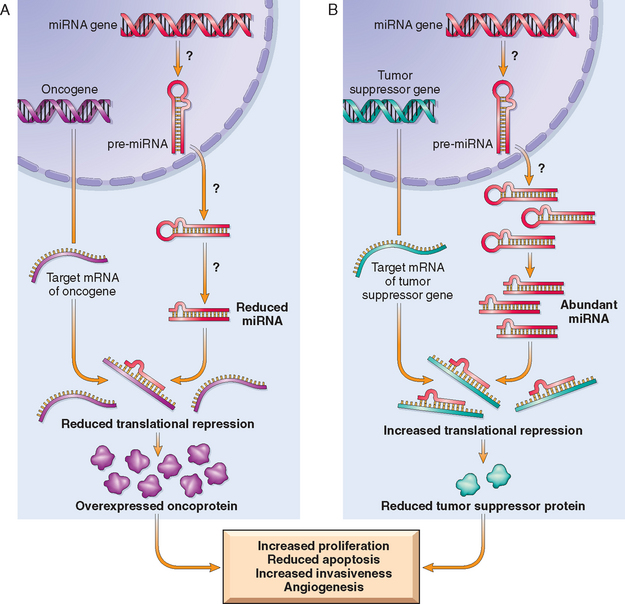
FIGURE 7-39 Role of miRNAs in tumorigenesis. A, Reduced activity of a miRNA that inhibits translation of an oncogene gives rise to an excess of oncoproteins. B, Overactivity of a miRNA that targets a tumor suppression gene reduces the production of the tumor suppressor protein. Question marks in A and B indicate that the mechanisms by which changes in the level or activity of miRNA are not entirely known.

These findings not only provide novel insights into carcinogenesis, they also have practical implications. For instance, drugs that inhibit or augment the functions of miRNAs could be useful in chemotherapy. Since miRNAs regulate normal cellular differentiation, the patterns of miRNA expression (“miRNA profiling”) can provide clues to the cell of origin and classification of tumors. Much remains to be learned about these oncogenic miRNAs, or so called “oncomirs.”

Molecular Basis of Multistep Carcinogenesis

The notion that malignant tumors arise from a protracted sequence of events is supported by epidemiologic, experimental, and molecular studies. The study of oncogenes and tumor suppressor genes has provided a firm molecular footing for the concept of multistep carcinogenesis.143

Given that malignant tumors must acquire several fundamental abnormalities, discussed above, it follows that each cancer must result from the accumulation of multiple mutations. Indeed, recently completed genome-wide sequencing analysis of breast and colon cancers has revealed that individual tumors accumulate an average of 90 mutant genes. A much smaller subset of these (11/tumor) were mutated at significant frequency.144 Included among the mutated genes are some known oncogenes and tumor suppressor genes, and others that were not previously known to be tumor-associated. It is not yet established which of these mutations establish the transformed state, contribute to tumor progression, or are “passengers” (neutral mutations) occurring in genomically unstable cells that are merely “along for the ride”. More directly, however, no single oncogene can fully transform nonimmortalized cells in vitro, but cells can generally be transformed by combinations of oncogenes. Such cooperation is required because each oncogene is specialized to induce part of the phenotype necessary for full transformation. For instance, the RAS oncogene induces cells to secrete growth factors and permits them to grow without anchorage to a normal substrate (anchorage independence), whereas the MYC oncogene renders cells more sensitive to growth factors and immortalizes cells. These two genes, acting in conjunction, can cause neoplastic transformation of mouse fibroblasts in culture.

Furthermore, it seems that evolution has installed a variety of “intrinsic tumor-suppressive mechanisms” such as apoptosis and senescence that thwart the actions of growth-promoting mutations. Indeed, in cells with competent checkpoints, oncogenic signaling through proteins like RAS leads not to transformation but to senescence or apoptosis.33 Thus, emergence of malignant tumors requires mutational loss of many genes, including those that regulate apoptosis and senescence.145 A classic example of incremental acquisition of the malignant phenotype is documented by the study of colon carcinoma. Many of these cancers are believed to evolve through a series of morphologically identifiable stages: colon epithelial hyperplasia followed by formation of adenomas that progressively enlarge and ultimately undergo malignant transformation (Chapter 17). The proposed molecular correlates of this adenoma-carcinoma sequence are illustrated in Figure 7-40. According to this scheme, inactivation of the APC tumor suppressor gene occurs first, followed by activation of RAS and, ultimately, loss of a tumor suppressor gene on 18q and loss of p53. Also depicted is the senescence pathway if p53 loss does not occur. Indeed, it has been shown that most cells in most adenomas are senescent. It is thought that mutation of a proto-oncogene such as RAS drives a cell into senescence instead of proliferation33 by activating the DNA-damage checkpoint, as discussed previously. The loss of p53 in adenomas prevents oncogene-induced senescence, allowing the adenomatous cells to continue to proliferate, generating a carcinoma. While multiple mutations, including gain of oncogenes and loss of tumor suppressors, are required for carcinogenesis, the precise temporal sequence of mutations may be different in each organ and tumor type.

image

FIGURE 7-40 Molecular model for the evolution of colorectal cancers through the adenoma-carcinoma sequence. Although APC mutation is an early event and loss of p53 occurs late in the process of tumorigenesis, the timing for the other changes may be variable. Note also that individual tumors may not have all of the changes listed. Top right, cells that gain oncogene signaling without loss of p53 eventually enter oncogene-induced senescence.

Carcinogenic Agents and Their Cellular Interactions

More than 200 years ago the London surgeon Sir Percival Pott correctly attributed scrotal skin cancer in chimney sweeps to chronic exposure to soot. Based on this observation, the Danish Chimney Sweeps Guild ruled that its members must bathe daily. No public health measure since that time has achieved so much in the control of a form of cancer. Subsequently, hundreds of chemicals have been shown to be carcinogenic in animals.

Some of the major agents are presented in Table 7-10. A few comments are offered on a handful of these.

TABLE 7-10 Major Chemical Carcinogens

DIRECT-ACTING CARCINOGENS
Alkylating Agents
β-Propiolactone
Dimethyl sulfate
Diepoxybutane
Anticancer drugs (cyclophosphamide, chlorambucil, nitrosoureas, and others)
Acylating Agents
1-Acetyl-imidazole
Dimethylcarbamyl chloride
PROCARCINOGENS THAT REQUIRE METABOLIC ACTIVATION
Polycyclic and Heterocyclic Aromatic Hydrocarbons
Benz[a]anthracene
Benzo[a]pyrene
Dibenz[a,h]anthracene
3-Methylcholanthrene
7,12-Dimethylbenz[a]anthracene
Aromatic Amines, Amides, Azo Dyes
2-Naphthylamine (β-naphthylamine)
Benzidine
2-Acetylaminofluorene
Dimethylaminoazobenzene (butter yellow)
Natural Plant and Microbial Products
Aflatoxin B1
Griseofulvin
Cycasin
Safrole
Betel nuts
Others
Nitrosamine and amides
Vinyl chloride, nickel, chromium
Insecticides, fungicides
Polychlorinated biphenyls

Steps Involved in Chemical Carcinogenesis

As discussed earlier, carcinogenesis is a multistep process. This is most readily demonstrated in experimental models of chemical carcinogenesis, in which the stages of initiation and progression during cancer development were first described.146 The classic experiments that allowed the distinction between initiation and promotion were performed on mouse skin and are outlined in Figure 7-41. The following concepts relating to the initiation-promotion sequence have emerged from these experiments:

Initiation results from exposure of cells to a sufficient dose of a carcinogenic agent (initiator); an initiated cell is altered, making it potentially capable of giving rise to a tumor (groups 2 and 3). Initiation alone, however, is not sufficient for tumor formation (group 1).
Initiation causes permanent DNA damage (mutations). It is therefore rapid and irreversible and has “memory.” This is illustrated by group 3, in which tumors were produced even if the application of the promoting agent was delayed for several months after a single application of the initiator.
Promoters can induce tumors in initiated cells, but they are nontumorigenic by themselves (group 5). Furthermore, tumors do not result when the promoting agent is applied before, rather than after, the initiating agent (group 4). This indicates that, in contrast to the effects of initiators, the cellular changes resulting from the application of promoters do not affect DNA directly and are reversible. As discussed later, promoters enhance the proliferation of initiated cells, an effect that may contribute to the development of additional mutations in these cells. That the effects of promoters are reversible is further documented in group 6, in which tumors failed to develop in initiated cells if the time between multiple applications of the promoter was sufficiently extended.
image

FIGURE 7-41 Experiments demonstrating the initiation and promotion phases of carcinogenesis in mice. Group 2: application of promoter repeated at twice-weekly intervals for several months. Group 3: application of promoter delayed for several months and then applied twice weekly. Group 6: promoter applied at monthly intervals.

Although the concepts of initiation and promotion have been derived largely from experiments involving induction of skin cancer in mice, these stages are also discernible in the development of cancers of the liver, urinary bladder, breast, colon, and respiratory tract. With this brief overview of two major steps in carcinogenesis, we can examine initiation and promotion in more detail (Fig. 7-42). All initiating chemical carcinogens are highly reactive electrophiles (have electron-deficient atoms) that can react with nucleophilic (electron-rich) sites in the cell. Their targets are DNA, RNA, and proteins, and in some cases these interactions cause cell death. Initiation, obviously, inflicts nonlethal damage on the DNA that cannot be repaired. The mutated cell then passes on the DNA lesions to its daughter cells. Chemicals that can cause initiation of carcinogenesis can be classified into two categories: direct acting and indirect acting.

image

FIGURE 7-42 General schema of events in chemical carcinogenesis. Note that promoters cause clonal expansion of the initiated cell, thus producing a preneoplastic clone. Further proliferation induced by the promoter or other factors causes accumulation of additional mutations and emergence of a malignant tumor.

Direct-Acting Agents

Direct-acting agents require no metabolic conversion to become carcinogenic. Most of them are weak carcinogens but are important because some are cancer chemotherapeutic drugs (e.g., alkylating agents) that have successfully cured, controlled, or delayed recurrence of certain types of cancer (e.g., leukemia, lymphoma, and ovarian carcinoma), only to evoke later a second form of cancer, usually acute myeloid leukemia. The risk of induced cancer is low, but its existence dictates judicious use of such agents.

Indirect-Acting Agents

The designation indirect-acting agent refers to chemicals that require metabolic conversion to an ultimate carcinogen before they become active. Some of the most potent indirect chemical carcinogens—the polycyclic hydrocarbons—are present in fossil fuels. Others, for example, benzo[a]pyrene and other carcinogens, are formed in the high-temperature combustion of tobacco in cigarette smoking. These products are implicated in the causation of lung cancer in cigarette smokers. Polycyclic hydrocarbons may also be produced from animal fats during the process of broiling meats and are present in smoked meats and fish. The principal active products in many hydrocarbons are epoxides, which form covalent adducts (addition products) with molecules in the cell, principally DNA, but also with RNA and proteins.

The aromatic amines and azo dyes are another class of indirect-acting carcinogens that were widely used in the past in the aniline dye and rubber industries.147 Many other occupational carcinogens are listed in Table 7-3.

Most chemical carcinogens require metabolic activation for conversion into ultimate carcinogens (see Fig. 7-42). Other metabolic pathways may lead to the inactivation (detoxification) of the procarcinogen or its derivatives. Thus, the carcinogenic potency of a chemical is determined not only by the inherent reactivity of its electrophilic derivative but also by the balance between metabolic activation and inactivation reactions.

Most of the known carcinogens are metabolized by cytochrome P-450–dependent mono-oxygenases. The genes that encode these enzymes are quite polymorphic, and the activity and inducibility of these enzymes have been shown to vary among different individuals. Because these enzymes are essential for the activation of procarcinogens, the susceptibility to carcinogenesis is regulated in part by polymorphisms in the genes that encode these enzymes. Thus, it may be possible to assess cancer risk in a given individual by genetic analysis of such enzyme polymorphisms.147

The metabolism of polycyclic aromatic hydrocarbons, such as benzo[a]pyrene by the product of the P-450 gene, CYP1A1, provides an instructive example. Approximately 10% of the white population has a highly inducible form of this enzyme that is associated with an increased risk of lung cancer in smokers.148,149 Light smokers with the susceptible genotype CYP1A1 have a sevenfold higher risk of developing lung cancer, compared with smokers without the permissive genotype. Not all variations in the activation or detoxification of carcinogens are genetically determined. Age, sex, and nutritional status also determine the internal dose of toxicants produced and hence influence the risk of cancer development in a particular individual.150

Molecular Targets of Chemical Carcinogens.

Because malignant transformation results from mutations, it comes as no surprise that the majority of initiating chemicals are mutagenic. Thus, DNA is the primary target for chemical carcinogens, but there is no single or unique alteration associated with initiation of chemical carcinogenesis. Although any gene may be the target of chemical carcinogens, the commonly mutated oncogenes and tumor suppressors, such as RAS and p53, are particularly important targets. An illustrative example of a chemical carcinogenesis is aflatoxin B1, a naturally occurring agent produced by some strains of Aspergillus, a mold that grows on improperly stored grains and nuts. There is a strong correlation between the dietary level of this food contaminant and the incidence of hepatocellular carcinoma in parts of Africa and the Far East. Interestingly, aflatoxin B1 produces mutations in the p53 gene; 90% or more of these mutations are a characteristic G : C→T : A transversion in codon 249 (called 249(ser) p53 mutation).151 By contrast, p53 mutations are much less frequent in liver tumors from areas where aflatoxin contamination of food is not a risk factor, and the 249(ser) mutation is uncommon. Thus, the detection of the “signature mutation” within the p53 gene establishes aflatoxin as the causative agent. These associations are proving to be useful tools in epidemiologic studies of chemical carcinogenesis.

Additionally, vinyl chloride, arsenic, nickel, chromium, insecticides, fungicides, and polychlorinated biphenyls are potential carcinogens in the workplace and at home. Finally, nitrites used as food preservatives have caused concern, since they cause nitrosylation of amines contained in the food. The nitrosoamines so formed are suspected to be carcinogenic.

Initiation and Promotion of Chemical Carcinogenesis

Unrepaired alterations in the DNA are essential first steps in the process of initiation. For the change to be heritable, the damaged DNA template must be replicated. Thus, for initiation to occur, carcinogen-altered cells must undergo at least one cycle of proliferation so that the change in DNA becomes fixed. In the liver, many chemicals are activated to reactive electrophiles, yet most of them do not produce cancers unless the liver cells proliferate within a few days of the formation of DNA adducts. In tissues that are normally quiescent, the mitogenic stimulus may be provided by the carcinogen itself, because many cells die as a result of toxic effects of the carcinogenic chemical, thereby stimulating regeneration in the surviving cells. Alternatively, cell proliferation may be induced by concurrent exposure to biologic agents such as viruses and parasites, dietary factors, or hormonal influences. Agents that do not cause mutation but instead stimulate the division of mutated cells are known as promoters.

The carcinogenicity of some initiators is augmented by subsequent administration of promoters (such as phorbol esters, hormones, phenols, and drugs) that by themselves are nontumorigenic. Application of promoters leads to proliferation and clonal expansion of initiated (mutated) cells. Such cells have reduced growth factor requirements and may also be less responsive to growth-inhibitory signals in their extracellular milieu. Driven to proliferate, the initiated clone of cells suffers additional mutations, developing eventually into a malignant tumor. Thus, the process of tumor promotion includes multiple steps: proliferation of preneoplastic cells, malignant conversion, and eventually tumor progression, which depends on changes in tumor cells and the tumor stroma—the process of multistep carcinogenesis highlighted above.

RADIATION CARCINOGENESIS

Radiant energy, whether in the form of the UV rays of sunlight or as ionizing electromagnetic and particulate radiation, is a well-established carcinogen. UV light is clearly implicated in the causation of skin cancers, and ionizing radiation exposure from medical or occupational exposure, nuclear plant accidents, and atomic bomb detonations has produced a variety of cancers. Although the contribution of radiation to the total human burden of cancer is probably small, the well-known latency of damage caused by radiant energy and its cumulative effect require extremely long periods of observation and make it difficult to ascertain its full significance. An increased incidence of breast cancer has become apparent decades later among women exposed during childhood to atomic bomb tests. The incidence peaked during 1988–1992 and then declined.152 Moreover, radiation’s possible additive or synergistic effects with other potential carcinogenic influences add another dimension to the picture.

Ultraviolet Rays

There is ample evidence from epidemiologic studies that UV rays derived from the sun cause an increased incidence of squamous cell carcinoma, basal cell carcinoma, and possibly melanoma of the skin.153 The degree of risk depends on the type of UV rays, the intensity of exposure, and the quantity of the light-absorbing “protective mantle” of melanin in the skin. Persons of European origin who have fair skin that repeatedly becomes sunburned but stalwartly refuses to tan and who live in locales receiving a great deal of sunlight (e.g., Queensland, Australia, close to the equator) have among the highest incidence of skin cancers (melanomas, squamous cell carcinomas, and basal cell carcinomas) in the world. Nonmelanoma skin cancers are associated with total cumulative exposure to UV radiation, whereas melanomas are associated with intense intermittent exposure—as occurs with sunbathing. The UV portion of the solar spectrum can be divided into three wavelength ranges: UVA (320–400 nm), UVB (280–320 nm), and UVC (200–280 nm). Of these, UVB is believed to be responsible for the induction of cutaneous cancers. UVC, although a potent mutagen, is not considered significant because it is filtered out by the ozone shield around the earth (hence the concern about ozone depletion).

The carcinogenicity of UVB light is attributed to its formation of pyrimidine dimers in DNA. This type of DNA damage is repaired by the nucleotide excision repair pathway. There are five steps in nucleotide excision repair, and in mammalian cells the process may involve 30 or more proteins. It is postulated that with excessive sun exposure, the capacity of the nucleotide excision repair pathway is overwhelmed, and error-prone nontemplated DNA-repair mechanisms become operative that provide for the survival of the cell at the cost of genomic mutations that in some instances, lead to cancer. The importance of the nucleotide excision repair pathway of DNA repair is most graphically illustrated by the high frequency of cancers in individuals with the hereditary disorder xeroderma pigmentosum (discussed previously).126

Ionizing Radiation

Electromagnetic (x-rays, γ rays) and particulate (α particles, β particles, protons, neutrons) radiations are all carcinogenic. The evidence is so voluminous that a few examples suffice.152,154 Many individuals pioneering the use of x-rays developed skin cancers. Miners of radioactive elements in central Europe and the Rocky Mountain region of the United States have a tenfold increased incidence of lung cancers compared to the rest of the population. Most telling is the follow-up of survivors of the atomic bombs dropped on Hiroshima and Nagasaki. Initially there was a marked increase in the incidence of leukemias—principally acute and chronic myelogenous leukemia—after an average latent period of about 7 years. Subsequently the incidence of many solid tumors with longer latent periods (e.g., breast, colon, thyroid, and lung) increased.

In humans there is a hierarchy of vulnerability of different tissues to radiation-induced cancers. Most frequent are the acute and chronic myeloid leukemia. Cancer of the thyroid follows closely but only in the young. In the intermediate category are cancers of the breast, lungs, and salivary glands. In contrast, skin, bone, and the gastrointestinal tract are relatively resistant to radiation-induced neoplasia, even though the gastrointestinal epithelial cells are vulnerable to the acute cell-killing effects of radiation, and the skin is in the pathway of all external radiation. Nonetheless, the physician dare not forget: practically any cell can be transformed into a cancer cell by sufficient exposure to radiant energy.

MICROBIAL CARCINOGENESIS

Many RNA and DNA viruses have proved to be oncogenic in animals as disparate as frogs and primates. Despite intense scrutiny, however, only a few viruses have been linked with human cancer. Our discussion focuses on human oncogenic viruses as well as the emerging role of the bacterium Helicobacter pylori in gastric cancer.

Oncogenic RNA Viruses

Human T-Cell Leukemia Virus Type 1.

Although the study of animal retroviruses has provided spectacular insights into the molecular basis of cancer, only one human retrovirus, human T-cell leukemia virus type 1 (HTLV-1), is firmly implicated in the causation of cancer in humans.

HTLV-1 causes a form of T-cell leukemia/lymphoma that is endemic in certain parts of Japan and the Caribbean basin but is found sporadically elsewhere, including the United States.155 Similar to the human immunodeficiency virus, which causes acquired immunodeficiency syndrome (AIDS), HTLV-1 has tropism for CD4+ T cells, and hence this subset of T cells is the major target for neoplastic transformation. Human infection requires transmission of infected T cells via sexual intercourse, blood products, or breastfeeding. Leukemia develops in only 3% to 5% of the infected individuals after a long latent period of 40 to 60 years.

There is little doubt that HTLV-1 infection of T lymphocytes is necessary for leukemogenesis, but the molecular mechanisms of transformation are not entirely clear. In contrast to several murine retroviruses, HTLV-1 does not contain an oncogene, and no consistent integration next to a proto-oncogene has been discovered. In leukemic cells, however, viral integration shows a clonal pattern. In other words, although the site of viral integration in host chromosomes is random (the viral DNA is found at different locations in different cancers), the site of integration is identical within all cells of a given cancer. This would not occur if HTLV-1 were merely a passenger that infects cells after transformation. The HTLV-1 genome contains the gag, pol, env, and long-terminal-repeat regions typical of other retroviruses, but, in contrast to other leukemia viruses, it contains another region, referred to as tax. It seems that the secrets of its transforming activity are locked in the tax gene.156 The product of this gene is essential for viral replication, because it stimulates transcription of viral mRNA by acting on the 5′ long terminal repeat. It is now established that the Tax protein can also activate the transcription of several host cell genes involved in proliferation and differentiation of T cells. These include the immediate early gene FOS, genes encoding interleukin-2 (IL-2) and its receptor, and the gene for the myeloid growth factor granulocyte-macrophage colony-stimulating factor. In addition, Tax protein inactivates the cell cycle inhibitor p16/INK4a and enhances cyclin D activation, thus dysregulating the cell cycle. Tax also activates NF-κb, a transcription factor that regulates a host of genes, including pro-survival/anti-apoptotic genes. Another mechanism by which Tax may contribute to malignant transformation is through genomic instability. Recent data show that Tax interferes with DNA-repair functions and inhibits ATM-mediated cell cycle checkpoints activated by DNA damage.156

The main steps that lead to the development of adult T-cell leukemia/lymphoma may be summarized as follows. Infection by HTLV-1 causes the expansion of a nonmalignant polyclonal cell population through stimulatory effects of Tax on cell proliferation. The proliferating T cells are at increased risk of mutations and genomic instability induced by Tax. This instability allows the accumulation of mutations and chromosomal abnormalities, and eventually a monoclonal neoplastic T-cell population emerges. The malignant cells replicate independently of IL-2 and contain molecular and chromosomal abnormalities.

Oncogenic DNA Viruses

As with RNA viruses, several oncogenic DNA viruses that cause tumors in animals have been identified. Of the various human DNA viruses, four—HPV, Epstein-Barr virus (EBV), hepatitis B virus (HBV), and Kaposi sarcoma herpesvirus, also called human herpesvirus 8—have been implicated in the causation of human cancer. A fifth virus, Merkel cell polyomavirus, has been identified in Merkel cell carcinomas and may soon join the rogue’s gallery; it is described in Chapter 25. Kaposi sarcoma herpesvirus is discussed in Chapters 6 and 11. Though not a DNA virus, HCV is also associated with cancer and is discussed briefly here.157

Human Papillomavirus.

At least 70 genetically distinct types of HPV have been identified. Some types (e.g., 1, 2, 4, and 7) cause benign squamous papillomas (warts) in humans (Chapters 19 and 22. By contrast, high-risk HPVs (e.g., types 16 and 18) have been implicated in the genesis of several cancers, particularly squamous cell carcinoma of the cervix and anogenital region.158,159 Thus, cervical cancer is a sexually transmitted disease, caused by transmission of HPV. In addition, at least 20% of oropharyngeal cancers are associated with HPV. In contrast to cervical cancers, genital warts have low malignant potential and are associated with low-risk HPVs, predominantly HPV-6 and HPV-11. Interestingly, in benign warts the HPV genome is maintained in a nonintegrated episomal form, while in cancers the HPV genome is integrated into the host genome, suggesting that integration of viral DNA is important for malignant transformation. As with HTLV-1, the site of viral integration in host chromosomes is random, but the pattern of integration is clonal. Cells in which the viral genome has integrated show significantly more genomic instability. Furthermore, since the integration site is random there is no consistent association with a host proto-oncogene. Rather, integration interrupts the viral DNA within the E1/E2 open reading frame, leading to loss of the E2 viral repressor and overexpression of the oncoproteins E6 and E7.

Indeed, the oncogenic potential of HPV can be related to the products of two viral genes, E6 and E7. Together, they interact with a variety of growth-regulating proteins encoded by proto-oncogenes and tumor suppressor genes (Fig. 7-43). The E7 protein binds to the RB protein and displaces the E2F transcription factors that are normally sequestered by RB, promoting progression through the cell cycle. Of note, E7 protein from high-risk HPV types has a higher affinity for RB than does E7 from low-risk HPV types. E7 also inactivates the CDKIs p21 and p27. E7 proteins from high-risk HPV types (types 16, 18, and 31) also bind and presumably activate cyclins E and A. The E6 protein has complementary effects. It binds to and mediates the degradation of p53 and BAX, a pro-apoptotic member of the BCL2 family, and it activates telomerase. Like E7, E6 from high-risk HPV types has a higher affinity for p53 than E6 from low-risk HPV types. Interestingly the E6-p53 interaction may offer some clues regarding polymorphisms and risk factors for development of cervical cancer. Human p53 is polymorphic at amino acid 72, encoding either a proline or arginine residue at that position. The p53 Arg72 variant is much more susceptible to degradation by E6. Not surprisingly, infected individuals with the Arg72 polymorphism are more likely to develop cervical carcinomas.160

image

FIGURE 7-43 Effect of HPV proteins E6 and E7 on the cell cycle. E6 and E7 enhance p53 degradation, causing a block in apoptosis and decreased activity of the p21 cell cycle inhibitor. E7 associates with p21 and prevents its inhibition of the cyclin-CDK4 complex; E7 can bind to RB, removing cell cycle restriction. The net effect of HPV E6 and E7 proteins is to block apoptosis and remove the restraints to cell proliferation (see Fig. 7-29).

(Modified from Münger K, Howley PM: Human papillomavirus immortalization and transformation functions. Virus Res 89:213–228, 2002.)

To summarize, high-risk HPV types express oncogenic proteins that inactivate tumor suppressors, activate cyclins, inhibit apoptosis, and combat cellular senescence. Thus, it is evident that many of the hallmarks of cancer discussed earlier are driven by HPV proteins. The primacy of HPV infection in the causation of cervical cancer is confirmed by the effectiveness of anti-HPV vaccines in preventing cervical cancer. However, infection with HPV itself is not sufficient for carcinogenesis. For example, when human keratinocytes are transfected with DNA from HPV types 16, 18, or 31 in vitro, they are immortalized but do not form tumors in experimental animals. Co-transfection with a mutated RAS gene results in full malignant transformation. In addition to such genetic co-factors, HPV in all likelihood also acts in concert with environmental factors (Chapter 22). These include cigarette smoking, coexisting microbial infections, dietary deficiencies, and hormonal changes, all of which have been implicated in the pathogenesis of cervical cancers. A high proportion of women infected with HPV clear the infection by immunological mechanisms, but some do not for unknown reasons.

Epstein-Barr Virus.

EBV, a member of the herpes family, has been implicated in the pathogenesis of several human tumors: the African form of Burkitt lymphoma; B-cell lymphomas in immunosuppressed individuals (particularly in those with HIV infection or undergoing immunosuppressive therapy after organ transplantation); a subset of Hodgkin lymphoma; nasopharyngeal and some gastric carcinomas and rare forms of T cell lymphomas and natural killer (NK) cell lymphomas.161 Except for nasopharyngeal carcinoma, all others are B-cell tumors. These neoplasms are reviewed elsewhere in this book; therefore, only their association with EBV is discussed here.

EBV infects B lymphocytes and possibly epithelial cells of the oropharynx. EBV uses the complement receptor CD21 to attach to and infect B cells. The infection of B cells is latent; that is, there is no viral replication and the cells are not killed, but the B cells latently infected with EBV are immortalized and acquire the ability to propagate indefinitely in vitro. The molecular basis of B-cell proliferations induced by EBV is complex, but as with other viruses it involves the “hijacking” of several normal signaling pathways.162 One EBV gene, latent membrane protein-1 (LMP-1), acts as an oncogene, in that its expression in transgenic mice induces B-cell lymphomas. LMP-1 behaves like a constitutively active CD40 receptor, a key recipient of helper T-cell signals that stimulate B-cell growth (Chapter 6). LMP-1 activates the NF-κB and JAK/STAT signaling pathways and promotes B-cell survival and proliferation, all of which occur autonomously (i.e., without T cells or other outside signals) in EBV-infected B cells. Concurrently, LMP-1 prevents apoptosis by activating BCL2. Thus, the virus “borrows” a normal B-cell activation pathway to expand the pool of latently infected cells. Another EBV gene, EBNA-2, encodes a nuclear protein that mimics a constitutively active Notch receptor. EBNA-2 transactivates several host genes, including cyclin D and the src family of proto-oncogenes. In addition, the EBV genome contains a viral cytokine, vIL-10, that was hijacked from the host genome. This viral cytokine can prevent macrophages and monocytes from activating T cells and is required for EBV-dependent transformation of B cells. In immunologically normal individuals EBV-driven polyclonal B-cell proliferation in vivo is readily controlled, and the individual either remains asymptomatic or develops a self-limited episode of infectious mononucleosis (Chapter 8). Evasion of the immune system seems to be a key step in EBV-related oncogenesis.

Burkitt lymphoma is a neoplasm of B lymphocytes that is the most common childhood tumor in central Africa and New Guinea. A morphologically identical lymphoma occurs sporadically throughout the world. The association between endemic Burkitt lymphoma and EBV is quite strong (Fig. 7-44):

More than 90% of African tumors carry the EBV genome.
One hundred percent of the patients have elevated antibody titers against viral capsid antigens.
Serum antibody titers against viral capsid antigens are correlated with the risk of developing the tumor.
image

FIGURE 7-44 Possible evolution of EBV-induced Burkitt lymphoma.

Although EBV is intimately involved in the causation of Burkitt lymphoma, several observations suggest that additional factors must also be involved.163,164 (1) EBV infection is not limited to the geographic locales where Burkitt lymphoma is found, but it is a ubiquitous virus that asymptomatically infects almost all humans worldwide. (2) The EBV genome is found in only 15% to 20% of sufferers of Burkitt lymphoma outside Africa. (3) There are significant differences in the patterns of viral gene expression in EBV-transformed (but not tumorigenic) B-cell lines and Burkitt lymphoma cells. Most notably, Burkitt lymphoma cells do not express LMP-1, EBNA2, and other EBV proteins that drive B-cell growth and immortalization.

Given these observations, how then does EBV contribute to the genesis of endemic Burkitt lymphoma? A plausible scenario is shown in Figure 7-44. In regions of the world where Burkitt lymphoma is endemic, concomitant infections such as malaria impair immune competence, allowing sustained B-cell proliferation. Eventually, however, T-cell immunity directed against EBV antigens such as EBNA2 and LMP-1 eliminates most of the EBV-infected B cells, but a small number of cells downregulate expression of these immunogenic antigens. These cells persist indefinitely, even in the face of normal immunity. Lymphoma cells may emerge from this population only with the acquisition of specific mutations, most notably translocations that activate the c-MYC oncogene. It should be noted that in nonendemic areas 80% of tumors do not harbor the EBV genome, but all tumors possess the t(8;14) or other translocations that dysregulate c-MYC. This observation suggests that, although non-African Burkitt lymphomas are triggered by mechanisms other than EBV, they develop through very similar oncogenic pathways.

In summary, in the case of Burkitt lymphoma, it seems that EBV is not directly oncogenic, but by acting as a polyclonal B-cell mitogen, it sets the stage for the acquisition of the t(8;14) translocation and other mutations, which ultimately release the cells from normal growth regulation. In normal individuals, EBV infection is readily controlled by effective immune responses directed against viral antigens expressed on the cell membranes. Hence, the vast majority of infected individuals remain asymptomatic or develop self-limited infectious mononucleosis. In regions of Africa where Burkitt lymphoma is endemic, poorly understood cofactors (e.g., chronic malaria) may favor the acquisition of genetic events (e.g., the t(8;14) translocation) that lead to transformation.

The role played by EBV is more direct in B-cell lymphomas in immunosuppressed patients. Some persons with AIDS and those who receive long-term immunosuppressive therapy for preventing allograft rejection present with multifocal B-cell tumors within lymphoid tissue or in the central nervous system. These tumors are polyclonal at the outset but can develop into monoclonal neoplasms. In contrast to Burkitt lymphoma, the tumors in immunosuppressed patients uniformly express LMP-1 and EBNA2, that are recognized by cytotoxic T cells. These potentially lethal proliferations can be subdued if the immunological status of the host improves, as may occur with withdrawal of immunosuppressive drugs in transplant recipients.

Nasopharyngeal carcinoma is also associated with EBV infection. This tumor is endemic in southern China, in some parts of Africa, and in the Inuit population of the Arctic. In contrast to Burkitt lymphoma, 100% of nasopharyngeal carcinomas obtained from all parts of the world contain EBV DNA.165 The viral integration in the host cells is clonal, thus ruling out the possibility that EBV infection occurred after tumor development. Antibody titers to viral capsid antigens are greatly elevated, and in endemic areas patients develop IgA antibodies before the appearance of the tumor. The 100% correlation between EBV and nasopharyngeal carcinoma suggests that EBV110 plays a role in the genesis of this tumor, but (as with Burkitt tumor) the restricted geographic distribution indicates that genetic or environmental cofactors, or both, also contribute to tumor development. LMP-1 is expressed in epithelial cells as well. In these cells, as in B cells, LMP-1 activates the NF-κB pathway. Furthermore, LMP-1 induces the expression of pro-angiogenic factors such as VEGF, FGF-2, MMP9, and COX2, which may contribute to oncogenesis. The relationship of EBV to the pathogenesis of Hodgkin lymphoma is discussed in Chapter 13.

Hepatitis B and C Viruses.

Epidemiologic studies strongly suggest a close association between HBV infection and the occurrence of liver cancer (Chapter 18). It is estimated that 70% to 85% of hepatocellular carcinomas worldwide are due to infection with HBV or HCV.111,166-168 HBV is endemic in countries of the Far East and Africa; correspondingly, these areas have the highest incidence of hepatocellular carcinoma. Despite compelling epidemiologic and experimental evidence, the mode of action of these viruses in liver tumorigenesis is not fully elucidated. The HBV and HCV genomes do not encode any viral oncoproteins, and although the HBV DNA is integrated within the human genome, there is no consistent pattern of integration in liver cells. Indeed, the oncogenic effects of HBV and HCV are multifactorial, but the dominant effect seems to be immunologically mediated chronic inflammation with hepatocyte death leading to regeneration and genomic damage. Although the immune system is generally thought to be protective, recent work has demonstrated that in the setting of unresolved chronic inflammation, as occurs in viral hepatitis or chronic gastritis caused by H. pylori (see below), the immune response may become maladaptive, promoting tumorigenesis.

As with any cause of hepatocellular injury, chronic viral infection leads to the compensatory proliferation of hepatocytes. This regenerative process is aided and abetted by a plethora of growth factors, cytokines, chemokines, and other bioactive substances that are produced by activated immune cells and promote cell survival, tissue remodeling, and angiogenesis (Chapter 3). The activated immune cells also produce other mediators, such as reactive oxygen species, that are genotoxic and mutagenic. One key molecular step seems to be activation of the NF-κB pathway in hepatocytes in response to mediators derived from the activated immune cells. Activation of the NF-κB pathway within hepatocytes blocks apoptosis, allowing the dividing hepatocytes to incur genotoxic stress and to accumulate mutations. Although this seems to be the dominant mechanism in the pathogenesis of viral-induced hepatocellular carcinoma, both HBV and HCV also contain proteins within their genomes that may more directly promote the development of cancer. The HBV genome contains a gene known as HBx that can directly or indirectly activate a variety of transcription factors and several signal transduction pathways. In addition, viral integration can cause secondary rearrangements of chromosomes, including multiple deletions that may harbor unknown tumor suppressor genes.

Though not a DNA virus, HCV is also strongly linked to the pathogenesis of liver cancer. The molecular mechanisms used by HCV are less well defined than are those of HBV. In addition to chronic liver cell injury and compensatory regeneration, components of the HCV genome, such as the HCV core protein, may have a direct effect on tumorigenesis, possibly by activating a variety of growth-promoting signal transduction pathways.

Helicobacter pylori

First incriminated as a cause of peptic ulcers, H. pylori now has acquired the dubious distinction of being the first bacterium classified as a carcinogen. Indeed, H. pylori infection is implicated in the genesis of both gastric adenocarcinomas and gastric lymphomas.169

The scenario for the development of gastric adenocarcinoma is similar to that of HBV- and HCV-induced liver cancer. It involves increased epithelial cell proliferation in a background of chronic inflammation. As in viral hepatitis, the inflammatory milieu contains numerous genotoxic agents, such as reactive oxygen species. There is an initial development of chronic gastritis, followed by gastric atrophy, intestinal metaplasia of the lining cells, dysplasia, and cancer. This sequence takes decades to complete and occurs in only 3% of infected patients. Like HBV and HCV, the H. pylori genome also contains genes directly implicated in oncogenesis. Strains associated with gastric adenocarcinoma have been shown to contain a “pathogenicity island” that contains cytotoxin-associated A (CagA) gene. Although H. pylori is noninvasive, CagA penetrates into gastric epithelial cells, where it has a variety of effects, including the initiation of a signaling cascade that mimics unregulated growth factor stimulation.

As mentioned above, H. pylori is associated with an increased risk for the development of gastric lymphomas as well. The gastric lymphomas are of B-cell origin, and because the tumors recapitulate some of the features of normal Peyer’s patches, they are often called lymphomas of mucosa-associated lymphoid tissue, or MALTomas (also discussed in Chapters 13 and 17. Their molecular pathogenesis is incompletely understood but seems to involve strain-specific H. pylori factors, as well as host genetic factors, such as polymorphisms in the promoters of inflammatory cytokines such as IL-1β and tumor necrosis factor (TNF). It is thought that H. pylori infection leads to the appearance of H. pylori–reactive T cells, which in turn stimulate a polyclonal B-cell proliferation. In chronic infections, currently unknown mutations may be acquired that give individual cells a growth advantage. These cells grow out into a monoclonal “MALToma” that nevertheless remains dependent on T-cell stimulation of B-cell pathways that activate the transcription factor NF-κB. At this stage, eradication of H. pylori by antibiotic therapy “cures” the lymphoma by removing the antigenic stimulus for T cells. At later stages, however, additional mutations may be acquired, such as an (11;18) translocation, that cause NF-κB to be activated constitutively. At this point, the MALToma no longer requires the antigenic stimulus of the bacterium for growth and survival and develops the capacity to spread beyond the stomach to other tissues.

Host Defense against Tumors—Tumor Immunity

The idea that tumors are not entirely self and may be recognized by the immune system was conceived by Paul Ehrlich, who proposed that immune recognition of autologous tumor cells may be capable of eliminating tumors. Subsequently, Lewis Thomas and Macfarlane Burnet formalized this concept by coining the term immune surveillance, which implies that a normal function of the immune system is to survey the body for emerging malignant cells and destroy them.170,171 This idea has been supported by many observations—the occurrence of lymphocytic infiltrates around tumors and in lymph nodes draining sites of cancer; experimental results, mostly with transplanted tumors; the increased incidence of some cancers in immunodeficient individuals; and the direct demonstration of tumor-specific T cells and antibodies in patients. The fact that cancers occur in immunocompetent individuals suggests that immune surveillance is imperfect; however, that some tumors escape such policing does not preclude the possibility that others may have been aborted.172 The concept of tumor immune surveillance has recently been expanded to encompass not only the protective role of the immune system in tumor development but also the effect of the immune system in selecting for tumor variants.173,174 These variants have reduced immunogenicity and can more easily escape immunological detection and rejection. The term cancer immunoediting is now being used to describe the effects of the immune system in preventing tumor formation and also in “sculpting” the immunogenic properties of tumors to select tumor cells that escape immune elimination.175

In the following section we explore some of the important questions about tumor immunity: What is the nature of tumor antigens? What host effector systems may recognize tumor cells? Is antitumor immunity effective against spontaneous neoplasms? Can immune reactions against tumors be exploited for immunotherapy?

TUMOR ANTIGENS

Antigens that elicit an immune response have been demonstrated in many experimentally induced tumors and in some human cancers.176 Initially, they were broadly classified into two categories based on their patterns of expression: tumor-specific antigens, which are present only on tumor cells and not on any normal cells, and tumor-associated antigens, which are present on tumor cells and also on some normal cells. This classification, however, is imperfect because many antigens thought to be tumor-specific turned out to be expressed by some normal cells as well. The modern classification of tumor antigens is based on their molecular structure and source.

The early attempts to purify and characterize tumor antigens relied on producing monoclonal antibodies specific for tumor cells and defining the antigens that these antibodies recognized. An important advance in the field was the development of techniques for identifying tumor antigens that were recognized by cytotoxic T lymphocytes (CTLs), because CTLs are the major immune defense mechanism against tumors. Recall that CTLs recognize peptides derived from cytoplasmic proteins that are displayed bound to class I major histocompatibility complex (MHC) molecules (Chapter 6). Below we describe the main classes of tumor antigens (Fig. 7-45).

image

FIGURE 7-45 Tumor antigens recognized by CD8+ T cells.

(Modified from Abbas AK, Lichtman AH: Cellular and Molecular Immunology, 5th ed. Philadelphia, WB Saunders, 2003.)

Products of Mutated Genes.

Neoplastic transformation, as we have discussed, results from genetic alterations in proto-oncogenes and tumor suppressor genes; these mutated proteins represent antigens that have never been seen by the immune system and thus can be recognized as non-self.177,178 Additionally, because of the genetic instability of tumor cells, many different genes may be mutated in these cells, including genes whose products are not related to the transformed phenotype and have no known function. Products of these mutated genes can also be potential tumor antigens. The products of altered proto-oncogenes, tumor suppressor genes, or other mutated genes not associated with transformation are synthesized in the cytoplasm of tumor cells, and like any cytoplasmic protein, they may enter the class I MHC antigenprocessing pathway and be recognized by CD8+ T cells. In addition, these proteins may enter the class II antigen-processing pathway in antigen-presenting cells that have phagocytosed dead tumor cells, and thus be recognized by CD4+ T cells also. Because these altered proteins are not present in normal cells, they do not induce self-tolerance. Some cancer patients have circulating CD4+ and CD8+ T cells that can respond to the products of mutated oncogenes such as RAS, p53, and BCR-ABL proteins. In animals, immunization with mutated RAS or p53 proteins induces CTLs and rejection responses against tumors expressing these mutants. However, these oncoproteins do not seem to be major targets of tumorspecific CTLs in most patients.

Overexpressed or Aberrantly Expressed Cellular Proteins.

Tumor antigens may be normal cellular proteins that are abnormally expressed in tumor cells and elicit immune responses. In a subset of human melanomas some tumor antigens are structurally normal proteins that are produced at low levels in normal cells and overexpressed in tumor cells. One such antigen is tyrosinase, an enzyme involved in melanin biosynthesis that is expressed only in normal melanocytes and melanomas.179 T cells from melanoma patients recognize peptides derived from tyrosinase, raising the possibility that tyrosinase vaccines may stimulate such responses to melanomas; clinical trials with these vaccines are ongoing. It may be surprising that these patients are able to respond to a normal self-antigen. The probable explanation is that tyrosinase is normally produced in such small amounts and in so few cells that it is not recognized by the immune system and fails to induce tolerance.

Another group, the “cancer-testis” antigens, are encoded by genes that are silent in all adult tissues except the testis—hence their name. Although the protein is present in the testis it is not expressed on the cell surface in an antigenic form, because sperm do not express MHC class I antigens. Thus, for all practical purposes these antigens are tumor specific. Prototypic of this group is the melanoma antigen gene (MAGE) family. Although originally described in melanomas, MAGE antigens are expressed by a variety of tumor types. For example, MAGE-1 is expressed on 37% of melanomas and a variable number of lung, liver, stomach, and esophageal carcinomas.180 Similar antigens called GAGE, BAGE, and RAGE have been detected in other tumors.

Tumor Antigens Produced by Oncogenic Viruses.

As we have discussed, several viruses are associated with cancers. Not surprisingly, these viruses produce proteins that are recognized as foreign by the immune system. The most potent of these antigens are proteins produced by latent DNA viruses; examples in humans include HPV and EBV. There is abundant evidence that CTLs recognize antigens of these viruses and that a competent immune system plays a role in surveillance against virus-induced tumors because of its ability to recognize and kill virus-infected cells. In fact, the concept of immune surveillance against tumors is best established for DNA virus–induced tumors. Indeed, vaccines against HPV antigens are effective in preventing cervical cancers in young females.

Oncofetal Antigens.

Oncofetal antigens are proteins that are expressed at high levels on cancer cells and in normal developing (fetal) but not adult tissues. It is believed that the genes encoding these proteins are silenced during development and are derepressed upon malignant transformation. Oncofetal antigens were identified with antibodies raised in other species, and their main importance is that they provide markers that aid in tumor diagnosis. As techniques for detecting these antigens have improved, it has become clear that their expression in adults is not limited to tumors. Amounts of these proteins are increased in tissues and in the circulation in various inflammatory conditions, and they are found in small quantities even in normal tissues. There is no evidence that oncofetal antigens are important inducers or targets of antitumor immunity. The two most thoroughly characterized oncofetal antigens are carcinoembryonic antigen (CEA) and α-fetoprotein (AFP). These are discussed in the section on “Tumor Markers”.

Altered Cell Surface Glycolipids and Glycoproteins.

Most human and experimental tumors express higher than normal levels and/or abnormal forms of surface glycoproteins and glycolipids, which may be diagnostic markers and targets for therapy. These altered molecules include gangliosides, blood group antigens, and mucins. Many antibodies have been raised in animals that recognize the carbohydrate groups or peptide cores of these molecules. Although most of the epitopes recognized by these antibodies are not specifically expressed on tumors, they are present at higher levels on cancer cells than on normal cells. This class of antigens is a target for cancer therapy with specific antibodies.

Among the glycolipids expressed at high levels in melanomas are the gangliosides GM2, GD2, and GD3. Clinical trials of anti-GM2 and anti-GD3 antibodies and immunization with vaccines containing GM2 are underway in melanoma patients. Mucins are high-molecular-weight glycoproteins containing numerous O-linked carbohydrate side chains on a core polypeptide. Tumors often have dysregulated expression of the enzymes that synthesize these carbohydrate side chains, which leads to the appearance of tumor-specific epitopes on the carbohydrate side chains or on the abnormally exposed polypeptide core. Several mucins have been the focus of diagnostic and therapeutic studies, including CA-125 and CA-19-9, expressed on ovarian carcinomas, and MUC-1, expressed on breast carcinomas. Unlike many mucins, MUC-1 is an integral membrane protein that is normally expressed only on the apical surface of breast ductal epithelium, a site that is relatively sequestered from the immune system. In ductal carcinomas of the breast, however, the molecule is expressed in an unpolarized fashion and contains new, tumor-specific carbohydrate and peptide epitopes detectable by mouse monoclonal antibodies. The peptide epitopes induce both antibody and T-cell responses in cancer patients and are therefore being considered as candidates for tumor vaccines.

Cell Type–Specific Differentiation Antigens.

Tumors express molecules that are normally present on the cells of origin. These antigens are called differentiation antigens because they are specific for particular lineages or differentiation stages of various cell types. Such differentiation antigens are typically normal self-antigens, and therefore they do not induce immune response in tumor-bearing hosts. Their importance is as potential targets for immunotherapy and for identifying the tissue of origin of tumors. For example, lymphomas may be diagnosed as B cell–derived tumors by the detection of surface markers characteristic of this lineage, such as CD20. Antibodies against CD20 are also used for tumor immunotherapy. These kill normal B cells as well but because hemopoeitic stem cells are spared, new B cells emerge eventually. The idiotypic determinants of the surface immunoglobulin of a clonal B-cell population are markers for that B-cell clone, because all other B cells express different idiotypes. Therefore, the immunoglobulin idiotype is a highly specific tumor antigen for B-cell lymphomas and leukemias.

ANTITUMOR EFFECTOR MECHANISMS

Cell-mediated immunity is the dominant antitumor mechanism in vivo. Although antibodies can be made against tumors, there is no evidence that they play a protective role under physiologic conditions. The cellular effectors that mediate immunity were described in Chapter 6, so it is necessary here only to characterize them briefly.

Cytotoxic T lymphocytes: The antitumor effect of cytotoxic T cells reacting against tumor antigens is well established in experimentally induced tumors. In humans, CD8+ CTLs play a protective role against virus-associated neoplasms (e.g., EBV- and HPV-induced tumors) and have been demonstrated in the blood and tumor infiltrates of cancer patients. In some cases, such CD8+ T cells do not develop spontaneously in vivo but can be generated by immunization with tumor antigen–pulsed dendritic cells.
Natural killer cells: NK cells are lymphocytes that are capable of destroying tumor cells without prior sensitization and thus may provide the first line of defense against tumor cells.181 After activation with IL-2 and IL-15, NK cells can lyse a wide range of human tumors, including many that seem to be nonimmunogenic for T cells. T cells and NK cells seem to provide complementary antitumor mechanisms. Tumors that fail to express MHC class I antigens cannot be recognized by T cells, but these tumors may trigger NK cells because the latter are inhibited by recognition of normal autologous class I molecules (Chapter 6). The triggering receptors on NK cells are extremely diverse and belong to several gene families. NKG2D proteins expressed on NK cells and some T cells are important activating receptors. They recognize stress-induced antigens that are expressed on tumor cells and cells that have incurred DNA damage and are at risk for neoplastic transformation.
Macrophages: Activated macrophages exhibit cytotoxicity against tumor cells in vitro. T cells, NK cells, and macrophages may collaborate in antitumor reactivity, because interferon-γ, a cytokine secreted by T cells and NK cells, is a potent activator of macrophages. Activated macrophages may kill tumors by mechanisms similar to those used to kill microbes (e.g., production of reactive oxygen metabolites; Chapter 2) or by secretion of TNF.
Antibodies: Although there is no evidence for the protective effects of antitumor antibodies against spontaneous tumors, administration of monoclonal antibodies against tumor cells can be therapeutically effective. A monoclonal antibody against CD20, a B-cell surface antigen, is widely used for treatment of lymphomas.

IMMUNE SURVEILLANCE AND ESCAPE

Given the many potential antitumor mechanisms, is there any evidence that they operate in vivo to prevent emergence of neoplasms? The strongest argument for the existence of immune surveillance is the increased frequency of cancers in immunodeficient hosts. About 5% of persons with congenital immunodeficiencies develop cancers, about 200 times the rate in immunocompetent individuals. Immunosuppressed transplant recipients and persons with AIDS also have an increased incidence of malignancies. Most (but not all) of these neoplasms are lymphomas, often diffuse large B-cell lymphomas. Particularly illustrative is the rare X-linked recessive immunodeficiency disorder termed XLP (X-linked lymphoproliferative syndrome), caused by mutations in the gene encoding an adapter protein (SAP), which participates in lymphocyte signaling pathways.182 When affected boys develop an EBV infection, such infection does not take the usual self-limited form of infectious mononucleosis but instead evolves into a chronic or sometimes fatal form of infectious mononucleosis or, even worse, B-cell lymphoma.

Most cancers occur in persons who do not suffer from any overt immunodeficiency. It is evident, then, that tumor cells must develop mechanisms to escape or evade the immune system in immunocompetent hosts. Several such mechanisms may be operative (Fig. 7-46).

Selective outgrowth of antigen-negative variants: During tumor progression, strongly immunogenic subclones may be eliminated.
Loss or reduced expression of MHC molecules: Tumor cells may fail to express normal levels of HLA class I molecules, thereby escaping attack by cytotoxic T cells. Such cells, however, may trigger NK cells.
Lack of costimulation: It may be recalled that sensitization of T cells requires two signals, one by a foreign peptide presented by MHC molecules and the other by costimulatory molecules (Chapter 6); although tumor cells may express peptide antigens with class I molecules, they often do not express costimulatory molecules. This not only prevents sensitization but also may render T cells anergic or, worse, cause them to undergo apoptosis. To bypass this problem, attempts are being made to immunize patients with autologous tumor cells that have been transfected with the gene for the costimulatory molecule B7-1 (CD 80). In another approach, autologous dendritic cells expanded in vitro and pulsed with tumor antigens (e.g., MAGE1) are infused into cancer patients. Because dendritic cells express high levels of costimulatory molecules, it is expected that such immunization will stimulate antitumor T cells.
Immunosuppression: Many oncogenic agents (e.g., chemicals and ionizing radiation) suppress host immune responses. Tumors or tumor products also may be immunosuppressive. For example, TGF-β, secreted in large quantities by many tumors, is a potent immunosuppressant. In some cases the immune response induced by the tumor may inhibit tumor immunity. Several mechanisms of such inhibition have been described. For instance, recognition of tumor cells may lead to engagement of the T-cell inhibitory receptor, CTLA4, or activation of regulatory T cells that suppress immune responses.
Antigen masking: The cell surface antigens of tumors may be hidden, or masked, from the immune system by glycocalyx molecules, such as sialic acid–containing mucopolysaccharides. This may be a consequence of the fact that tumor cells often express more of these glycocalyx molecules than normal cells do.
Apoptosis of cytotoxic T cells: Some melanomas and hepatocellular carcinomas express FasL. It has been postulated that these tumors kill Fas-expressing T lymphocytes that come in contact with them, thus eliminating tumor-specific T cells.183
image

FIGURE 7-46 Mechanisms by which tumors evade the immune system.

(Reprinted from Abbas AK, Lichtman AH: Cellular and Molecular Immunology, 5th ed. Philadelphia, WB Saunders, 2003)

Thus, it seems that there is no dearth of mechanisms by which tumor cells can outwit the host and thrive despite an intact immune system.

It is worth mentioning that although much of the focus in the field of tumor immunity has been on the mechanisms by which the host immune system defends against tumors, there is some recent evidence that, paradoxically, the immune system may promote the growth of tumors.184 It is possible that activated lymphocytes and macrophages produce growth factors for tumor cells, and regulatory T-cells and certain subtypes of macrophages may suppress the host response to tumors. Enzymes, such as MMPs, that enhance tumor invasion, may also be produced. Harnessing the protective actions of the immune system and abolishing its ability to increase tumor growth are obviously important goals of immunologists and oncologists.

Clinical Aspects of Neoplasia

Ultimately the importance of neoplasms lies in their effects on patients. Although malignant tumors are of course more threatening than benign tumors, any tumor, even a benign one, may cause morbidity and mortality. Indeed, both malignant and benign tumors may cause problems because of (1) location and impingement on adjacent structures, (2) functional activity such as hormone synthesis or the development of paraneoplastic syndromes, (3) bleeding and infections when the tumor ulcerates through adjacent surfaces, (4) symptoms that result from rupture or infarction, and (5) cachexia or wasting.

Local and Hormonal Effects

Location is crucial in both benign and malignant tumors. A small (1-cm) pituitary adenoma, though benign and possibly nonfunctional, can compress and destroy the surrounding normal gland and thus lead to serious hypopituatarism. Cancers arising within or metastatic to an endocrine gland may cause an endocrine insufficiency by destroying the gland. Neoplasms in the gut, both benign and malignant, may cause obstruction as they enlarge. Infrequently, peristaltic movement telescopes the neoplasm and its affected segment into the downstream segment, producing an obstructing intussusception (Chapter 17).

Hormone production is seen with benign and malignant neoplasms arising in endocrine glands. Such functional activity is more typical of benign than of malignant tumors, which may be sufficiently undifferentiated to have lost such capability. A benign beta-cell adenoma of the pancreatic islets less than 1 cm in diameter may produce sufficient insulin to cause fatal hypoglycemia. In addition, nonendocrine tumors may elaborate hormones or hormone-like products and give rise to paraneoplastic syndromes (discussed later). The erosive and destructive growth of cancers or the expansile pressure of a benign tumor on any natural surface, such as the skin or mucosa of the gut, may cause ulcerations, secondary infections, and bleeding. Melena (blood in the stool) and hematuria, for example, are characteristic of neoplasms of the gut and urinary tract. Neoplasms, benign as well as malignant, may cause problems in varied ways, but all are far less common than the cachexia of malignancy.

Cancer Cachexia

Individuals with cancer commonly suffer progressive loss of body fat and lean body mass accompanied by profound weakness, anorexia, and anemia, referred to as cachexia. Unlike starvation, the weight loss seen in cachexia results equally from loss of fat and lean muscle. There is some correlation between the tumor burden and the severity of the cachexia. However, cachexia is not caused by the nutritional demands of the tumor. In persons with cancer, the basal metabolic rate is increased, despite reduced food intake. This is in contrast to the lower metabolic rate that occurs as an adaptational response in starvation. Although patients with cancer are often anorexic, cachexia probably results from the action of soluble factors such as cytokines produced by the tumor and the host rather than reduced food intake. The basis of these metabolic abnormalities is not fully understood. It is suspected that TNF produced by macrophages in response to tumor cells or by the tumor cells themselves mediates cachexia. TNF at high concentrations may mobilize fats from tissue stores and suppress appetite; both activities would contribute to cachexia. Other cytokines, such as IL-1, interferon-γ, and leukemia inhibitory factor, synergize with TNF. Additionally, other soluble factors produced by tumors, such as proteolysis-inducing factor and a lipid-mobilizing factor, increase the catabolism of muscle and adipose tissue.185 These factors reduce protein synthesis by decreasing m-RNA translation and by stimulating protein catabolism through the activation of the ATP-dependent ubiquitin-proteasome pathway. It is now thought that there is a balance between factors that regulate muscle hypertrophy, such as IGF, and factors that regulate muscle catabolism. In cachexia these homeostatic mechanisms are disrupted, tilting the scales toward cachectic factors. There is currently no satisfactory treatment for cancer cachexia other than removal of the underlying cause, the tumor. However, cachexia clearly hampers effective chemotherapy, by reducing the dosages that can be given. Furthermore, it has been estimated that a third of deaths of cancer are attributable to cachexia, rather than directly due to the tumor burden itself. Identification of the molecular mechanisms involved in cancer cachexia may allow treatment of cachexia itself.

Paraneoplastic Syndromes

Symptom complexes in cancer-bearing individuals that cannot readily be explained, either by the local or distant spread of the tumor or by the elaboration of hormones indigenous to the tissue from which the tumor arose, are known as paraneoplastic syndromes.186 These occur in about 10% of persons with malignant disease. Despite their relative infrequency, paraneoplastic syndromes are important to recognize, for several reasons:

They may represent the earliest manifestation of an occult neoplasm.
In affected patients they may represent significant clinical problems and may even be lethal.
They may mimic metastatic disease and therefore confound treatment.

A classification of paraneoplastic syndromes and their presumed origins is presented in Table 7-11. A few comments on some of the more common and interesting syndromes follow.

TABLE 7-11 Paraneoplastic Syndromes

Clinical Syndromes Major Forms of Underlying Cancer Causal Mechanism
ENDOCRINOPATHIES
Cushing syndrome Small-cell carcinoma of lung ACTH or ACTH-like substance
Pancreatic carcinoma
Neural tumors
Syndrome of inappropriate antidiuretic hormone secretion Small-cell carcinoma of lung; intracranial neoplasms Antidiuretic hormone or atrial natriuretic hormones
Hypercalcemia Squamous cell carcinoma of lung Parathyroid hormone–related protein (PTHRP), TGF-α, TNF, IL-1
Breast carcinoma
Renal carcinoma
Adult T-cell leukemia/lymphoma
Hypoglycemia Ovarian carcinoma  
Fibrosarcoma Insulin or insulin-like substance
Other mesenchymal sarcomas
Carcinoid syndrome Hepatocellular carcinoma  
Bronchial adenoma (carcinoid) Serotonin, bradykinin
Pancreatic carcinoma
Polycythemia Gastric carcinoma  
Renal carcinoma Erythropoietin
Cerebellar hemangioma
Hepatocellular carcinoma
NERVE AND MUSCLE SYNDROMES
Myasthenia Bronchogenic carcinoma Immunological
Disorders of the central and peripheral nervous system Breast carcinoma  
DERMATOLOGIC DISORDERS
Acanthosis nigricans Gastric carcinoma Immunological; secretion of epidermal growth factor
Lung carcinoma
Uterine carcinoma
Dermatomyositis Bronchogenic, breast carcinoma Immunological
OSSEOUS, ARTICULAR, AND SOFT-TISSUE CHANGES
Hypertrophic osteoarthropathy and clubbing of the fingers Bronchogenic carcinoma Unknown
VASCULAR AND HEMATOLOGIC CHANGES
Venous thrombosis (Trousseau phenomenon) Pancreatic carcinoma Tumor products (mucins that activate clotting)
Bronchogenic carcinoma
Other cancers
Nonbacterial thrombotic endocarditis Advanced cancers Hypercoagulability
Red cell aplasia Thymic neoplasms Unknown
OTHERS
Nephrotic syndrome Various cancers Tumor antigens, immune complexes

ACTH, adrenocorticotropic hormone; IL, interleukin; TGF, transforming growth factor; TNF, tumor necrosis factor.

The endocrinopathies are frequently encountered paraneoplastic syndromes.187 Because the cancer cells are not of endocrine origin, the functional activity is referred to as ectopic hormone production. Cushing syndrome is the most common endocrinopathy. Approximately 50% of individuals with this endocrinopathy have carcinoma of the lung, chiefly the small-cell type. It is caused by excessive production of corticotropin or corticotropin-like peptides. The precursor of corticotropin is a large molecule known as pro-opiomelanocortin. Lung cancer patients with Cushing syndrome have elevated serum levels of pro-opiomelanocortin and of corticotropin. The former is not found in serum of patients with excess corticotropin produced by the pituitary.

Hypercalcemia is probably the most common paraneoplastic syndrome; overtly symptomatic hypercalcemia is most often related to some form of cancer rather than to hyperparathyroidism. Two general processes are involved in cancer-associated hypercalcemia: (1) osteolysis induced by cancer, whether primary in bone, such as multiple myeloma, or metastatic to bone from any primary lesion, and (2) the production of calcemic humoral substances by extraosseous neoplasms. Hypercalcemia due to skeletal metastases is not a paraneoplastic syndrome.

Several humoral factors have been associated with paraneoplastic hypercalcemia of malignancy. The most important, parathyroid hormone–related protein (PTHRP), is a molecule related to, but distinct from, parathyroid hormone (PTH). PTHRP resembles the native hormone only in its N terminus.188 It has some biologic actions similar to those of PTH, and both hormones share a G protein–coupled receptor, known as PTH/PTHRP receptor (often referred to as PTH-R or PTHRP-R). In contrast to PTH, PTHRP is produced in small amounts by many normal tissues, including keratinocytes, muscles, bone, and ovary. It regulates calcium transport in the lactating breast and across the placenta, and seems to regulate development and remodeling in the lung. Tumors most often associated with paraneoplastic hypercalcemia are carcinomas of the breast, lung, kidney, and ovary. In breast cancers, PTHRP production is associated with osteolytic bone disease, bone metastasis, and humoral hypercalcemia. The most common lung neoplasm associated with hypercalcemia is squamous cell bronchogenic carcinoma. In addition to PTHRP, several other factors, such as IL-1, TGF-α, TNF, and dihydroxyvitamin D, have also been implicated in causing the hypercalcemia of malignancy.

The neuromyopathic paraneoplastic syndromes take diverse forms, such as peripheral neuropathies, cortical cerebellar degeneration, a polymyopathy resembling polymyositis, and a myasthenic syndrome similar to myasthenia gravis (Chapter 27). The cause of these syndromes is poorly understood. In some cases, antibodies, presumably induced against tumor cell antigens (Chapter 28) that cross-react with neuronal cell antigens, have been detected. It is postulated that some neural antigens are ectopically expressed by visceral cancers. For some unknown reason, the immune system recognizes these antigens as foreign and mounts an immune response.

Acanthosis nigricans is characterized by gray-black patches of verrucous hyperkeratosis on the skin. This disorder occurs rarely as a genetically determined disease in juveniles or adults (Chapter 25). In addition, in about 50% of the cases, particularly in those over age 40, the appearance of such lesions is associated with some form of cancer. Sometimes the skin changes appear before discovery of the cancer.

Hypertrophic osteoarthropathy is encountered in 1% to 10% of patients with bronchogenic carcinomas. Rarely, other forms of cancer are involved. This disorder is characterized by (1) periosteal new bone formation, primarily at the distal ends of long bones, metatarsals, metacarpals, and proximal phalanges; (2) arthritis of the adjacent joints; and (3) clubbing of the digits. Although the osteoarthropathy is seldom seen in noncancer patients, clubbing of the fingertips may be encountered in liver diseases, diffuse lung disease, congenital cyanotic heart disease, ulcerative colitis, and other disorders. The cause of hypertrophic osteoarthropathy is unknown.

Several vascular and hematologic manifestations may appear in association with a variety of forms of cancer. As mentioned in the discussion of thrombosis (Chapter 4), migratory thrombophlebitis (Trousseau syndrome) may be encountered in association with deep-seated cancers, most often carcinomas of the pancreas or lung. Disseminated intravascular coagulation may complicate a diversity of clinical disorders (Chapter 14). Acute disseminated intravascular coagulation is most commonly associated with acute promyelocytic leukemia and prostatic adenocarcinoma. Bland, small, nonbacterial fibrinous vegetations sometimes form on the cardiac valve leaflets (more often on left-sided valves), particularly in individuals with advanced mucin-secreting adenocarcinomas. These lesions, called nonbacterial thrombotic endocarditis, are described further in Chapter 12. The vegetations are potential sources of emboli that can further complicate the course of cancer.

GRADING AND STAGING OF TUMORS

Methods to quantify the probable clinical aggressiveness of a given neoplasm and its apparent extent and spread in the individual patient are necessary for making an accurate prognosis and for comparing end results of various treatment protocols. For instance, the results of treating well-differentiated thyroid adenocarcinoma that is localized to the thyroid gland will be different from those obtained from treating highly anaplastic thyroid cancers that have invaded the neck organs. Systems have been developed to express, at least in semiquantitative terms, the level of differentiation, or grade, and extent of spread of a cancer within the patient, or stage, as parameters of the clinical gravity of the disease.

Grading of a cancer is based on the degree of differentiation of the tumor cells and, in some cancers, the number of mitoses or architectural features. Grading schemes have evolved for each type of malignancy, and generally range from two categories (low grade and high grade) to four categories. Criteria for the individual grades vary with each form of neoplasia and so are not detailed here, but all attempt, in essence, to judge the extent to which the tumor cells resemble or fail to resemble their normal counterparts. Although histologic grading is useful, the correlation between histologic appearance and biologic behavior is less than perfect. In recognition of this problem and to avoid spurious quantification, it is common practice to characterize a particular neoplasm in descriptive terms, for example, well-differentiated, mucin-secreting adenocarcinoma of the stomach, or poorly differentiated pancreatic adenocarcinoma. In general, with a few exceptions, such as soft-tissue sarcomas, grading of cancers has proved of less clinical value than has staging.

The staging of cancers is based on the size of the primary lesion, its extent of spread to regional lymph nodes, and the presence or absence of blood-borne metastases. The major staging system currently in use is the American Joint Committee on Cancer Staging. This system uses a classification called the TNM systemT for primary tumor, N for regional lymph node involvement, and M for metastases. The TNM staging varies for each specific form of cancer, but there are general principles. With increasing size the primary lesion is characterized as T1 to T4. T0 is used to indicate an in situ lesion. N0 would mean no nodal involvement, whereas N1 to N3 would denote involvement of an increasing number and range of nodes. M0 signifies no distant metastases, whereas M1 or sometimes M2 indicates the presence of metastases and some judgment as to their number.

LABORATORY DIAGNOSIS OF CANCER

Every year the approach to laboratory diagnosis of cancer becomes more complex, more sophisticated, and more specialized. For virtually every neoplasm mentioned in this text, the experts have characterized several subcategories; we must walk, however, before we can run. Each of the following sections attempts to present the state of the art, avoiding details of method.

Histologic and Cytologic Methods.

The laboratory diagnosis of cancer is, in most instances, not difficult. The two ends of the benign-malignant spectrum pose no problems; however, in the middle lies a gray zone that the novices dread and where experts tread cautiously. The focus here is on the roles of the clinician (often a surgeon) and the pathologist in facilitating the correct diagnosis.

Clinical data are invaluable for optimal pathologic diagnosis, but often clinicians underestimate its value. Radiation changes in the skin or mucosa can be similar to those associated with cancer. Sections taken from a healing fracture can mimic an osteosarcoma. Moreover, the laboratory evaluation of a lesion can be only as good as the specimen made available for examination. It must be adequate, representative, and properly preserved. Several sampling approaches are available: (1) excision or biopsy, (2) needle aspiration, and (3) cytologic smears. When excision of a small lesion is not possible, selection of an appropriate site for biopsy of a large mass requires awareness that the periphery may not be representative and the center largely necrotic. Appropriate preservation of the specimen is obvious, yet it involves such actions as prompt immersion in a usual fixative (commonly formalin solution, but other fluids can be used), preservation of a portion in a special fixative (e.g., glutaraldehyde) for electron microscopy, or prompt refrigeration to permit optimal hormone, receptor, or other types of molecular analysis. Requesting “quick-frozen section” diagnosis is sometimes desirable, for example, in determining the nature of a mass lesion or in evaluating the margins of an excised cancer to ascertain that the entire neoplasm has been removed. This method permits histologic evaluation within minutes. In experienced, competent hands, frozen-section diagnosis is highly accurate, but there are particular instances in which the better histologic detail provided by the more time-consuming routine methods is needed—for example, when extremely radical surgery, such as the amputation of an extremity, may be indicated. Better to wait a day or two despite the drawbacks, than to perform inadequate or unnecessary surgery.

Fine-needle aspiration of tumors is another approach that is widely used. The procedure involves aspirating cells and attendant fluid with a small-bore needle, followed by cytologic examination of the stained smear. This method is used most commonly for the assessment of readily palpable lesions in sites such as the breast, thyroid, and lymph nodes. Modern imaging techniques permit extension of the method to lesions in deep-seated structures, such as pelvic lymph nodes and pancreas. Fine-needle aspiration is less invasive and more rapidly performed than are needle biopsies. It obviates surgery and its attendant risks. Although it entails some difficulties, such as small sample size and sampling errors, in experienced hands it is extremely reliable, rapid, and useful.

Cytologic (Pap) smears provide yet another method for the detection of cancer (Chapter 22). This approach is widely used to screen for carcinoma of the cervix, often at an in situ stage, but it is also used with many other forms of suspected malignancy, such as endometrial carcinoma, bronchogenic carcinoma, bladder and prostatic tumors, and gastric carcinomas; for the identification of tumor cells in abdominal, pleural, joint, and cerebrospinal fluids; and, less commonly, with other forms of neoplasia.

As pointed out earlier, cancer cells have lowered cohesiveness and exhibit a range of morphologic changes encompassed by the term anaplasia. Thus, shed cells can be evaluated for the features of anaplasia indicative of their origin from a tumor (Figs. 7-47 and 7-48). In contrast to the histologist’s task, judgment here must be rendered based on the features of individual cells or, at most, a clump of cells, without the supporting evidence of loss of orientation of one cell to another, and (most importantly) evidence of invasion. This method permits differentiation among normal, dysplastic, and malignant cells and, in addition, permits the recognition of cellular changes characteristic of carcinoma in situ. The gratifying control of cervical cancer is the best testament to the value of the cytologic method.

image

FIGURE 7-47 A normal cervicovaginal smear shows large, flattened squamous cells and groups of metaplastic cells; interspersed are some neutrophils. There are no malignant cells.

(Courtesy of Dr. P.K. Gupta, University of Pennsylvania, Philadelphia, PA.)

image

FIGURE 7-48 An abnormal cervicovaginal smear shows numerous malignant cells that have pleomorphic, hyperchromatic nuclei; interspersed are some normal polymorphonuclear leukocytes.

(Courtesy of Dr. P.K. Gupta, University of Pennsylvania, Philadelphia, PA.)

Although histology and exfoliative cytology remain the most commonly used methods in the diagnosis of cancer, new techniques are being constantly added to the tools of the surgical pathologist. Some, such as immunohistochemistry, are already well established and widely used; others, including molecular methods, are rapidly finding their way into the “routine” category. Only some highlights of these diagnostic modalities are presented.

Immunohistochemistry.

The availability of specific antibodies has greatly facilitated the identification of cell products or surface markers. Some examples of the utility of immunohistochemistry in the diagnosis or management of malignant neoplasms follow.

Categorization of undifferentiated malignant tumors: In many cases malignant tumors of diverse origin resemble each other because of limited differentiation. These tumors are often quite difficult to distinguish on the basis of routine hematoxylin and eosin (H&E)–stained tissue sections. For example, certain anaplastic carcinomas, lymphomas, melanomas, and sarcomas may look quite similar, but they must be accurately identified because their treatment and prognosis are different. Antibodies specific to intermediate filaments have proved to be of particular value in such cases, because solid tumor cells often contain intermediate filaments characteristic of their cell of origin. For example, the presence of cytokeratins, detected by immunohistochemistry, points to an epithelial origin (carcinoma) (Fig. 7-49), whereas desmin is specific for neoplasms of muscle cell origin.
Determination of site of origin of metastatic tumors: Many cancer patients present with metastases. In some the primary site is obvious or readily detected on the basis of clinical or radiologic features. In cases in which the origin of the tumor is obscure, immunohistochemical detection of tissue-specific or organ-specific antigens in a biopsy specimen of the metastatic deposit can lead to the identification of the tumor source. For example, prostate-specific antigen (PSA) and thyroglobulin are markers of carcinomas of the prostate and thyroid, respectively.
Detection of molecules that have prognostic or therapeutic significance: Immunohistochemical detection of hormone (estrogen/progesterone) receptors in breast cancer cells is of prognostic and therapeutic value because these cancers are susceptible to anti-estrogen therapy (Chapter 23). In general, receptor-positive breast cancers have a better prognosis. Protein products of oncogenes such as ERBB2 in breast cancers can also be detected by immunostaining. Breast cancers with overexpression of ERBB2 protein generally have a poor prognosis. In general practice, the overexpression of ERBB2 is confirmed by fluorescent in situ hybridization (FISH) to confirm amplification of the genomic region containing the ERBB2 gene.
image

FIGURE 7-49 Anti-cytokeratin immunoperoxidase stain of a tumor of epithelial origin (carcinoma).

(Courtesy of Dr. Melissa Upton, University of Washington, Seattle, WA.)

Flow Cytometry.

Flow cytometry can rapidly and quantitatively measure several individual cell characteristics, such as membrane antigens and the DNA content of tumor cells. Flow cytometry has also proved useful in the identification and classification of tumors arising from T and B lymphocytes and from mononuclear-phagocytic cells. Monoclonal antibodies directed against various lymphohematopoietic cells are listed in Chapter 13.

Molecular Diagnosis.

Several molecular techniques—some established, others emerging—have been used for diagnosis and, in some cases, for predicting behavior of tumors.

Diagnosis of malignant neoplasms: Although molecular methods are not the primary modality of cancer diagnosis, they are of considerable value in selected cases. Molecular techniques are useful in differentiating benign (polyclonal) proliferations of T or B cells from malignant (monoclonal) proliferations. Because each T and B cell has unique rearrangements of its antigen receptor genes (Chapter 6), PCR–based detection of T-cell receptor or immunoglobulin genes allows distinction between monoclonal (neoplastic) and polyclonal (reactive) proliferations. Many hematopoietic neoplasms (leukemias and lymphomas) are associated with specific translocations that activate oncogenes. Detection of such translocations, usually by routine cytogenetic analysis or by FISH technique (Chapter 5), is often extremely helpful in diagnosis.189 In some cases, molecular techniques, such as PCR, can detect residual disease in cases that appear negative by conventional analysis. Diagnosis of sarcomas (Chapter 26) with characteristic translocations is also aided by molecular techniques, because chromosome preparations are often difficult to obtain from solid tumors. For example, many sarcomas of childhood, so-called round blue cell tumors (Chapter 10), can be difficult to distinguish from each other on the basis of morphology. However, the presence of the characteristic [t(11;22)(q24;q12)] translocation, established by PCR, in one of these tumors confirms the diagnosis of Ewing sarcoma.190 A molecular cytogenetic technique called spectral karyotyping has great sensitivity and allows the examination of all chromosomes in a single experiment.191 This technique, which is based on 24-color chromosomal painting with a mixture of fluorochromes, can detect all types of chromosomal rearrangements in tumor cells, even small, cryptic translocations and insertions (Chapter 5; see Fig. 5-35). It can also detect the origin of unidentified chromosomes, called marker chromosomes, seen in many hematopoietic malignancies. Another available technique is comparative genomic hybridization, now more conveniently converted to microarray format, which allows the analysis of chromosomal gains and losses in tumor cells. The use of DNA microarrays (discussed later), either tiling arrays, which cover the entire human genome, or single-nucleotide polymorphism arrays (SNP chips), also allows analysis of genomic amplifications and deletions at very high resolution.
Prognosis of malignant neoplasms: Certain genetic alterations are associated with poor prognosis, and hence their detection allows stratification of patients for therapy. For example, amplification of the N-MYC gene and deletions of 1p bode poorly for patients with neuroblastoma, while amplification of HER-2/NEU in breast cancer is an indication that therapy with antibodies against the ERBB2 receptor may be effective. These can be detected by routine cytogenetics and also by FISH or PCR assays. Oligodendrogliomas in which the only genomic abnormality is the loss of chromosomes 1p and 19q respond well to therapy and are associated with long-term survival when compared to tumors with intact 1p and 19q but with EGF receptor amplification.192
Detection of minimal residual disease: After treatment of patients with leukemia or lymphoma, the presence of minimal disease or the onset of relapse can be monitored by PCR-based amplification of nucleic acid sequences unique to the malignant clone. For example, detection of BCR-ABL transcripts by PCR gives a measure of the residual leukemia cells in treated patients with CML. Similarly, detection of specific KRAS mutations in stool samples of persons previously treated for colon cancer can alert the clinician to the possible recurrence of the tumor. The prognostic importance of minimal disease has been established in acute lymphoblastic leukemia, and is being evaluated in other neoplasms.
Diagnosis of hereditary predisposition to cancer: As was discussed earlier, germ-line mutations in several tumor suppressor genes, including BRCA1, BRCA2, and the RET proto-oncogene, are associated with a high risk of developing specific cancers. Thus, detection of these mutated alleles may allow the patient and physician to devise an aggressive screening program, consider the option of prophylactic surgery, and counseling of relatives at risk. Such analysis usually requires detection of a specific mutation (e.g., RET gene) or sequencing of the entire gene. The latter is necessitated when several different cancer-associated mutations are known to exist. Although the detection of mutations in such cases is relatively straightforward, the ethical issues surrounding such presymptomatic diagnosis are complex.

Molecular Profiles of Tumors

Until recently, studies of gene expression in tumors involved the analysis of individual genes. These studies have been revolutionized by the introduction of methods that can measure the expression of essentially all the genes in the genome simultaneously.193,194 The most common method for large-scale analysis of gene expression in use today is based on DNA microarray technology. There are essentially two methods for expression analysis. Either PCR products from cloned genes or oligonucleotides homologous to genes of interest are spotted onto a glass slide. Each method has its advantages and disadvantages. Chips can be purchased from commercial suppliers or produced on the premises, and high-density oligonucleotide arrays can contain more than 2 million elements. The gene chip is then hybridized to “probes” prepared from tumor and control samples (the probes are usually complementary DNA copies of RNAs extracted from tumor and uninvolved tissues) that have been labeled with a fluorochrome. After hybridization the chip is read using a laser scanner (Fig. 7-50); sophisticated software has been developed to measure the intensity of the fluorescence for each spot. A variety of analyses can then be performed with these data; one of the most useful for cancer research has been hierarchical clustering, which can be used in many ways to understand the molecular heterogeneity and biologic behavior of cancer. One can determine the expression profiles of many different individual tumors that have different outcomes, for example, breast cancers that relapsed and those that did not. Using a hierarchical clustering, a (hopefully) short list of genes that are differentially expressed in these two groups can be generated. This “signature” may then be used to predict the behavior of tumors. In this way it is hoped that gene expression profiles will improve our ability to stratify patients’ risk and guide treatment beyond the limits of histology and pathologic staging. Indeed, analysis of phenotypically identical large B-cell lymphomas (Chapter 13) from different individuals shows that these tumors are heterogeneous with respect to their gene expression profiles. Importantly, gene expression signatures have been identified that allow segregation of morphologically similar lymphomas into distinct subcategories with markedly different survival rates.195

image

FIGURE 7-50 Steps required for the analysis of global gene expression by DNA microarray. RNA is extracted from tumor and normal tissue. Complementary DNA (cDNA) synthesized from each preparation is labeled with fluorescent dyes (in the example shown, normal tissue cDNA is labeled with a green dye; tumor cDNA is labeled with a red dye). The array consists of a solid support in which DNA fragments from many thousands of genes are spotted. The labeled cDNAs from tumor and normal tissue are combined and hybridized to the genes contained in the array. Hybridization signals are detected using a confocal laser scanner and downloaded to a computer for analysis (red squares, expression of the gene is higher in tumor; green squares, expression of the gene is higher in normal tissue; black squares, no difference in the expression of the gene between tumor and normal tissue). In the display the horizontal rows correspond to each gene contained in the array; each vertical row corresponds to single samples.

A major problem in the analysis of gene expression in tumors is the heterogeneity of the tissue. In addition to the heterogeneity of the tumor cells, samples may contain variable amounts of stromal connective tissue, inflammatory infiltrates, and normal tissue cells. One way to overcome this problem is to obtain nearly pure tumor cells or small tumors free from associated stroma using laser capture microdissection. In this technique, the dissection of tumor cells is made under a microscope through a focused laser. The dissected material is then captured or “catapulted” into a small cap and processed for RNA and DNA isolation.

The applications of molecular profiling technology keep expanding and being refined, but much has already been accomplished. The work that has received the most publicity involves gene expression profiling of breast cancers. In addition to identifying new subtypes of breast cancers, a 70-gene prognosis signature has been established.196 It has been reported that the signature is a powerful predictor of disease prognosis for young patients and is particularly accurate for predicting metastasis during the first 5 years after diagnosis. Prognosis determined by gene expression profile correlates highly with histologic grade and estrogen receptor status but not with lymphatic spread of the tumor. A smaller panel of 21 genes is currently being used to assess the risk or recurrence and likely benefit of chemotherapy in a subset of breast cancer patients.197

The development of new microarray platforms and new technologies, such as high-throughput sequencing, make the methodical categorization of all the genomic changes present in a cancer cell a realistic possibility. Array-based comparative genomic hybridization can be used to look for alterations in genomic structure, such as amplifications and deletions. These changes can then be correlated to changes in gene expression. So-called single nucleotide polymorphism (SNP) chips, which include SNPs that span the entire genome, have been used in genome-wide linkage analysis (Chapter 5) and association studies to identify genes associated with increased risk of cancer.198-200 Arrays tiled across the entire genome can be used to look for novel transcripts, novel promoters, and novel splice variants. These tiling arrays can also be used to identify epigenetic events, such as DNA methylation, and, when combined with a technique called chromatin immunoprecipitation, can map the genomic site of chromatin marks, as well as genomic binding sites of transcription factors. High-throughput resequencing methods, which can generate hundreds of millions to billions of base pairs in a single run, may allow identification of unknown fusion gene products, as well as efficient resequencing of entire cancer genomes.201

Next on the horizon of molecular techniques for the global analysis of cancers is proteomics, a technique used to obtain profiles of proteins contained in tissues, serum, or other body fluids. Indeed, with the realization that mRNA levels are regulated post-transcriptionally, it is not clear how closely the levels of proteins, the molecules that execute cellular processes, actually correlate with mRNA levels. Technologies to achieve global protein measurements, such as mass spectroscopy and antibody arrays, are currently being developed.

The excitement created by the development of new techniques for the global molecular analysis of tumors has led some scientists to predict that the end of histopathology is in sight, and to consider existing approaches to tumor diagnosis as the equivalent of magical methods of divination. Indeed, it is hard to escape the excitement generated by the development of entirely new and powerful methods of molecular analysis. However, what lies ahead is not the replacement of one set of techniques by another. On the contrary, the most accurate diagnosis and prognosis of cancer will be arrived at by a combination of morphologic and molecular techniques.

Tumor Markers

Biochemical assays for tumor-associated enzymes, hormones, and other tumor markers in the blood cannot be used for definitive diagnosis of cancer; however, they contribute to the detection of cancer and in some instances are useful in determining the effectiveness of therapy or the appearance of a recurrence.

A host of tumor markers have been described, and new ones are identified every year. Only a few have stood the test of time and proved to have clinical usefulness.

The application of several markers, listed in Table 7-12, is considered in the discussion of specific forms of neoplasia in other chapters, so only a few widely used examples suffice here. PSA, used to screen for prostatic adenocarcinoma, may be one of the most used, and most successful, tumor markers in clinical practice.202 Prostatic carcinoma can be suspected when elevated levels of PSA are found in the blood. However, PSA screening also highlights problems encountered with virtually every tumor marker. Although PSA levels are often elevated in cancer, PSA levels also may be elevated in benign prostatic hyperplasia (Chapter 18). Furthermore, there is no PSA level that ensures that a person does not have prostate cancer. Thus, the PSA test suffers from both low sensitivity and low specificity. Other tumor markers occasionally used in clinical practice include CEA, which is elaborated by carcinomas of the colon, pancreas, stomach, and breast, and AFP, which is produced by hepatocellular carcinomas, yolk sac remnants in the gonads, and occasionally teratocarcinomas and embryonal cell carcinomas. Unfortunately, like PSA, both of these markers can be produced by a variety of non-neoplastic conditions as well. Thus, as with PSA levels, CEA and AFP assays lack both specificity and sensitivity required for the early detection of cancers. They are still useful in the detection of recurrences after excision. With successful resection of the tumor, these markers disappear from the serum; their reappearance almost always signifies the beginning of the end.

TABLE 7-12 Selected Tumor Markers

HORMONES
Human chorionic gonadotropin Trophoblastic tumors, nonseminomatous testicular tumors
Calcitonin Medullary carcinoma of thyroid
Catecholamine and metabolites Pheochromocytoma and related tumors
Ectopic hormones See “Paraneoplastic Syndromes” (Table 7-11)
ONCOFETAL ANTIGENS
α-Fetoprotein Liver cell cancer, nonseminomatous germ cell tumors of testis
Carcinoembryonic antigen Carcinomas of the colon, pancreas, lung, stomach, and heart
ISOENZYMES
Prostatic acid phosphatase Prostate cancer
Neuron-specific enolase Small-cell cancer of lung, neuroblastoma
SPECIFIC PROTEINS
Immunoglobulins Multiple myeloma and other gammopathies
Prostate-specific antigen and prostate-specific membrane antigen Prostate cancer
MUCINS AND OTHER GLYCOPROTEINS
CA-125 Ovarian cancer
CA-19-9 Colon cancer, pancreatic cancer
CA-15-3 Breast cancer
NEW MOLECULAR MARKERS
p53, APC, RAS mutants in stool and serum Colon cancer
p53 and RAS mutants in stool and serum Pancreatic cancer
p53 and RAS mutants in sputum and serum Lung cancer
p53 mutants in urine Bladder cancer

Other widely used markers include human chorionic gonadotropin for testicular tumors, CA-125 for ovarian tumors, and immunoglobulins in multiple myeloma and other secretory plasma cell tumors. The development of tests to detect cancer markers in blood and body fluids is an active area of research. Some of the markers being evaluated include the detection of mutated APC, p53, and RAS in the stool of individuals with colorectal carcinomas; the presence of mutated p53 and of hypermethylated genes in the sputum of persons with lung cancer and in the saliva of persons with head and neck cancers; and the detection of mutated p53 in the urine of patients with bladder cancer.

REFERENCES

1 Jemal A, et al. Cancer Statistics, 2008. CA Cancer J Clin. 2008;58:71.

2 Willis R. The Spread of Tumors in the Human Body. London: Butterworth, 1952.

3 Fusco A, Fedele M. Roles of HMGA proteins in cancer. Nat Rev Cancer. 2007;7:899.

4 Morrison SJ, Spradling C. Stem cells and niches: mechanisms that promote stem cell maintenance throughout life. Cell. 2008;132:598.

5 Jordan C, Guzman M, Noble M. Cancer stem cells. N Engl J Med. 2006;355:1253.

6 Ward R, Dirks P. Cancer stem cells: at the headwaters of tumor development. Annu Rev Pathol. 2007;2:175.

7 Al-Hajj M, et al. Prospective identification of tumorigenic breast cancer cells. Proc Natl Acad Sci U S A. 2003;100:3983.

8 O’Brien CA, et al. A human colon cancer cell capable of initiating tumour growth in immunodeficient mice. Nature. 2007;445:106.

9 Quintana E, et al. Efficient tumor formation by single human melanoma cells. Nature. 2008;456:593.

10 Park IK, et al. Bmi-1 is required for maintenance of adult self-renewing haematopoietic stem cells. Nature. 2003;423:302.

11 Padera T, et al. Lymphatic metastasis in the absence of functional intratumor lymphatics. Science. 2002;296:1883.

12 Choi S-H, et al. Clinicopathologic analysis of sentinel lymph node mapping in early breast cancer. Breast J. 2003;9:153.

13 Covens A. Sentinel lymph nodes. Cancer. 2003;97:2945.

14 Ghafoor A, et al. Cancer statistics for African Americans. CA Cancer J Clin. 2002;52:326.

15 O’Brien K, et al. Cancer statistics for Hispanics, 2003. CA Cancer J Clin. 2003;53:208.

16 Parkin DM. Global cancer statistics in the year 2000. Lancet Oncol. 2001;2:533.

17 Parkin DM, et al. Global cancer statistics, 2002. CA Cancer J Clin. 2005;55:74.

18 Calle E, Kaaks R. Overweight, obesity and cancer: epidemiological evidence and proposed mechanisms. Nat Rev Cancer. 2004;4:579.

19 Knudson AG. Cancer genetics. Am J Med Genet. 2002;111:96.

20 Narod S. Modifiers of risk of hereditary breast cancer. Oncogene. 2005;25:5832.

21 Rustgi A. The genetics of hereditary colon cancer. Genes Dev. 2007;21:2525.

22 Easton D, et al. Genome-wide association study identifies novel breast cancer susceptibility loci. Nature. 2007;447:1087.

23 Pho LG, et al. Melanoma genetics: a review of genetic factors and clinical phenotypes in familial melanoma. Curr Opin Oncol. 2006;18:173.

24 Tlsty TD, Coussens LM. Tumor stroma and regulation of cancer development. Annu Rev Pathol. 2006;1:119.

25 Sinicrope FA. Targeting cyclooxygenase-2 for prevention and therapy of colorectal cancer. Mol Carcinog. 2006;45:447.

26 Howe LR, Dannenberg AJ. A role for cyclooxygenase-2 inhibitors in the prevention and treatment of cancer. Semin Oncol. 2002;29:111.

27 Gale RE. Evaluation of clonality in myeloid stem-cell disorders. Semin Hematol. 1999;36:361.

28 Santarosa M, Ashworth A. Haploinsufficiency for tumour suppressor genes: when you don’t need to go all the way. Biochim Biophys Acta. 2004;1654:105.

29 Zhang W, et al. MicroRNAs in tumorigenesis: a primer. Am J Pathol. 2007;171:728.

30 Rana TM. Illuminating the silence: understanding the structure and function of small RNAs. Nat Rev Mol Cell Biol. 2007;8:23.

31 Loeb LA, et al. Multiple mutations and cancer. Proc Natl Acad Sci U S A. 2003;100:776.

32 Weinberg RA, Hanahan D. The hallmarks of cancer. Cell. 2000;100:57.

33 Halazonetis TD, et al. An oncogene-induced DNA damage model for cancer development. Science. 2008;319:1352.

34 Kern SE. Progressive genetic abnormalities in human neoplasia. In: Mendelsohn J, Howley PM, Israel MA, et al, editors. The Molecular Basis of Cancer. 2nd ed. Philadelphia: WB Saunders; 2001:41.

35 Plaza-Menacho I, et al. Current concepts in RET-related genetics, signaling and therapeutics. Trends Genet. 2006;22:627.

36 Lakhani VT, et al. The multiple endocrine neoplasia syndromes. Annu Rev Med. 2007;58:253.

37 Badalamenti G, et al. Gastrointestinal stromal tumors (GISTs): focus on histopathological diagnosis and biomolecular features. Ann Oncol. 2007;18(Suppl 6):vi36.

38 Rowinsky EK. The erbB family: targets for therapeutic development against cancer and therapeutic strategies using monoclonal antibodies and tyrosine kinase inhibitors. Annu Rev Med. 2004;55:433.

39 Hudis C. Trastuzumab—mechanism of action and use in clinical practice. N Engl J Med. 2007;357:39.

40 Malumbres M, Barbacid M. RAS oncogenes: the first 30 years. Nat Rev Cancer. 2003;3:459.

41 Jaffee EM, et al. Focus on pancreas cancer. Cancer Cell. 2002;2:25.

42 Minna JD, et al. Focus on lung cancer. Cancer Cell. 2002;1:49.

43 Hingorani SR, Tuveson DA. Ras redux: rethinking how and where Ras acts. Curr Opin Genet Dev. 2003;13:6.

44 Michaloglou C, et al. BRAFE600 in benign and malignant human tumours. Oncogene. 2007;27:877.

45 Pollock P, et al. High frequency of BRAF mutations in nevi. Nat Genet. 2003;33:19.

46 Krause DS, Van Eetten RA. Tyrosine kinases as targets for cancer therapy. N Engl J Med. 2005;353:172.

47 Goldman JM, Melo JV. Chronic myeloid leukemia—advances in biology and new approaches to treatment. N Engl J Med. 2003;349:1451.

48 Kurzrock R, et al. Philadelphia chromosome-positive leukemias: from basic mechanisms to molecular therapeutics. Ann Intern Med. 2003;138:819.

49 Sattler M, Griffin JD. Molecular mechanisms of transformation by the BCR-ABL oncogene. Semin Hematol. 2003;40:4.

50 Sharma SV, Settleman J. Oncogene addiction: setting the stage for molecularly targeted cancer therapy. Genes and Development. 2007;21:3214.

51 Campbell PJ, Green AR. The myeloproliferative disorders. N Engl J Med. 2006;355:2452.

52 Patel JH, et al. Analysis of genomic targets reveals complex functions of MYC. Nat Rev Cancer. 2004;4:562.

53 Adhikary S, Eilers M. Transcriptional regulation and transformation by Myc proteins. Nat Rev Mol Cell Biol. 2005;6:635.

54 Dominguez-Sola D, et al. Non-transcriptional control of DNA replication by c-Myc. Nature. 2007;448:445.

55 Meyer N, et al. The Oscar-worthy role of Myc in apoptosis. Semin Cancer Biol. 2006;16:275.

56 Chu I, et al. The Cdk inhibitor p27 in human cancer: prognostic potential and relevance to anticancer therapy. Nat Rev Cancer. 2008;8:253.

57 Kim WY, Sharpless NE. The regulation of INK4/ARF in cancer and aging. Cell. 2006;127:265.

58 Bartek J, Lukas J. Mammalian G1- and S-phase checkpoints in response to DNA damage. Curr Opin Cell Biol. 2001;13:738.

59 Kastan MB, Bartek J. Cell cycle checkpoints and cancer. Nature. 2004;432:316.

60 Knudson A. Two genetic hits (more or less) to cancer. Nat Rev Cancer. 2001;1:157.

61 Kaelin WG. von Hippel-Lindau disease. Annu Rev Pathol. 2007;2:145.

62 Massague J. G1 cell-cycle control and cancer. Nature. 2004;432:298.

63 Ji P, et al. An Rb–Skp2–p27 pathway mediates acute cell cycle inhibition by Rb and is retained in a partial-penetrance Rb mutant. Mol Cell. 2004;16:47.

64 Binne UK, et al. Retinoblastoma protein and anaphase-promoting complex physically interact and functionally cooperate during cell-cycle exit. Nature Cell Biol. 2007;9:225.

65 Skapek SX, et al. Regulation of cell lineage specification by the retinoblastoma tumor suppressor. Oncogene. 2006;25:5268.

66 Macaluso M, et al. Rb family proteins as modulators of gene expression and new aspects regarding the interaction with chromatin remodeling enzymes. Oncogene. 2006;25:5263.

67 Ajioka I, et al. Differentiated horizontal interneurons clonally expand to form metastatic retinoblastoma in mice. Cell. 2007;131:378.

68 Sherr CJ, McCormick F. The RB and p53 pathways in cancer. Cancer Cell. 2002;2:103.

69 Vousden K, Lane D. p53 in health and disease. Nat Rev Mol Cell Biol. 2007;8:275.

70 Frebourg T, et al. Germ-line p53 mutations in 15 families with Li-Fraumeni syndrome. Am J Hum Genet. 1995;56:608.

71 Nichols KE, et al. Germ-line p53 mutations predispose to a wide spectrum of early-onset cancers. Cancer Epidemiol Biomarkers Prev. 2001;10:83.

72 Onel K, Cordon-Cardo C. MDM2 and prognosis. Mol Cancer Res. 2004;2:1.

73 Shmueli A, Oren M. Regulation of p53 by Mdm2: fate is in the numbers. Mol Cell. 2004;13:4.

74 Wei CL, et al. A global map of p53 transcription-factor binding sites in the human genome. Cell. 2006;124:207.

75 Riley T, et al. Transcriptional control of human p53-regulated genes. Nature Rev Mol Cell Biol. 2008;402:402.

76 He L, et al. microRNAs join the p53 network—another piece in the tumour-suppression puzzle. Nat Rev Cancer. 2007;7:819.

77 Shiloh Y. The ATM-mediated DNA-damage response: taking shape. Trends Biochem Sci. 2006;31:402.

78 Cimprich KA, Cortez D. ATR: an essential regulator of genome integrity. Nature Rev Med. 2008;9:616.

79 Di Micco R, et al. Breaking news: high-speed race ends in arrest—how oncogenes induce senescence. Trends Cell Biol. 2007;17:529.

80 Murray-Zmijewski, et al. A complex barcode underlies the heterogenous response of p53 to stress. Nature Rev Med. 2008;9:702.

81 Deyoung M, Ellisen L. p63 and p73 in human cancer: defining the network. Oncogene. 2007;26:5169.

82 Ratovitski E, et al. p63 and p73: teammates or adversaries? Cancer Cell. 2006;9:1.

83 Leong C, et al. The p63/p73 network mediates chemosensitivity to cisplatin in a biologically defined subset of primary breast cancers. J Clin Invest. 2007;117:1370.

84 Shibata H, et al. Rapid colorectal adenoma formation initiated by conditional targeting of the Apc gene. Science. 1997;278:120.

85 Polakis P. The many ways of Wnt in cancer. Curr Opin Genet Dev. 2007;17:45.

86 Wei Y, et al. Activation of β-catenin in epithelial and mesenchymal hepatoblastomas. Oncogene. 2000;19:498.

87 Hirohashi S, Kanai Y. Cell adhesion system and human cancer morphogenesis. Cancer Sci. 2003;94:575.

88 Thiery J, Sleeman J. Complex networks orchestrate epithelial-mesenchymal transitions. Nat Rev Mol Cell Biol. 2006;7:131.

89 Bierie B, Moses H. Tumour microenvironment: TGFβ: the molecular Jekyll and Hyde of cancer. Nat Rev Cancer. 2006;6:506.

90 Jiang B-H, Liu L-Z. PI3K/PTEN signaling in tumorigenesis and angiogenesis. Biochim Biophys Acta. 2008;1784:150.

91 Chaloub N, Baker SJ. PTEN and PI3-kinase pathway in cancer. Ann Rev Path Mech Dis. 2009;4:97.

92 Neurofibromatosis 1. In: Gutmann D, Collins F, Vogelstein B, Kinzler K, editors. The Genetic Basis of Human Cancer. 2nd ed. New York: McGraw-Hill; 2002:417-437.

93 Neurofibromatosis 2. In: MacCollin M, Gusella J, Vogelstein B, Kinzler K, editors. The Genetic Basis of Human Cancer. New York: McGraw-Hill; 2002:439.

94 Harvey K, Tapon N. The Salvador-Warts-Hippo pathway—an emerging tumour-suppressor network. Nat Rev Cancer. 2007;7:182.

95 Wilms tumor. In: Haber D, Vogelstein B, Kinzler K, editors. The Genetic Basis of Human Cancer. New York: McGraw-Hill; 2002:403.

96 Beachy PA, et al. Tissue repair and stem cell renewal in carcinogenesis. Nature. 2004;432:324.

97 Evan GI, Vousden KH. Proliferation, cell cycle and apoptosis in cancer. Nature. 2001;411:342.

98 Korsmeyer SJ. Programmed cell death and the regulation of homeostasis. Harvey Lect. 1999;95:21.

99 Igney FH, Krammer PH. Death and anti-death: tumour resistance to apoptosis. Nat Rev Cancer. 2002;2:277.

100 Green D, Kroemer G. The pathophysiology of mitochondrial cell death. Science. 2004;305:626.

101 Danial NN, Korsmeyer SJ. Cell death: critical control points. Cell. 2004;116:205.

102 Deng Y, et al. Telomere dysfunction and tumor suppression: the senescence connection. Nature Rev. Cancer. 2008;8:450.

103 Sharpless N, DePinho R. Telomeres, stem cells, senescence, and cancer. J Clin Invest. 2004;113:160.

104 Nagy J, et al. VEGF-A and the induction of pathological angiogenesis. Annu Rev Pathol. 2007;2:251.

105 Bergers G, Benjamin L. Tumorigenesis and the angiogenic switch. Nat Rev Cancer. 2003;3:401.

106 Sonpavde G, et al. Bevacizumab in colorectal cancer. N Engl J Med. 2004;351:1690.

107 Noguera-Troise I, et al. Blockade of Dll4 inhibits tumour growth by promoting non-productive angiogenesis. Nature. 2006;444:1032.

108 Ridgway J, et al. Inhibition of Dll4 signalling inhibits tumour growth by deregulating angiogenesis. Nature. 2006;444:1083.

109 Fidler IJ. The pathogenesis of cancer metastasis: the “seed and soil” hypothesis revisited. Nat Rev Cancer. 2003;3:453.

110 Bissell MJ, Radisky D. Putting tumours in context. Nat Rev Cancer. 2001;1:46.

111 Radisky D, Muschler J, Bissell MJ. Order and disorder: the role of extracellular matrix in epithelial cancer. Cancer Invest. 2002;20:139.

112 Overall CM, Kleifeld O. Validating matrix metalloproteinases as drug targets and anti-targets for cancer therapy. Nature Rev Cancer. 2006;6:227.

113 Sahai E. Illuminating the metastatic cascade. Nature Rev Cancer. 2007;7:737.

114 Epstein RJ. The CXCL12-CXCR4 chemotactic pathway as a target of adjuvant breast cancer therapies. Nat Rev Cancer. 2004;4:901.

115 Steeg P. Tumor metastasis: mechanistic insights and clinical challenges. Nat Med. 2006;12:895.

116 Ramaswamy S, et al. A molecular signature of metastasis in primary solid tumors. Nat Genet. 2003;33:49.

117 Nguyen D, Massague J. Genetic determinants of cancer metastasis. Nat Rev Genet. 2007;8:341.

118 Steeg PZ. Metastasis suppressors alter the signal transduction of cancer cells. Nat Rev Cancer. 2003;3:55.

119 Tavazoie SF, et al. Endogenous human microRNAs that suppress breast cancer metastases. Nature. 2008;451:157.

120 Ma L, et al. Tumor invasions and metastases initiated by microRNA-10b in breast cancer. Nature. 2008;449:682.

121 Peindao H, et al. Snail, ZEB and bHLH factors in tumour progression; an alliance against the epithelial phenotype? Nature Rev Cancer. 2007;7:415.

122 Hoeijmakers JH. Genome maintenance mechanisms for preventing cancer. Nature. 2001;411:366.

123 Lynch HT, de la Chapelle A. Hereditary colorectal cancer. N Engl J Med. 2003;348:919.

124 Jiricny J, Marra G. DNA repair defects in colon cancer. Curr Opin Genet Dev. 2003;13:61.

125 Friedberg EC. How nucleotide excision repair protects against cancer. Nat Rev Cancer. 2001;1:22.

126 Wang W. Emergence of a DNA-damage response network consisting of Fanconi anaemia and BRCA proteins. Nat Rev Genet. 2007;8:735.

127 Hickson ID, et al. Role of the Bloom’s syndrome helicase in maintenance of genome stability. Biochem Soc Trans. 2001;29:201.

128 Venkatiraman AR. Linking the cellular functions of BRCA gene to cancer pathogenesis and treatment. Ann Rev Path Mech Dis. 2009;4:435.

129 Condeelis J, Pollard JW. Macrophages: obligate partners for tumor cell migration, invasion, and metastasis. Cell. 2006;124:263.

130 Cunha GR, et al. Role of stromal microenvironment in carcinogenesis of prostate. Int J Cancer. 2003;107:1.

131 Finak G, et al. Stromal gene expression predicts clinical outcome in breast cancer. Nat Med. 2008;14:518.

132 Yeung SJ, et al. Roles of p53, Myc, and HIF1 in regulating glycolysis—the seventh hallmark of cancer. Cell Mol Life Sci. 2008. Advance Online Publication.

133 DeBerardinis RJ, et al. Brick by brick: metabolism and tumor cell growth. Curr Opin Gen Devel. 2008;18:54.

134 Hsu PP, Sabatini DM. Cancer cell metabolism: warburg and beyond. Cell. 2008;134:703.

135 Dang CV, et al. The interplay between MYC and HIF in cancer. Nature Rev Cancer. 2008;8:51.

136 Denko NC. Hypoxia, HIF1 and glucose metabolism in the solid tumor. Nature Rev Cancer. 2008;8:705.

137 Tomlins SA, et al. Recurrent fusion of TMPRSS2 and ETS transcription factor genes in prostate cancer. Science. 2005;310:644.

138 Kumar-Sinha C, et al. Recurrent gene fusions in prostate cancer. Nature Rev Cancer. 2008;8:497.

139 Hogarty MD, Brodeur GM. Gene amplification in human cancers: biological and clinical significance. In: Vogelstein B, Kinzler KW, editors. The Genetic Basis of Human Cancer. 2nd ed. New York: McGraw-Hill; 2002:115-128.

140 Ting A, et al. The cancer epigenome—components and functional correlates. Genes Dev. 2006;20:3215.

141 Esteller M. Epigenetics in cancer. N Engl J Med. 2008;358:1148.

142 Dutta A, Lee YS. MicroRNA in cancer. Ann Rev Path Mech Dis. 2009;4:175.

143 Hahn W, Weinberg R. Rules for making human tumor cells. N Engl J Med. 2002;347:1593.

144 Wood LD, et al. The genomic landscapes of human breast and colorectal cancers. Science. 2007;318(5853):1108.

145 Cichowski K, Hahn WC. Unexpected pieces of the senescence puzzle. Cell. 2008;133:958.

146 Tennant R. Chemical carcinogenesis. In: Franks LM, Teich NM, editors. An Introduction to the Cellular and Molecular Biology of Cancer. 3rd ed. Oxford: Oxford University Press; 1997:106-125.

147 Perera F. Environment and cancer: who are susceptible? Science. 1997;278:1068.

148 Vineis P, et al. CYP1A1 T3801 C polymorphism and lung cancer: a pooled analysis of 2,451 cases and 3,358 controls. Int J Cancer. 2003;104:650.

149 Hecht SS. Cigarette smoking and lung cancer: chemical mechanisms and approaches to prevention. Lancet Oncol. 2002;3:461.

150 Palli D, et al. Biomarkers of dietary intake of micronutrients modulate DNA adduct levels in healthy adults. Carcinogenesis. 2003;24:739.

151 Hussain S, et al. TP53 mutations and hepatocellular carcinoma: insights into the etiology and pathogenesis of liver cancer. Oncogene. 2007;26:2166.

152 Preston DL, et al. Radiation effects on breast cancer risk: a pooled analysis of eight cohorts. Radiat Res. 2002;158:220.

153 Cleaver JE, Crowley E. UV damage, DNA repair and skin carcinogenesis. Front Biosci. 2002;7:1024.

154 Neronova E, et al. Chromosome alterations in cleanup workers sampled years after the Chernobyl accident. Radiat Res. 2003;160:46.

155 Matsuoka M, Jeang K.-T. Human T-cell leukaemia virus type 1 (HTLV-1) infectivity and cellular transformation. Nat Rev Cancer. 2007;7:270.

156 Grassmann R, et al. Molecular mechanisms of cellular transformation by HTLV-1 Tax. Oncogene. 2005;24:5976.

157 McLaughlin-Drubin ME, Munger K. Viruses associated with human cancer. Biochim Biophys Acta. 2008;1782:127.

158 Woodman C, et al. The natural history of cervical HPV infection: unresolved issues. Nat Rev Cancer. 2007;7:11.

159 zur Hausen H. Papillomaviruses and cancer: from basic studies to clinical application. Nat Rev Cancer. 2002;2:342.

160 Zehbe I, et al. Codon 72 polymorphism of and its association with cervical cancer. The Lancet. 1999;354:218.

161 Kutok J, Wang F. Spectrum of Epstein-Barr virus–associated diseases. Annu Rev Pathol. 2006;1:375.

162 Thorley-Lawson D. Epstein-Barr virus: exploiting the immune system. Nat Rev Immunol. 2001;1:75.

163 Thorley-Lawson DA, Gross A. Mechanism of disease: persistence of Epstein-Barr virus and the origins of associated lymphomas. N Engl J Med. 2004;350:1328.

164 Lindstrom MS, Wiman KG. Role of genetic and epigenetic changes in Burkitt lymphoma. Semin Cancer Biol. 2002;12:381.

165 Raab-Traub N. Epstein-Barr virus in the pathogenesis of NPC. Semin Cancer Biol. 2002;12:431.

166 Tang H, et al. Molecular functions and biological roles of hepatitis B virus x protein. Cancer Sci. 2006;97:977.

167 Kremsdorf D, et al. Hepatitis B virus–related hepatocellular carcinoma: paradigms for viral-related human carcinogenesis. Oncogene. 2006;25:3823.

168 Levrero M. Viral hepatitis and liver cancer: the case of hepatitis C. Oncogene. 2006;25:3834.

169 Peek RMJr, Crabtree JE. Helicobacter infection and gastric neoplasia. J Pathol. 2006;208:233.

170 Burnet FM. The concept of immunological surveillance. Prog Exper Tumor Res. 1970;13:1.

171 Dunn GP, et al. Cancer immunoediting: from immunosurveillance to tumor escape. Nat Immunol. 2002;3:991.

172 Aptsiauri N, et al. MHC class I antigens and immune surveillance in transformed cells. Int Rev Cytol. 2007;256:139.

173 Zitvogel L, et al. Cancer despite immunosurveillance: immunoselection and immunosubversion. Nat Rev Immunol. 2006;6:715.

174 Kim R, et al. Cancer immunoediting from immune surveillance to immune escape. Immunology. 2007;121:1.

175 Dunn GP, et al. Interferons, immunity and cancer immunoediting. Nat Rev Immunol. 2006;6(11):836.

176 Coulie PG, Hanagiri T, Takenoyama M. From tumor antigens to immunotherapy. Int J Clin Oncol. 2001;6:163.

177 Pardoll D. Does the immune system see tumors as foreign or self? Annu Rev Immunol. 2003;21:807.

178 Boon T, Van den Eynde B. Tumour immunology. Curr Opin Immunol. 2003;15:129.

179 Castelli C, et al. T-cell recognition of melanoma-associated antigens. J Cell Physiol. 2000;182:323.

180 Barker PA, Salehi A. The MAGE proteins: emerging roles in cell cycle progression, apoptosis, and neurogenetic disease. J Neurosci Res. 2002;67:705.

181 Cerwenka A, Lanier LL. Natural killer cells, viruses and cancer. Nat Rev Immunol. 2001;1:41.

182 Latour S, Veillette A. Molecular and immunological basis of X-linked lymphoproliferative disease. Immunol Rev. 2003;192:212.

183 Strand S, Galle PR. Immune evasion by tumours: involvement of the CD95 (APO-1/Fas) system and its clinical implications. Mol Med Today. 1998;4:63.

184 Hanahan D, Lanzavecchia A, Mihich E. The novel dichotomy of immune interactions with tumors. Cancer Res. 2003;63:3005.

185 Acharyya S, Guttridge D. Cancer cachexia signaling pathways continue to emerge yet much still points to the proteasome. Clin Cancer Res. 2007;13:1356.

186 Darnell R, Posner J. Paraneoplastic syndromes involving the nervous system. N Engl J Med. 2003;349:1543.

187 Mazzone PJ, Arroliga AC. Endocrine paraneoplastic syndromes in lung cancer. Curr Opin Pulm Med. 2003;9:313.

188 Hoey RP, et al. The parathyroid hormone-related protein receptor is expressed in breast cancer bone metastases and promotes autocrine proliferation in breast carcinoma cells. Br J Cancer. 2003;88:567.

189 Swansbury J. Some difficult choices in cytogenetics. Methods Mol Biol. 2003;220:245.

190 Rowland JM. Molecular genetic diagnosis of pediatric cancer: current and emerging methods. Pediatr Clin North Am. 2002;49:1415.

191 Bayani J, Squire JA. Advances in the detection of chromosomal aberrations using spectral karyotyping. Clin Genet. 2001;59:65.

192 Louis DN, Pomeroy SL, Cairncross JG. Focus on central nervous system neoplasia. Cancer Cell. 2002;1:125.

193 Lakhani SR, Ashworth A. Microarray and histopathological analysis of tumours: the future and the past? Nat Rev Cancer. 2001;1:151.

194 Riggins GJ, Morin PJ. Gene expression profiling in cancer. In: Vogelstein B, Kinzler KW, editors. The Genetic Basis of Human Cancers. 2nd ed. New York: McGraw-Hill; 2002:131-141.

195 Rosenwald A, et al. The use of molecular profiling to predict survival after chemotherapy for diffuse large-B-cell lymphoma. New Engl J Med. 2002;346:1937.

196 van de Vijver MJ, et al. A gene-expression signature as a predictor of survival in breast cancer. N Engl J Med. 2002;347:1999.

197 Paik S, et al. A Multigene assay to predict recurrence of tamoxifen-treated, node-negative breast cancer. N Engl J Med. 2004;351(27):2817.

198 Eeles RA, et al. Multiple newly identified loci associated with prostate cancer susceptibility. Nat Genet. 2008;40(3):316.

199 Thomas G, et al. Multiple loci identified in a genome-wide association study of prostate cancer. Nat Genet. 2008;40(3):310.

200 Hunter DJ, et al. A genome-wide association study identifies alleles in FGFR2 associated with risk of sporadic postmenopausal breast cancer. Nat Genet. 2007;39(7):870.

201 Campbell PJ, et al. Identification of somatically acquired rearrangements in cancer using genome-wide massively parallel paired-end sequencing. Nat Genet. 2008;40(6):722.

202 Lilja H, et al. Prostate-specific antigen and prostate cancer: prediction, detection and monitoring. Nature Rev Cancer. 2008;8(4):268.