See the discussion on the myocardium in the section on Responses to Injury.
Developmental Errors: Congenital Anomalies: The complex events involved in the embryologic development of the heart and great vessels allow substantial opportunities for congenital anomalies to develop (see Fig. 10-1). The functional significance of these anomalies varies widely. Animals with the most extreme defects are unable to survive in utero, and those with the mildest lesions could have no clinical signs of disease during life. However, animals with defects of intermediate severity are most likely to be presented to a veterinarian because of gradually developing signs of cardiac failure, including poor exercise tolerance, cyanosis, and stunted body growth. Ectopia cordis is a congenital development of the heart at an abnormal site outside of the thoracic cavity. In cattle, cases in healthy adult animals have been described in which the heart was located subcutaneously in the caudoventral neck area. The most frequently observed cardiovascular anomalies in domestic animals are listed in Box 10-11.
The causes of congenital cardiovascular anomalies are varied. Most animal species have a low background frequency of spontaneous cardiac malformations. In many species, especially in dogs, these defects are heritable and can be attributed to either single or multiple gene effects. Under experimental conditions, cardiovascular congenital defects can be elicited by exposure of pregnant dams to various chemicals and drugs, physical agents, toxins, or nutritional deficiencies. Chemical compounds implicated include thalidomide, ethanol, salicylates, griseofulvin, and cortisone. Prenatal exposure to x-irradiation or fetal hypoxia can induce defects. Maternal nutritional deficiencies of vitamin A, pantothenic acid, riboflavin, or zinc and excess intake of vitamin A, retinoic acid, or copper can result in cardiovascular anomalies in newborn animals. Infectious diseases have been incriminated, but not confirmed, in cardiovascular defects; they include bluetongue infections in sheep, bovine virus diarrhea in cattle, and parvoviral infections in dogs and cats.
Sites of the major cardiovascular anomalies in the dog are shown in Fig. 10-46.
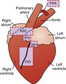
Fig. 10-46 Schematic diagram of the sites of the major cardiovascular anomalies of the dog.
AS, Aortic stenosis; ASD, atrial septal defect; PDA, patent ductus arteriosus; PS, pulmonic stenosis; VSD, ventricular septal defect. (Redrawn with permission from School of Veterinary Medicine, Purdue University.)
Failure of Closure of Fetal Cardiovascular Shunts:
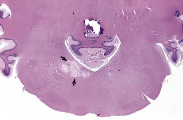
Interventricular Septal Defect: A ventricular septal defect indicates failure of complete development of the interventricular septum and allows the shunting of blood between the ventricles (Fig. 10-47). The defect occurs in many species and more commonly in the upper, membranous portion of the interventricular septum rather than in the lower, muscular septum. Among breeds of dogs, the greatest frequency has been observed in the English bulldog, English springer spaniel, and West Highland white terrier.
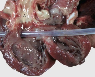
Fig. 10-47 Ventricular septal defect (high defect), heart, opened left side, calf.
Note the large opening in the basal portion of the ventricular septum (arrow) immediately below the aortic valve through which the tube has been passed. A, Aorta; LV, left ventricle. (Courtesy Dr. M. D. McGavin, College of Veterinary Medicine, University of Tennessee.)
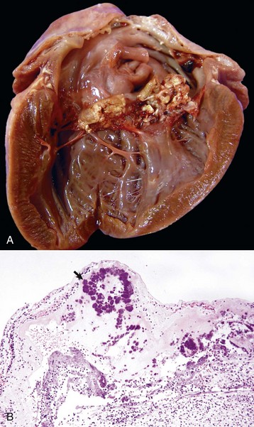
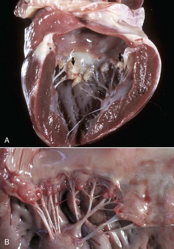
Atrial Septal Defect: An atrial septal defect could represent the failure of closure of the foramen ovale, which is an interatrial septal shunt that allows blood to bypass the lungs of the fetus, or it can be the result of true septal defects at another site because of faulty development of the interatrial septum (Fig. 10-48). Although this defect occurs in all domestic animal species, dog breeds with greatest frequency of this defect are the Boxer, Doberman pinscher, and Samoyed.
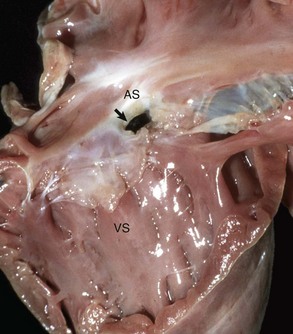
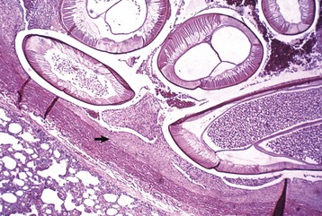
Fig. 10-48 Endocardial cushion defect and tricuspid dysplasia, heart, opened right side, pig.
The endocardial cushion defect (prominent opening [arrow]) can be mistaken as an atrial septal defect but not the location and presence of abnormal valves incorporated in the defect. AS, Atrial septum; VS, Ventricular septum. (Courtesy School of Veterinary Medicine, Purdue University.)
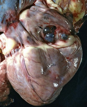
Tetralogy of Fallot: Tetralogy of Fallot is a complicated cardiac anomaly with four lesions (Fig. 10-49). The three primary defects are a ventricular septal defect located high in the septum, pulmonic stenosis (see later discussion), and dextroposition of the aorta (see later discussion). The fourth defect, which develops secondarily, is hypertrophy of the right ventricular myocardium. This complex anomaly is inherited in Keeshond dogs and is frequent in English bulldogs. Cyanosis is often an associated clinical sign. The anomaly is one of the most common cardiac abnormalities seen in hearts of humans (so-called blue babies). In genetic and pathologic studies of Keeshond dogs, the basic defect has been determined to be hypoplasia and malpositioning of the conotruncal septum. Wide variability in the severity of the lesions has been observed. The inheritance pattern in Keeshonds is a simple autosomal locus with partial penetration in heterozygotes and complete penetrance in homozygotes.
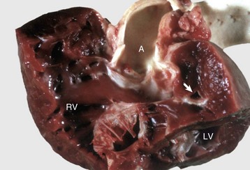
Fig. 10-49 Tetralogy of Fallot, heart, dissected, dog.
Above the large membranous ventricular septal defect is an overlying, straddling aorta (A). There is also severe pulmonic stenosis (arrow) with massive right ventricular hypertrophy. LV, Left ventricle; RV, right ventricle. (Courtesy School of Veterinary Medicine, Purdue University.)
Cardiomyopathies: Primary and secondary cardiomyopathies (Box 10-12) represent important, sometimes inherited, generalized myocardial diseases of either idiopathic or known causation that can result in either hypertrophy or atrophy of the affected myocardium. Primary or idiopathic cardiomyopathies are progressive cardiac diseases. These diseases affect cats, dogs, and cattle and resemble some diseases of humans. These diseases are divided into three morphologic types: hypertrophic, dilated, and restrictive cardiomyopathies. Secondary cardiomyopathies (also termed specific heart muscle diseases) are generalized myocardial diseases of known cause.
Hypertrophic cardiomyopathy (hypertrophy): Hypertrophic cardiomyopathy occurs frequently in cats, especially in young adult to middle-aged males (1 to 3 years old), and is seen infrequently in dogs, usually affecting males of large breeds. Cats usually have congestive heart failure, and approximately 10% to 20% have posterior paresis from concurrent thromboembolism of the caudal abdominal aorta (“saddle thrombosis”) secondary to left atrial thrombosis. Hypertrophic cardiomyopathy is inherited as an autosomal dominant trait with complete penetrance in Maine coon cats. The occurrence of clusters of cases in Persians, American shorthairs, and ragdoll cat breeds strongly suggests heritability of the disease in some cases. Some cats and dogs die unexpectedly as the only clinical expression of the disease. This clinical presentation also is often seen in humans with the disease. In both cats and dogs, the hearts are enlarged and have prominent hypertrophy of the left ventricle and interventricular septum (Fig. 10-50). The left ventricular cavity is small and the left atrium is dilated. In a few cases, the interventricular septum is disproportionately hypertrophied in relation to the remainder of the myocardium. Microscopically, the lesions of the myocardium are prominent disarrays or disorganizations of myocytes, with interweaving rather than parallel arrangement of fibers (Fig. 10-51; Web Fig. 10-20). Myocyte hypertrophy, various degenerative alterations in myocytes, and interstitial fibrosis also are present.
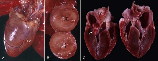
Fig. 10-50 Hypertrophic cardiomyopathy, heart, cat.
A, Note the thickened left ventricular wall (LV). B, The thickened left ventricular free wall and septum have markedly reduced the lumen of the left ventricle (LV). C, There is severe diffuse concentric hypertrophy of the left ventricular free wall, interventricular septum, and papillary muscles. Left atrial dilation is present. There is a fibrotic plaque (arrow) of the midinterventricular septum where the hypertrophied papillary muscle contacts the septum. (A and B courtesy Dr. W. Crowell, College of Veterinary Medicine, The University of Georgia; and Noah’s Arkive, College of Veterinary Medicine, The University of Georgia. C courtesy Ettinger SJ, Feldman EC (eds): Textbook of veterinary internal medicine. Diseases of the dog and cat, vol 2, ed 7, Philadelphia, 2010, Saunders.)
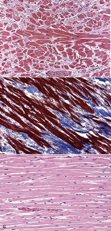
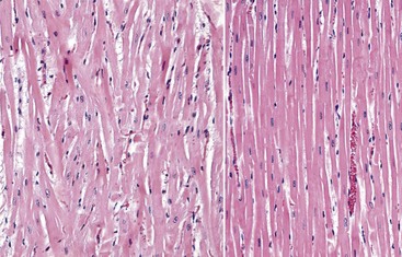
Fig. 10-51 Hypertrophic cardiomyopathy, heart, ventricular myocardium, cat.
A, Cardiac myocytes are hypertrophied and in disarray. H&E stain. B, Masson’s trichrome stain demonstrates abundant amounts of interstitial collagen (blue) produced by fibroblasts. C, Normal cardiac myocytes arranged in parallel bundles. H&E stain. (A and B courtesy Atlantic Veterinary College, University of Prince Edward Island. C courtesy Dr. L. Borst, College of Veterinary Medicine.)
Web Fig. 10-20 Hypertrophic cardiomyopathy, heart, ventricular myocardium, cat.
A, Note the pattern of interwoven cardiac myocytes, indicating myofiber disarray, and the hypertrophic myocytes (compare with Figs. 10-20 and 10-51). The number of fibroblasts is increased in the interstitium. H&E stain. B, Normal cardiac myocytes arranged in parallel bundles. (Courtesy Dr. L Borst, College of Veterinary Medicine, University of Illinois.)
Dilated or congestive cardiomyopathy: Dilated or congestive cardiomyopathy is an important cause of congestive heart failure in cats and dogs. Many affected cats and some dogs have low tissue concentrations of taurine, and supplementation of cats with taurine has reversed the clinical signs of cardiac failure. Routine taurine supplementation of feline commercial diets has resulted in a dramatic reduction in cases of dilated cardiomyopathy. Taurine-deficient foxes also develop cardiac failure. Cattle with dilated cardiomyopathy in Switzerland and Japan have an autosomal recessive mode of inheritance.
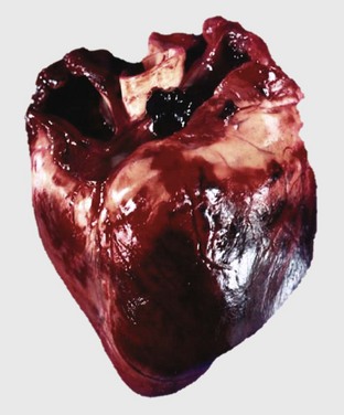
Affected cats often are middle-aged males, and affected dogs often are males of large breeds such as Doberman pinschers, Portuguese water dogs, dalmatians, Scottish deerhounds, Irish wolfhounds, Saint Bernards, Afghan hounds, Newfoundland dogs, Old English sheepdogs, Great Danes, and boxers, although smaller breeds, such as English cocker spaniels, may be affected. The disease often has a familial pattern in the affected breeds and appears to be inherited as an autosomal recessive or X-linked recessive trait. Some cats also develop aortic thromboembolism. At necropsy, lesions of congestive heart failure are present and the hearts are rounded because of biventricular dilation (Figs. 10-52 and 10-53). The dilated cardiac chambers often have a diffusely white, thickened endocardium. Microscopic and ultrastructural alterations are nonspecific, can be either mild or absent, and may include interstitial fibrosis and fatty infiltration and changes of myocyte degeneration, including the occurrence of so-called attenuated wavy fibers.
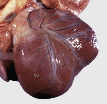
Fig. 10-52 Dilated (congestive) cardiomyopathy, heart, left ventricle (LV) and right ventricle (RV), dog.
Biventricular dilation has resulted in the heart having a double apex. (Courtesy Dr. T. Boosinger, College of Veterinary Medicine, Auburn University; and Noah’s Arkive, College of Veterinary Medicine, The University of Georgia.)
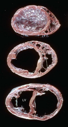
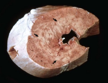
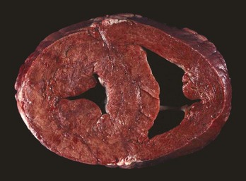
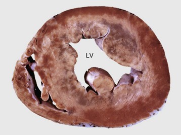
Fig. 10-53 Dilated (congestive) cardiomyopathy, heart, ventricles, cross-section, dog.
The left ventricle (LV) and right ventricle have thin walls, dilated chambers, and white fibrotic endocardium. (Courtesy Dr. Y. Niyo, College of Veterinary Medicine, Iowa State University; and Noah’s Arkive, College of Veterinary Medicine, The University of Georgia.)
Restrictive cardiomyopathy: Restrictive cardiomyopathy occurs infrequently. It occurs in cats as one of two types of endocardial lesions that result in impaired ventricular filling. In one type, the left ventricular endocardium has diffuse notable fibrosis. Available evidence suggests that the fibrotic lesion is preceded by endomyocarditis. The second type results from excessive moderator bands that traverse the left ventricular cavity. Other examples of restrictive cardiomyopathy in animals include endocardial fibrosis in certain strains of aged rats and congenital endocardial fibroelastosis in Burmese cats (Fig. 10-54).
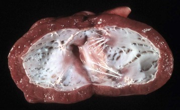
Fig. 10-54 Subendocardial fibroelastosis, heart, left ventricle, dog.
The endocardium is opaque because increased amounts of collagen and elastic fibers were deposited in the subendocardium secondary to turbulence of blood flow within the ventricles. This dog had a persistent ductus arteriosus. This lesion may have a hereditary basis in Burmese cats and is often a sequela to turbulence within ventricles in cardiac disease. (Courtesy College of Veterinary Medicine, University of Illinois.)
Molecular mechanisms of hereditary cardiomyopathies: Our understanding of the molecular mechanisms of the hereditary cardiomyopathies is developing rapidly. In humans with familial hypertrophic cardiomyopathy inherited in an autosomal dominant manner, a variety of single-gene mutations have been documented. The mutations affect genes that encode sarcomere proteins of cardiac myocytes. Altered cardiac proteins include cardiac α- and β-myosin heavy chain, cardiac troponin T and I, and Ca-tropomyosin, actin, titin, ventricular myosin essential light chain, ventricular myosin regulatory light chain, and cardiac myosin-binding protein C. It remains unclear how these mutant proteins result in functional and structural alterations of cardiac muscle cells. However, recent studies suggest that shortening of telomeres (structures that cap the ends of chromosomes) triggers apoptosis of cardiac muscle cells and may explain the end-stage finding of myocardial fibrosis in heart failure of various causes, including cardiomyopathy. Some similar gene mutations and altered proteins were recently discovered in the various heritable cardiomyopathies of animals (e.g. cardiac myosin-binding protein C in Maine Coon cats and Ragdoll cats with hypertrophic cardiomyopathy). Also, a portion of cases of dilated cardiomyopathy in humans appears to be inherited. In these patients, alterations in several myocytic proteins, including dystrophin, actin, desmin, troponin T, β-myosin heavy chain, lamin, and taffazin, and alterations in the cardiac calcium regulating protein phospholamban have been documented.
Neoplastic Transformation: Various primary and secondary neoplasms develop either in or near the heart. Primary neoplasms include rhabdomyoma, rhabdomyosarcoma, schwannoma, and hemangiosarcoma. Rhabdomyomas and rhabdomyosarcomas are rare in animals and form white to gray nodules in the myocardium that often project into the cardiac chambers. Congenital rhabdomyomatosis in pigs and guinea pigs is a nonneoplastic hamartoma (i.e., malformation often resembling a neoplasm that is composed of an overgrowth of mature cells and tissues that normally occur in the affected organ). Single or multiple, pale, poorly circumscribed areas are scattered in the myocardium and are composed of large glycogen-laden myocytes.
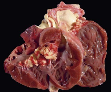
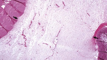
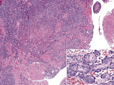
Malignant lymphoma (lymphosarcoma) is the most common secondary neoplasm occurring in the heart and often causes lesions in the hearts of cattle, which can be severe enough to cause death from cardiac failure. Cardiac lesions may be present in dogs and cats with malignant lymphoma. The neoplastic cell infiltration can be diffuse or nodular and involve the myocardium, endocardium, and pericardium. Lymphomatous tissue appears as white masses that may resemble deposits of fat (Fig. 10-55). Microscopically, extensive infiltrations of neoplastic lymphocytes are present between myocytes (Fig. 10-56). Other neoplasms, such as malignant melanomas, occasionally have metastatic lesions in the heart.
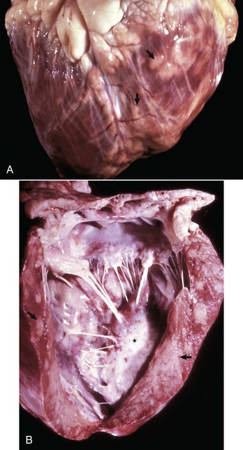
Fig. 10-55 Lymphosarcoma, heart, myocardium, cow.
A, Sites of infiltrating neoplastic lymphocytes in the ventricular myocardium are evident as numerous white areas and nodules (arrows). B, Similar white areas of tumor are visible in the section of the left ventricular wall (arrows) and subendocardially (asterisk) in the ventricular septum. (Courtesy College of Veterinary Medicine, University of Illinois.)
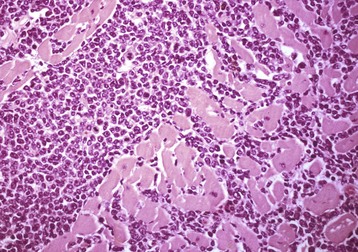
Fig. 10-56 Lymphosarcoma, heart, section of myocardium, cow.
Neoplastic lymphocytes have extensively infiltrated between the cardiomyocytes. Extensive infiltration can result in myocyte atrophy and loss. H&E stain. (Courtesy School of Veterinary Medicine, Purdue University.)
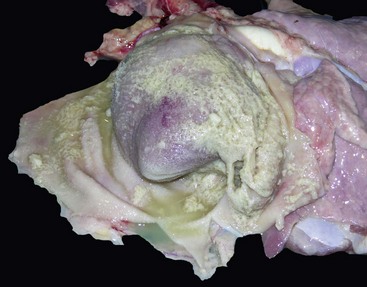
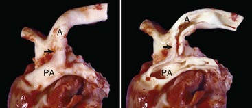
Heart-based tumors are primary neoplasms of extracardiac tissues in dogs and rarely cats. They arise at the base of the heart and can produce vascular obstruction and cardiac failure. The most common neoplasm arising at this location is the aortic body tumor or paraganglioma (chemodectoma), but occasionally, ectopic thyroid or parathyroid tissue gives origin to neoplasms in this area. The aortic body is a chemoreceptor organ. In some cases, aortic body tumors become large, white, firm masses that surround and compress the great vessels and atria (Fig. 10-57). Brachycephalic dog breeds are most frequently affected. Microscopically, the neoplastic cells are polyhedral with vacuolated cytoplasm and are supported by a fine connective tissue stroma (see Fig. 12-55).
Toxicoses: The mechanisms of cardiotoxicities include (1) exaggerated pharmacologic action of drugs acting on cardiovascular tissues, (2) exposure to substances that depress myocardial function, (3) direct injury of cardiac muscle cells by chemicals, and (4) hypersensitivity reactions.
White snakeroot–induced myocardial degeneration: See the discussion on white snakeroot–induced myocardial degeneration in the section on Disorders of Horses.
Ionophore-induced myocardial degeneration: See the discussion on ionophore-induced myocardial degeneration in the section on Disorders of Horses.
Gossypol-induced myocardial degeneration: See the discussion on gossypol-induced myocardial degeneration in the section on Disorders of Ruminants.
Chemotherapeutic agent-induced myocardial degeneration: Cardiotoxicity has emerged as a significant clinical entity in veterinary medicine in recent years with the growing use of antineoplastic drugs in small animal practice and the widespread use of growth promotants in ruminants (see Fig. 10-69).
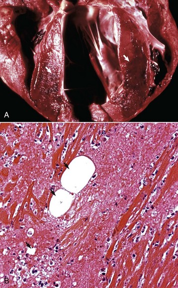
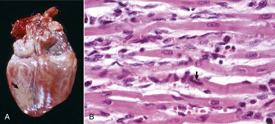
Myocardial Necrosis and Mineralization: Myocardial necrosis and mineralization can result from a number of causes, including nutritional deficiencies, chemical and plant toxins, ischemia, metabolic disorders, heritable diseases, and physical injuries (see Box 10-5). From this large list of causes of myocardial injury, some of the most frequently observed current examples are ionophore toxicity in horses and ruminants, vitamin E–selenium deficiency in the young of all species, “heart-brain syndrome” of dogs (see Fig. 10-83), anthracycline toxicity in dogs, and gossypol toxicosis in pigs. In various localized areas throughout the world, numerous deaths in ruminants have resulted from consumption of poisonous plants such as Acacia georginae and Dichapetalum cymosum.
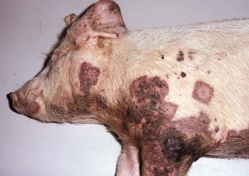
Mulberry Heart Disease: See the discussion of mulberry heart disease in the section on Disorders of Pigs.
Brain-Heart Syndrome: See the discussion on brain-heart syndrome in Chapter 14.
Myocarditis: See the discussion on inflammation in the section on Myocardium, Responses to Injury.
Bacterial Septicemias: See the discussion on bacterial septicemias in Chapter 4.
See the discussion on the cardiac conduction system in the section on Responses to Injury.
Schwannomas: Schwannomas involve cardiac nerves in cattle and appear as single or multiple white nodules detected as incidental findings at slaughter (see Fig. 14-115).
Conduction system diseases have been described mainly in dogs and horses, probably because clinical cardiac evaluations are done most frequently in these species. Secondary conduction system disorders result from myocardial disease (inflammation, neoplasia, or degeneration) near the conduction system. Specific presumably inherited diseases in dogs include (1) syncope in pug dogs with lesions of the bundle of His; (2) intermittent sinus arrest in deaf dalmatian dogs, presumably associated with lesions in the sinus node; (3) sinoatrial syncope (sick sinus dysfunction) in female miniature schnauzers, West Highland white terriers, cocker spaniels, and dachshunds; (4) inherited ventricular arrhythmia and sudden unexpected death in German shepherds, and (5) widespread conduction pathology in Alaskan sled dogs that died suddenly and unexpectedly in a race. Other arrhythmias in dogs and horses are atrial fibrillation and heart block. Dogs with atrial fibrillation often have concurrent congestive heart failure and have atrial dilation with AV valve insufficiency, but most horses live a normal or near-normal lifespan, may respond to cardioversion, and at necropsy have atrial myocardial fibrosis. Heart block of the first degree (delay of impulse through AV node), second degree (intermittent failure to conduct through the AV node with dropped beats), and third degree (complete) has been associated with myocardial lesions, such as areas of scarring, in horses and dogs. Second-degree heart block is considered to be a normal phenomenon in horses.
Persistent atrial standstill (silent atria, AV myopathy) is a progressive cardiac disease of English springer spaniels and cats characterized by notable atrial dilation and fibrosis.
Atrial fibrillation occurs in cattle in association with right atrial dilation and fibrosis and alterations in the SA mode. Also, sudden (unexpected) cardiac death is described in racehorses with right atrial myocardial fibrosis, fibrosis of the upper ventricular septum, and arteriosclerosis of intramyocardial arteries.
See the discussion of the endocardium and heart valves in the section on Responses to Injury. The major types of AV valvular diseases are shown in Fig. 10-58.
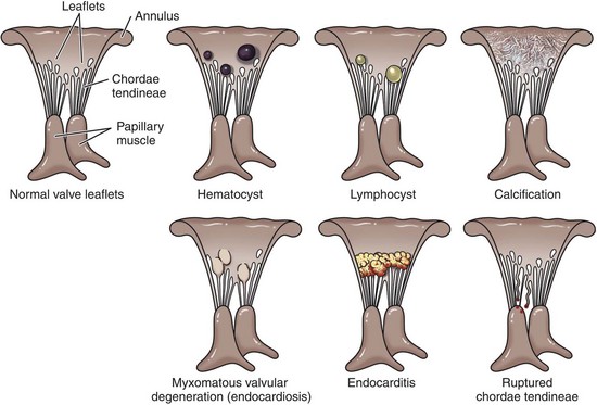
Fig. 10-58 Schematic diagram of the major types of cardiac atrioventricular valvular disease. (Redrawn with permission from School of Veterinary Medicine, Purdue University.)
Developmental Errors: Congenital Anomalies:
Failure of Normal Valvular Development:
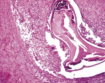
Pulmonic stenosis: Pulmonic stenosis has been recognized as a frequently occurring anomaly in dogs and is inherited in the beagle (Fig. 10-59). Other breeds in which this lesion is frequent are basset hound, boxer, Chihuahua, Chow chow, cocker spaniel, English bulldog, Labrador retriever, mastiff, Newfoundland, Samoyed, schnauzer, and terrier. Several types of valvular lesions have been described and include formation of a circumferential band of fibrous or muscular tissue beneath the valve (subvalvular stenosis) or malformation of the valve (valvular stenosis), with a small central orifice in a dome of thickened valvular tissue. Notable concentric hypertrophy (see the discussion on hypertrophy in the section on Disturbances of Growth, Myocardium, Responses to Injury) of the right ventricle develops from the resulting pressure overload.
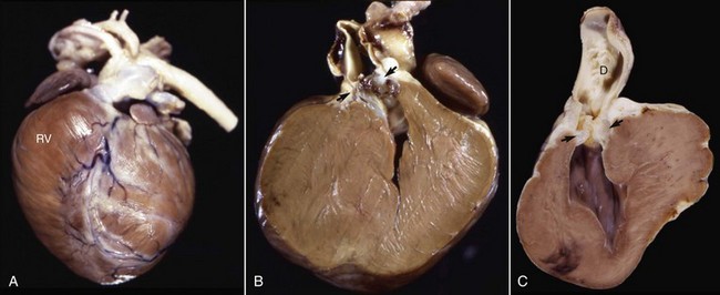
Fig. 10-59 Pulmonic stenosis, heart, pulmonary artery, dogs.
A, Closed heart, and B, sectioned heart. Note the prominent concentric right ventricular (RV) hypertrophy resulting from pressure overload. The orifice of the pulmonic valve (arrows) is markedly narrowed. C, sectioned heart, there is poststenotic dilation (D) of the pulmonary artery with irregular intimal thickenings (jet lesions). (Courtesy Atlantic Veterinary College, University of Prince Edward Island.)
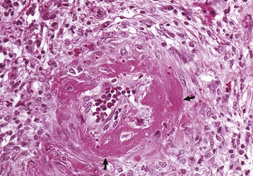
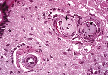
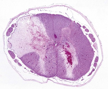
Aortic and subaortic stenoses: True stenoses of the aortic valve are uncommon. Subaortic stenosis is a cardiac anomaly frequently observed in pigs and dogs. It apparently is inherited in Newfoundland, boxer, and German shepherd dogs (Fig. 10-60). The lesion is also observed in the German shorthair pointer, golden retriever, Great Dane, Rottweiler, Samoyed, and bull terrier breeds. In clinical cases, the stenosis is produced by the presence of a thick zone of endocardial fibrous tissue that encircles the left ventricular outflow tract below the valve. In mild cases, often subclinical, the lesion is limited to white nodules on the ventricular septum immediately below the valve. Microscopically, the altered endocardial tissue can contain proliferated mesenchymal cells, mucinous ground substance, and foci of metaplastic cartilage. Other cardiac lesions develop as a result of the altered left ventricular outflow; these include left ventricular concentric hypertrophy, disseminated foci of myocardial necrosis, fibrosis in the inner left ventricular wall, and thickening of the walls of intramyocardial arteries.
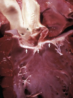
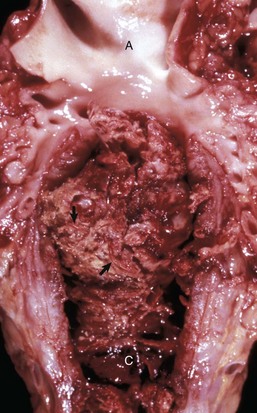
Fig. 10-60 Subaortic stenosis, heart, opened left side, dog.
A thick, white, broad band of fibrous connective tissue (arrows) encircles the left ventricular outflow tract below the aortic valve. The force of the blood ejected through the stenotic lesion is responsible for the “jet lesions” in the overlying aorta (A) (right half: roughened surface; left half: dilation [note the gray area]). (Courtesy College of Veterinary Medicine, University of Illinois.)
Valvular dysplasias: endocardial cushion defects: Other valvular developmental anomalies include endocardial cushion defects (persistent AV canal) in pigs, sheep, and cats; mitral dysplasia in cats and dogs; and tricuspid dysplasia in cats and dogs (see Fig. 10-48). Tricuspid dysplasia is inherited as an autosomal dominant trait with reduced penetrance in Labrador retrievers. The tricuspid valve is abnormal with shortened and thickened chordae tendineae resulting in an incompetent valve and right-sided heart failure.
Endocardial fibroelastosis: Endocardial fibroelastosis in animals was historically recognized as a primary cardiac defect in Burmese and Siamese cats. Affected animals had prominent, white, thickened endocardium, especially of the left ventricle, because of the proliferation of fibroelastic tissue (see Fig. 10-54). Endocardial fibroelastosis is a reaction of the endocardium to hypoxia and often associated with heart disease, which results in dilated cardiac chambers. It is unclear whether this is a true congenital anomaly or a response to left atrial dilation.
Valvular hematomas: See the discussion on valvular hematomas in the section on Disorders of Ruminants.
Valvular lymphocysts: See the discussion on valvular lymphocysts in the section on Disorders of Ruminants.
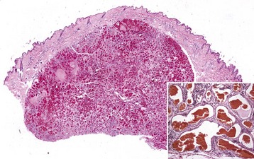
Uremic Endocarditis: Uremic endocarditis is most commonly a disorder in dogs that follows acute or repeated episodes of uremia. These episodes cause ulcerative endocarditis (injury of the endothelium) of the left atrium that is resolved via healing characterized by fibrosis, with or without mineralization and chronically dilated atria (Fig. 10-61).
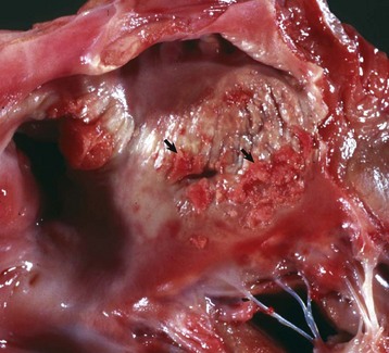
Fig. 10-61 Ulcerative endocarditis (uremia), heart, endocardium of left atrium, dog.
Note the white-red, thick, wrinkled area (arrows) of endocarditis, mineralization, and fibrous tissue (scar) formation caused by uremia in this dog with chronic renal failure. (Courtesy Dr. K. Read, College of Veterinary Medicine, Texas A&M University; and Noah’s Arkive, College of Veterinary Medicine, The University of Georgia.)
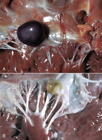
Myxomatous Valvular Degeneration (Valvular Endocardiosis): Degenerative changes in the valves are frequently seen in older dogs and the process that leads to this lesion is termed myxomatous valvular degeneration (valvular endocardiosis). Also see the section on Disorders of Dogs; also see Fig. 10-84).
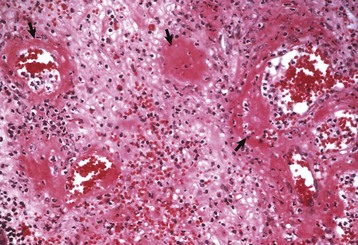
Endocardial Mineralization: Endocardial mineralization occurs from intake of excessive amounts of vitamin D and from intoxication by calcinogenic plants (Cestrum diurnum, Trisetum flavescens, Solanum malacoxylon, Solanum torvum) that contain vitamin D analogs. These plant-induced syndromes of cattle have been called by different names in various areas of the world, such as “Manchester wasting disease” in Jamaica, “enzootic calcinosis” in Europe, “Naalehu disease” in Hawaii, “enteque seco” in Argentina, and “espichamento” in Brazil. Multiple, large, white, rough, firm plaques of mineralized fibroelastic tissue are present in the endocardium and intima of large elastic arteries. Fibrosis, with or without mineralization, occurs in chronically dilated hearts, in hearts of debilitated cattle with Johne’s disease (see Figs. 10-32 and 10-39), in dogs with healed lesions of left atrial ulcerative endocarditis associated with a prior uremic episode (see Fig. 10-61), and in the so-called jet lesions produced by the trauma of refluxed blood in valvular insufficiencies.
Valvular Endocarditis: See the discussion on vegetative valvular and mural endocarditis in the section on Inflammation, Endocardium, Responses to Injury, as well as the discussion on valvular endocarditis in Chapter 3.
See the discussion on blood and lymphatic vascular systems in the section on Responses to Injury. The major arterial diseases are shown in Fig. 10-62.
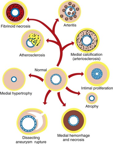
Fig. 10-62 Schematic diagram of the major arterial diseases. (Redrawn with permission from School of Veterinary Medicine, Purdue University.)
Effusions: See the discussion on effusions in the section on Disturbances of Circulation, Pericardium and Epicardium, Responses to Injury and Disorders of Domestic Animals.
Aortic rupture and rupture of large arteries: Aortic rupture and rupture of large arteries can be the sequela of severe trauma or occur spontaneously (see Figs. 10-34 and 10-35). Sudden rupture of the ascending aorta or pulmonary artery near the pulmonic valve in horses is associated with notable exertion and severe trauma to the ventral thorax from falling. Death ensues rapidly from cardiac tamponade, as the tear is in that portion of the aorta or pulmonary artery within the pericardial sac. In horses, the internal carotid artery can rupture into the adjacent guttural pouch, with subsequent epistaxis. This is a consequence of deep mycotic infection of the guttural pouch. Rupture of the middle uterine artery may occur during parturition in mares and with uterine torsion or prolapse in cows. Aortic rupture, with or without dissection, is an important cause of death in male turkeys. The most common vascular diseases with rupture are listed in Box 10-13.
Gastric Dilation and Volvulus: See the discussion on gastric dilation and volvulus in Chapter 7.
Developmental Errors: Congenital Anomalies:
Failure of closure of fetal cardiovascular shunts:
Patent ductus arteriosus: See the discussion of patent ductus arteriosus in the section on Diseases of Dogs.
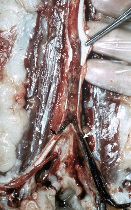
Portacaval shunts: Portacaval shunts occur in animals, particularly in the dog. The normal flow from the portal vein is diverted, either partially or completely, to the systemic circulation, thus bypassing the liver (Fig. 10-63). Normal hepatic detoxification of portal flow is incomplete and may result in neurologic signs and elevated circulating bile acids. The resulting nervous system syndrome is termed hepatic encephalopathy. Specifically, the shunts represent retained fetal vascular structures, as in persistent ductus venosus, or arise from prominent dilation of various portosystemic shunts that normally are quite small vessels. See Chapter 8 on diseases of the liver for further details.
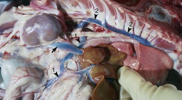
Fig. 10-63 Portacaval shunt, dog.
Note that the branch of the portal vein (arrowhead 1) passes under the caudal vena cava (arrow) and anastomoses with the azygous vein (arrowhead 2). The azygous vein returns the blood to the caudal vena cava near the heart and thus this blood and its ammoniacal and protein metabolites are shunted away from processing to blood urea nitrogen (BUN) in the liver. The liver is normal color but extremely small, which is typical of these types of shunts (see Chapter 8). (Courtesy Dr. M.D. McGavin, College of Veterinary Medicine, University of Tennessee.)
Malpositioning of great vessels:
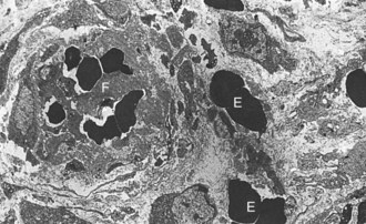
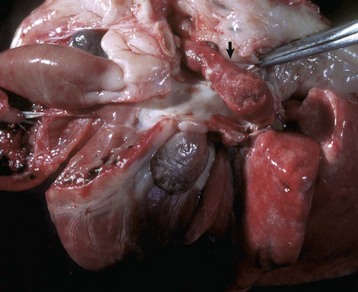
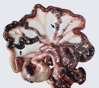
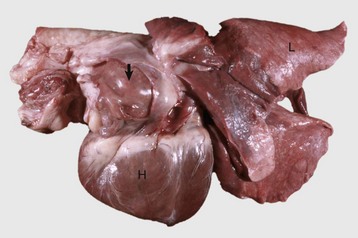
Persistent right aortic arch: Persistent right aortic arch occurs in dogs; German shepherd, Irish setter, and Great Dane dogs are predisposed (Fig. 10-64). This defect arises because the right fourth aortic arch, rather than the normal left fourth aortic arch, develops and ascends on the right side of the midline so that the ligamentum arteriosum forms a vascular ring over the esophagus and trachea. This arrangement eventually results in esophageal obstruction and proximal dilation (megaesophagus), which often results in aspiration pneumonia as the animal matures and consumes solid feed.
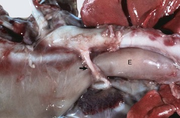
Fig. 10-64 Persistent right aortic arch, ligamentum arteriosum, megaesophagus, calf.
During embryogenesis the aorta was formed from the right aortic arch instead of the left one, thus the aorta is now on the right. For the ligamentum arteriosum (arrow) to connect the aorta with the pulmonary artery, it has to pass dorsally over the esophagus and trachea. The ligamentum, together with the aorta and pulmonary artery, form a vascular ring that constricts the esophagus (E), which is dilated cranial to the constriction. (Courtesy Dr. S. Snyder, College of Veterinary Medicine, Colorado State University; and Noah’s Arkive, College of Veterinary Medicine, The University of Georgia.)
Transposition of the aorta and pulmonary artery: Transposition of the aorta and pulmonary artery are severe anomalies, of which there are several types. In complete transposition, the aorta serves as the outflow from the right ventricle and the pulmonary artery is the left ventricle primary outflow. Other congenital anomalies, including ventricular septal defect, often accompany this anomaly.
Vascular Melanosis: See the discussion on vascular melanosis in Chapters 1 and 2.
Neoplastic Transformation: See the discussion on neoplastic transformation in the section on Disorders of Dogs.
Cell Degeneration and Death: Toxicants that affect vessels are listed in Web Box 10-1.
Vitamin E–Selenium Deficiency: See the discussion on vitamin E–selenium deficiency in the section on Disorders of Pigs.
Omphalophlebitis (“Navel Ill”): Omphalophlebitis (“navel ill”) is inflammation of the umbilical vein that often occurs in neonatal farm animals because of bacterial contamination of the umbilicus immediately after parturition. Bacteria from this site can cause septicemia, suppurative polyarthritis, hepatic abscesses (the umbilical vein drains into the liver), and umbilical abscesses.
Jugular Thrombophlebitis: Jugular thrombophlebitis may be associated with indwelling jugular catheters and is reported to be increased with several concurrent disease conditions such as hypoproteinemia, salmonellosis, endotoxemia, and large intestinal disease (see Fig. 10-43).
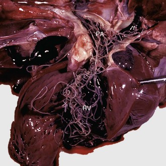
Rupture of the thoracic duct: Rupture of the thoracic duct, either as a result of trauma or from spontaneous disruption, causes chylothorax in dogs and cats (see Fig. 9-101). However, many cases of chylothorax occur without injury to the thoracic duct and have been attributed to lesions that interfere with central venous return or produce obstruction of the thoracic duct (right-sided heart failure, neoplasms, granulomas, cranial vena cava thrombosis, or dirofilariasis) or that are idiopathic.
Developmental Errors: Congenital Anomalies:
Lymphangiectasia: Lymphangiectasia is dilation of lymphatic vessels. The cause may be a congenital anomaly (Fig. 10-65) or caused by obstruction of lymph drainage by invading masses of malignant neoplasms or inflammation (Fig. 10-66).
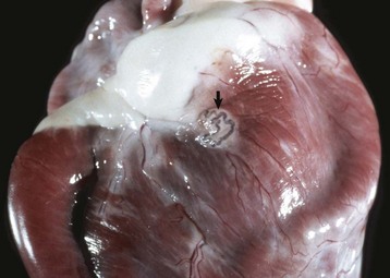
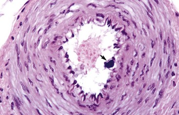
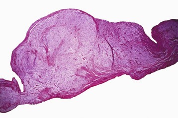
Fig. 10-65 Congenital lymphangiectasia, epicardium, young horse.
Note the tortuous appearance of the epicardial lymphatic vessel (arrow). In congenital lymphangiectasia, lymphatic vessels fail to make connections with other vessels or are obstructed because of anomalous development. (Courtesy College of Veterinary Medicine, University of Illinois.)
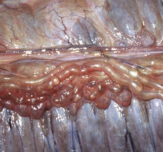
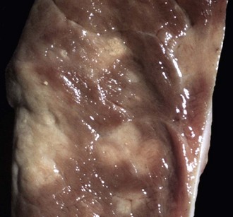
Fig. 10-66 Acquired lymphangiectasia, lymphoma (lymphosarcoma), mesoccolon, horse.
Note the distended lymphatics on the serosal surface of the large colon, the result of impeded lymph flow through the colic lymph nodes, caused by compression of their cortical and medullary sinuses by proliferating neoplastic lymphocytes. (Courtesy College of Veterinary Medicine, University of Illinois.)
Hereditary lymphedema: Hereditary lymphedema has been described in dogs, Ayrshire and Angus calves, and pigs. Affected animals have prominent subcutaneous edema that, in calves, often causes severe swelling of the tips of the ears. Interference with lymph drainage results from defective development of the lymphatic vessels that are aplastic or hypoplastic.
Neoplastic Transformation: Lymphangioma is a rare benign neoplasm composed of lymphatic channels. Lymphangiosarcoma, the malignant counterpart, occurs more often than the benign neoplasm. Vascular spaces formed by neoplastic lymphatic endothelial cells contain lymph rather than blood. Lymphatic vessels are frequently invaded by primary carcinomas and are a common route of metastasis (see the section on Disorders of Dogs).
Inflammation: The most common diseases with thrombosis and embolism are listed in Box 10-14.
Lymphangitis: Lymphangitis is a feature of many diseases (see Box 10-8). The affected vessels are often located in the distal limbs and are thick, cordlike structures (Fig. 10-67). Lymphedema can be present. Nodular suppurative lesions of lymphangitis often ulcerate and discharge pus onto the surface of the skin. In Johne’s disease, the mesenteric lymphatic vessels are often prominent because of granulomatous lymphangitis, an extension of the enteric infection producing a granulomatous enteritis and lymphangitis (Fig. 10-68).
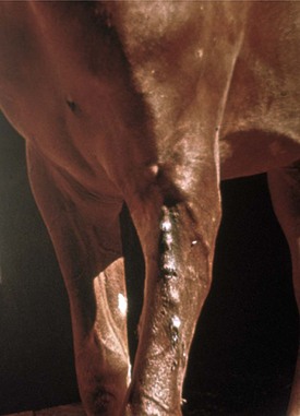
Fig. 10-67 Lymphangitis, forelimb, lymphatic vessels, horse.
Note the multiple swellings (cordlike) of the afferent lymphatics in the skin. These lymphatics lie in the subcutis and empty into the caudal superficial cervical (prescapular) lymph node. (Courtesy School of Veterinary Medicine, Purdue University.)
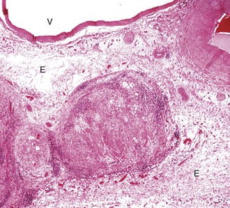
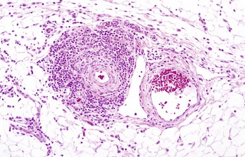
Fig. 10-68 Granulomatous lymphangitis, Johne’s disease, mesenteric lymphatic vessel, sheep.
The lymphatic is occluded by a fibrinous thrombus secondary to the destruction of the endothelium by inflammatory cells including macrophages. Early proliferating fibrous tissue and extensive edema (E) surround the lymphatic vessel. The adjacent artery (upper right) and vein (V) are unaffected. H&E stain. (Courtesy School of Veterinary Medicine, Purdue University.)
Ionophore-Induced Myocardial Degeneration
Ionophores, such as monensin, lasalocid, salinomycin, and narasin, are toxic to horses at extremely low concentrations. They are used as feed additives to increase feed efficiency and weight gain of beef and dairy cattle and control coccidiosis in poultry. Horses gain access to ionophores when they consume (1) ruminant feed containing ionophores, (2) horse feed accidentally mixed with ionophores, and (3) horse feed accidentally contaminated in a mill producing poultry, cattle, and horse feeds. Ionophores cause acute cardiac rhabdomyocyte degeneration and necrosis; this type of injury is discussed in detail in the section on Responses to Injury. Ionophores form complexes with monovalent cations, such as Na+ and Ca+, and cross cell membranes and enter the cell via ion transport systems in exchange for H+ and K+ ions. Increases in the concentrations of intracellular Ca+ and possible Na+ are thought to cause cell membrane injury and dysfunction resulting in mitochondrial swelling and decreased adenosine triphosphate (ATP) production. Additionally, they cause lipid peroxidation of cell membranes leading to loss of cell membrane integrity, fluid and ion shifts, and oncotic necrosis. Macroscopic and microscopic lesions are similar to those that occur with white snakeroot–induced myocardial degeneration described next (Fig. 10-69).
After ingestion, white snakeroot (Eupatorium rugosum) causes myocardial degeneration and necrosis (acute injury) followed by fibrosis (reparative response). Tremetol is the toxic compound in white snakeroot; it becomes toxic after microsomal activation of a precursor compound by cytochrome P-450 enzymes in the liver. The mechanism used by tremetol to cause injury is unclear, but dysfunction of mitochondrial oxidative phosphorylation by inhibiting the tricarboxylic acid cycle has been suggested. Acute cardiac rhabdomyocyte degeneration and necrosis are discussed in detail in the section on Responses to Injury and also in Chapter 15. Reparative responses are discussed in Chapter 3. Macroscopic lesions include pale white to tan areas and linear streaks throughout the myocardium; microscopic lesions include multifocal myocardial degeneration and necrosis with vacuolation of myocardial cytoplasm; loss of cross striations; fragmentation of rhabdomyocytes; hypereosinophilia, coagulation, and clumping of the sarcoplasm; and nuclear pyknosis and karyolysis. The pericardium may contain a modified transudate with fibrin.
Gossypol-induced myocardial degeneration can follow the ingestion of cottonseed or cottonseed products that contain excess free gossypol. Gossypol is a potentially toxic pigment in the cotton plant; however, it is toxic only when in a free form (not bound to protein). Gossypol causes myocardial degeneration and necrosis and cardiac conduction failure (see later). The macroscopic and microscopic characteristics of the lesions are in many ways similar to those caused by ionophore-induced and white snakeroot–induced myocardial degeneration in horses. Additionally, acute rhabdomyocytic degeneration and necrosis are discussed in detail in the section on Responses to Injury and also in Chapter 15. Horses are more sensitive to gossypol-induced myocardial degeneration and necrosis than ruminants and pigs. In summary, macroscopic lesions include pale white to tan areas throughout a “flabby” myocardium; microscopic lesions include multifocal myocardial degeneration and necrosis.
Equine Viral Arteritis: Equine viral arteritis is a systemic viral infection with a tropism for vascular endothelial cells. In this disease, affected small muscular arteries have lesions of fibrinoid necrosis, extensive edema, and leukocytic infiltration (Fig. 10-70). Grossly the vascular injury is reflected by hemorrhage and severe edema of the intestinal wall and mesentery accompanied by notable accumulation of serous fluids in body cavities and pulmonary edema (see Chapter 14).
Fig. 10-70 Acute arteritis, equine viral arteritis, small intestine, submucosa, horse.
Small arteries have fibrinoid degeneration (circumferential eosinophilic material [arrows]) with leukocytic infiltration of the tunica media. The surrounding loose connective tissue is edematous and infiltrated by numerous leukocytes. H&E stain. (Courtesy School of Veterinary Medicine, Purdue University.)
African Horse Sickness: Subacute Cardiac Form: African horse sickness is an insect-borne (Culicoides spp.) viral disease of Equidae that is endemic in Africa, the Middle East, India, and Spain. The occurrence is seasonal because the insect vectors thrive in hot wet conditions. The febrile disease may produce high mortality (up to 95%) and appears in several clinical forms, including the subacute cardiac form described here, as well as an acute respiratory form with massive pulmonary edema. The pathogenesis is initiated by the introduction of the virus by bites of the insect vector. The virus proliferates in local lymph node, and viremia ensues. The virus has a tropism for endothelial cells, monocytes, and macrophages and increased vascular permeability, edema, hemorrhage, and microthrombosis are produced. The gross lesions are extensive subcutaneous and intermuscular edematous swelling of the head and neck. Massive hydropericardium is present with accompanying epicardial and endocardial ecchymotic hemorrhages. Histopathologically, endothelial degeneration and necrosis occur with the edema. In the myocardium, hemorrhage, edema, and focal myocardial necrosis with inflammatory cell infiltrates are present (see Chapter 4 and Fig. 4-44).
Cranial Mesenteric Arteritis and Thrombosis: Cranial mesenteric arteritis and thrombosis results from fourth stage larval migration from Strongylus vulgaris (Fig. 10-71). Infection of horses by Strongylus vulgaris is now less common because of widespread use of highly efficacious antiparasitic drugs. During its larval development, the parasite migrates through the intestinal arteries, and the most severe lesions are generally found in the cranial mesenteric artery near its origin. The affected vessel is enlarged, and its wall is firm and fibrotic. The intimal surface often has an adhering thrombus admixed with larvae. Microscopically, the affected vessel has extensive infiltration of inflammatory cells and proliferation of fibroblasts throughout the wall. As a consequence, thromboembolism of the intestinal arteries frequently occurs and can produce colic, but the abundant collateral circulation to the equine intestinal tract makes intestinal infarction an unusual event.
Fig. 10-71 Verminous arteritis and mural thrombosis, strongylosis, abdominal aorta (A) and cranial mesenteric artery, horse.
A pale friable thrombotic mass, in which several Strongylus vulgaris larvae (arrows) are embedded, is attached to the wall of the cranial mesenteric artery (C). (Courtesy College of Veterinary Medicine, University of Illinois.)
Aortoiliac Thrombosis: Aortoiliac thrombosis is thought to be caused by migration of fourth stage larval migration from Strongylus vulgaris. See the previous section on cranial mesenteric arteritis and thrombosis.
Jugular Thrombophlebitis: See the discussion on jugular thrombophlebitis in the section on Inflammation, Blood and Lymphatic Vessels, Disorders of Domestic Animals.
Arterial Intimal Calcification: Arterial intimal calcification (intimal bodies) are distinctively small, mineralized masses within the subendothelium in small muscular arteries and arterioles of horses (Web Fig. 10-21). They have no deleterious effect.
Web Fig. 10-21 Intimal body (arrow), intestine, muscular artery, horse.
Intimal bodies are distinctive small, mineralized masses within the subendothelium of small muscular arteries and arterioles of horses. They are an incidental finding and have no pathologic significance. H&E stain. (Courtesy Dr. M.D. McGavin, College of Veterinary Medicine, University of Tennessee.)
Glanders Disease (Farcy): Cutaneous Form: Glanders disease (Farcy) is a contagious disease of horses caused by infection with Burkholderia (Pseudomonas) mallei. Once worldwide in distribution, it is now seen in Eastern Europe, Asia, and northern Africa. The disease occurs in several clinical forms, and the cutaneous form with involvement of lymphatic vessels is described here. The pathogenesis is initiated by ingestion of contaminated food and water. The organisms enter through the pharynx and are disseminated to the skin hematogenously. The gross lesions of the skin appear as multiple ulcerated nodules that follow infected lymphatic vessels. Most frequent in the limbs, the ulcerated lesions discharge suppurative exudate onto the skin surface. Swollen tortuous cutaneous lymphatic vessels are visible between the ulcerative lesions. Microscopically, the skin nodules represent pyogranulomatous inflammation extending from cutaneous lymphatic vessels with suppurative lymphangitis (see Fig. 4-24).
Miscellaneous Cutaneous Lymphangitides (See Box 10-8): The cutaneous lesions affecting lymphatic vessels in the diseases are as follows:
1. Ulcerative (likely caused by Corynebacterium pseudotuberculosis and other cutaneous bacteria)
These lesions mimic those of Glanders disease, and differentiation occurs by impression smears and microbiologic cultures and analyses. The skin of the legs, head, neck, and/or flanks has raised firm nodules (≈1 to 2 cm in diameter), draining nodules, and draining fistulous tracts, often arranged in linear bands (beaded appearance) that follow the flow of lymphatic vessels. These lesions contain or drain pus, which is often thick and white-yellow in color. Microscopically, lesions are characterized by suppurative to pyogranulomatous inflammation. Infectious microorganisms are often present in the exudate (see Fig. 4-24).
Suppurative pericarditis is seen mainly in cattle as a complication of traumatic reticuloperitonitis (“hardware disease”). Foreign bodies, such as nails or pieces of wire that accumulate in the reticulum, occasionally penetrate the reticular wall and diaphragm, enter the adjacent pericardial sac, and introduce infection. Some affected cattle survive for weeks to months until death ensues from congestive heart failure and septicemia. Grossly the pericardial surfaces are notably thickened by white, often rough, shaggy-appearing masses of fibrous connective tissue that enclose an accumulation of white to gray, thick, foul-smelling, purulent exudate (see Fig. 10-16). See the discussion on pericarditis in the section on Epicardium and Pericardium, Disorders of Domestic Animals and also Chapters 7 and 9.
Hematogenous spread of specific organisms may result in fibrinous pericarditis. Mannheimiosis, blackleg, coliform septicemias, contagious bovine pleuropneumonia, sporadic bovine encephalomyelitis, and in utero Brucella spp. and Arcanobacter pyogenesis fetal septicemias may all produce fibrinous pericarditis. In sheep, Mannheimiosis and streptococcal infections most commonly result in fibrinous pericarditis. Sterile swabs of the pericardial exudates are recommended to identify the causative organism. See the discussion on pericarditis in the section on Epicardium and Pericardium, Disorders of Domestic Animals and also Chapters 7, 9, 14, 18, and 19.
Gossypol-Induced Myocardial Degeneration
See the discussion on gossypol-induced myocardial degeneration in Disorders of Horses.
Dilated cardiomyopathy in red Holstein-Friesian and Simmental-Red Holsteins occurs frequently in Switzerland. Affected cattle develop right-sided heart failure with cardiac dilation. Histopathologic findings are loss of cardiac muscle cells and replacement fibrosis. Occasional cases in Holsteins were also reported in Japan, Canada, Australia, Denmark, Sweden, and the Netherlands. Congenital cardiomyopathy occurs in Poll Hereford cattle in association with wooly haircoat syndrome. Inherited as an autosomal recessive trait, affected calves usually die from congestive heart failure by 12 weeks of age (also see the discussion on cardiomyopathies in the section on Myocardium, Disorders of Domestic Animals).
Hemorrhagic myocarditis occurs together with the hemorrhagic inflammation typically found in skeletal muscle of cattle with blackleg (Clostridium chauvoei) (Fig. 10-72). See the discussion on myocarditis in the section on Myocardium, Disorders of Domestic Animals and also Chapter 15.
Fig. 10-72 Necrohemorrhagic myocarditis, “blackleg,” heart, steer.
A, Note the area of hemorrhagic myocarditis (arrows) in the wall of the ventricular myocardium. This disease is caused by Clostridium chauvoei, and lesions are most common in skeletal muscle. B, Necrohemorrhagic myocarditis, heart, cow. Note the myocardial necrosis, serocellular interstitial debris, and the clear spaces (arrows) representative of gas bubbles. (A courtesy Dr. J. Simon, College of Veterinary Medicine, University of Illinois. B courtesy Atlantic Veterinary College, University of Prince Edward Island.)
East Coast fever is a tick-transmitted protozoal disease of cattle in Africa caused by Theileria parva, which causes myocardial necrosis and inflammation (Web Fig. 10-22).
Eosinophilic myocarditis and the accumulation of eosinophils in the inflammatory response are the result of some parasitic infections such as sarcocystosis (see Chapter 15).
See the discussion on mineralization in the section on Cell Degeneration, Myocardium, Responses to Injury and also Chapters 1 and 15.
See the discussion on neoplastic transformation in the section on Disturbances of Growth, Myocardium, Disorders of Domestic Animals.
Vegetative Valvular Endocarditis
Endocarditis is usually the result of bacterial infections in which the affected valves have large, adhering, friable, yellow-to-gray masses of fibrin termed vegetations, which can largely occlude the valvular orifice (Fig. 10-73, A). Microscopically, the lesion consists of accumulated layers of fibrin and numerous embedded bacterial colonies underlain by a zone of infiltrated leukocytes and granulation tissue (Fig. 10-73, B). See the discussion on vegetative valvular and mural endocarditis in the section on Inflammation, Endocardium, Responses to Injury and also Chapter 3.
Fig. 10-73 Vegetative valvular endocarditis.
A, Mitral valve, heart, calf. Multiple, large, raised, friable, yellow-red thrombotic masses are attached to cusps of the mitral valve. The roughened and granular surface of the valve leaflets is attributable to fibrin, platelets, and trapped bacteria and erythrocytes. B, Bacterial infection, heart, tricuspid valve, cow. Note abundant masses of fibrin and bacterial colonies (arrow). H&E stain. (A courtesy Atlantic Veterinary College, University of Prince Edward Island. B courtesy Dr. M.D. McGavin, College of Veterinary Medicine, University of Tennessee.)
See the discussion on mineralization in the section on Cell Degeneration, Endocardium, Responses to Injury and also Chapters 1 and 15.
Valvular hematomas (hematocysts) frequently are observed on the AV valves of postnatal ruminants (Fig. 10-74, A). These lesions, which generally regress spontaneously by the time the animals are several months of age, do not produce any functional abnormalities. Lesions are bulging, blood-filled cysts, several millimeters in diameter, on the AV valves.
Fig. 10-74 Hematocysts and lymphocysts, calf.
A, Valvular hematocyst, heart, opened left side, mitral valve, postnatal calf. A dark, blood-filled cyst protrudes from a cusp of the mitral valve. Arrows indicate chordae tendineae. Hematocysts usually occur in ruminants, do not cause any functional abnormality, and usually regress within a few months of birth. B, Valvular lymphocyst, heart. A lymph-filled cyst is on a cusp of the atrioventricular valve. Like hematocysts, lymphocysts usually occur in ruminants, do not cause any functional abnormality, and usually regress within a few months of birth. (A courtesy Dr. M.D. McGavin, College of Veterinary Medicine, University of Tennessee. B courtesy College of Veterinary Medicine, University of Illinois.)
Valvular lymphocysts may also occur and appear as yellow serum-filled cysts on the AV valve cusps (Fig. 10-74, B).
Thrombotic Meningoencephalitis: Histophilus somni (formerly Haemophilus somnus) causes a systemic vasculitis in cattle resulting in meningoencephalitis. Mural thrombi from local vascular injury rather than thromboemboli from distal sites of vascular injury, such as the lungs, are the major type of thrombus in this disease. Gross lesions in the central nervous system (CNS) are characteristic of infarcts (see Fig. 14-89, A). Microscopic lesions are initially vasculitis and vascular necrosis, which are followed by thrombosis and infarction (see Fig. 14-89, B). Septic vasculitis, the initial event, is followed by edema and an influx of neutrophils and macrophages in and around vessel walls and adjacent parenchyma. Colonies of small Gram-negative bacilli are frequent in thrombi, in and around affected vessels, and in areas of necrosis. Histophilus somni can also cause a necrotizing myocarditis.
Thrombosis of the Caudal Vena Cava: Thrombosis of the caudal vena cava occurs in association with rupture of hepatic abscesses into either the hepatic vein or the caudal vena cava. The hepatic abscess inflammation extends into the adjacent large hepatic veins and results in formation of a septic thrombus in the caudal vena cava. Rupture and release of the contents of the abscess into the lumen can cause multiple septic emboli in the pulmonary capillaries and unexpected death of the affected animal, often preceded by severe hemoptysis.
Foreign Parasitic Diseases: Foreign parasitic diseases important in tropical regions of the world are characterized by presence of parasites in the lumens of veins. These diseases in cattle include schistosomiasis (blood fluke infection—Schistosoma spp.) in which adult parasites are present in the mesenteric and portal veins, and the resulting phlebitis is characterized by intimal proliferation and thrombosis.
Omphalophlebitis (“Navel Ill”): See the discussion on omphalophlebitis (“navel ill”) in the section on Inflammation, Blood and Lymphatic Vessels, Disorders of Domestic Animals, and also Chapters 8 and 16.
Johne’s Disease: Lesions of Johne’s disease affecting blood and lymphatic vessels and characterized by intimal mineralization and granulomatous lymphangitis, respectively, are discussed in greater detail in Chapters 4, 7, and 13.
Anthrax: Anthrax in herbivores often occurs as an acute febrile highly fatal septicemic disease. Although global in distribution, the disease is enzootic in certain areas. The etiology is Bacillus anthracis, a large rod-shaped spore-forming bacterium. The pathogenesis of the disease in affected herbivores is exposure by ingestion of contaminated food (especially bone meal) and water. The organisms produce a variety of lethal toxins that provoke local edema and tissue necrosis and increased vascular permeability associated with lymphangitis and lymphadenitis. Herbivores that die from septicemic anthrax have dark thick unclotted blood oozing from body orifices. These cases should NOT be subjected to necropsy to avoid massive contamination of the environment by the spores of the organism. Instead, a blood smear should be collected and examined for the presence of the diagnostic bacilli. If a case is necropsied by mistake, the diagnostic findings are massive splenomegaly (so-called blackberry jam spleen), disseminated serosal hemorrhages, and swollen edematous lymph nodes. Microscopic findings (histopathologic evaluation is NOT recommended) include massive numbers of typical large rod-shaped organisms in the blood, congestion, hemorrhage, lymphangitis, and lymphadenitis (see Chapter 4, Fig. 4-23, and Chapter 7, Fig. 7-135).
Neospora caninum: See the discussion on Neospora caninum in Chapter 4.
Malignant Catarrhal Fever: See the discussion on malignant catarrhal fever in Chapter 4.
Bovine Virus Diarrhea: See the discussion on bovine virus diarrhea in Chapters 4 and 7.
Bluetongue: See the discussion on bluetongue in Chapters 4 and 7.
Fibrinous pericarditis may accompany Glasser’s disease (Haemophilus parasuis) (Fig. 10-75), streptococcal infections, enzootic mycoplasma pneumonia, and salmonellosis. Sterile swabs of the pericardial exudates are recommended to identify the causative organism. See the discussion of pericarditis in the section on Pericardium and Epicardium, Disorders of Domestic Animals and also Chapters 3, 7, and 9.
Fig. 10-75 Fibrinous porcine polyserositis, Glasser’s disease, pericardium and epicardium (pericardial cavity), pig.
A fibrinous pericarditis is typical of Glasser’s disease (Haemophilus parasuis). Streptococcal infections, enzootic mycoplasma pneumonia, and salmonellosis can also cause this lesion. (Courtesy Dr. D. Driemeier, Federal University of Rio Grande do Sul, Brazil.)
See Fig. 10-75; also see the discussion in Chapters 4 and 7.
Endocarditis is commonly found in pigs and results from a bacterial septicemia. The most commonly isolated organisms are Streptococcus spp. and Erysipelothrix rhusiopathiae. Confirmation of the definitive organism requires bacterial isolation. Affected valves have large, adhering, friable, yellow-to-gray masses of fibrin termed vegetations, which can largely occlude the valvular orifice (Fig. 10-76). Microscopically, the lesion consists of accumulated layers of fibrin and numerous embedded bacterial colonies underlain by a zone of infiltrated leukocytes and granulation tissue (see Fig. 10-73, B). See the discussion on valvular and mural endocarditis in the section on Inflammation, Endocardium, Responses to Injury and also Chapter 3.
Fig. 10-76 Valvular endocarditis, Streptococcus suis, heart mitral and pulmonic valves, pig.
Note the friable, yellow material adhering to and replacing some of the normal left atrioventricular valve. The left ventricular chamber is dilated due to failure of normal function of the valve (eccentric hypertrophy). (Courtesy Atlantic Veterinary College, University of Prince Edward Island.)
See the discussion on mulberry heart disease in the next section on Dietary Microangiopathy.
Dietary Microangiopathy: Mulberry Heart Disease: Dietary microangiopathy of pigs or “mulberry heart disease” is produced by a deficiency of vitamin E and/or selenium and the resultant effect on the microvasculature, which is characterized by fibrinoid necrosis, and thromboses of small vessels resulting in microhemorrhages. The hemorrhage results in major discoloration of the epicardial surface of the heart, particularly the right atrium said to resemble a mulberry (Figs. 10-77 and 10-78). In addition to the epicardial hemorrhages, hydropericardium often ensues. Massive hemorrhagic hepatic necrosis (hepatosis dietetica) is also produced with vitamin E and/or selenium deficiency (see Fig. 8-69). With either form of the disease, fibrinoid necrosis of small muscular arteries and arterioles is widespread and is accompanied by endothelial damage and fibrin thrombi in capillaries, especially capillaries of the myocardium (Fig. 10-79; Web Fig. 10-23). This complex of vascular lesions has been termed dietary microangiopathy.
Fig. 10-77 Mulberry heart disease, suffusive hemorrhage, epicardium, right ventricle, heart, pig.
Red areas of suffusive hemorrhage (“mulberry-like”) are present on the epicardial surface of the right ventricle. (Courtesy Dr. M.A. Miller, College of Veterinary Medicine, University of Missouri; and Noah’s Arkive, College of Veterinary Medicine, The University of Georgia.)
Fig. 10-78 “Mulberry heart disease,” hemorrhage and necrosis, left and right ventricular myocardium, transverse section, pig.
Red and pale mottled areas are caused by hemorrhage and necrosis, respectively. (Courtesy Dr. M.A. Miller, College of Veterinary Medicine, University of Missouri; and Noah’s Arkive, College of Veterinary Medicine, The University of Georgia.)
Fig. 10-79 Vitamin E–Selenium deficiency (“mulberry heart disease”), fibrinoid necrosis, myocardial arteriole, heart, pig.
Note the circumferential eosinophilic deposits (arrows) in the wall of the arteriole. H&E stain. (Courtesy Dr. J. Simon, College of Veterinary Medicine, University of Illinois.)
Web Fig. 10-23 Fibrinoid necrosis, “mulberry heart disease,” vitamin E–selenium deficiency, heart, section of myocardium, pig.
The affected arteriole (left) has intraluminal masses of fibrin (F) and entrapped erythrocytes. Fibrin masses are present in the vessel wall, and the adjacent interstitium has edema and hemorrhage. Note scattered erythrocytes (E). TEM. Uranyl acetate and lead citrate stain. (Courtesy School of Veterinary Medicine, Purdue University.)
Cerebrospinal Angiopathy (Edema Disease): Cerebrospinal angiopathy in pigs is characteristically sporadic in pigs with signs of nervous system disease. Fibrinoid necrosis is consistently present in arteries of the CNS. Similar lesions occur in the arteries of the gastric submucosa of pigs with edema disease, a form of colibacillosis, and many researchers believe that cerebrospinal angiopathy represents a subacute form of edema disease (Fig. 10-80; Web Fig. 10-24; also see Fig. 10-42; Chapters 4, 7, and 11; and Figs. 7-126 and 7-127).
Fig. 10-80 Submucosal edema, edema disease, stomach, submucosa, pig.
The submucosa (between arrows) is distended with edema fluid. H&E stain. (Courtesy School of Veterinary Medicine, Purdue University.)
Web Fig. 10-24 Vasculitis with infarction (arrows), neuroparenchyma, edema disease (enterotoxemic colibacillosis), brainstem, pig.
Some strains of Escherichia coli produce a Shiga-like toxin that causes necrosis of smooth muscle cells in small arteries and arterioles in the central nervous system (CNS), leading to vasculitis and infarction. The toxin binds to specific receptors on endothelial cells (see Chapter 4), and this binding can initiate a chain of inflammatory and immunologic reactions that lead to vascular damage. H&E stain. (Courtesy Dr. M.D. McGavin, College of Veterinary Medicine, University of Tennessee.)
Fibrinoid Necrosis of Blood Vessels: Fibrinoid necrosis of arteries and veins (see Fig. 10-42) is particularly frequent in pigs and is an important diagnostic feature in cases of vitamin E–selenium deficiency (heart), edema disease (gastric submucosa), cerebrospinal angiopathy, porcine circovirus II vasculopathy, and organic mercury toxicosis (meninges). See the previous section on Dietary Microangiopathy: Mulberry Heart Disease.
Erysipelosis (Erysipelelothrix rhusiopathiae): Cutaneous lesions in erysipelosis are caused by Erysipelothrix rhusiopathiae and are the result of bacterial embolization to the skin during sepsis. Lesions consist of square to rhomboidal, firm, raised, pink to dark purple areas (Fig. 10-81, see Fig. 17-51) caused by vasculitis, thrombosis, and ischemia (infarction). The rhomboidal shape likely represents an area of skin supplied by a thrombosed vessel (see Chapter 17).
Fig. 10-81 Cutaneous infarcts, diamond skin disease, Erysipelothrix rhusiopathiae septicemia, skin, pig.
Emboli of Erysipelothrix rhusiopathiae have lodged in cutaneous vessels and caused a localized vasculitis, which has resulted in thrombosis followed by ischemia and cutaneous infarction. (Courtesy Dr. M.D. McGavin, College of Veterinary Medicine, University of Tennessee.)
Porcine Polyserositis (Streptococcus suis II): Streptococcus suis II is one of several bacteria that can cause the disease porcine polyserositis. Gross lesions include vasculitis leading to variable quantities of a gray-white friable material (fibrin) on serosal surfaces (fibrinous polyserositis) of the lungs (fibrinous pleuritis), heart (fibrinous pericarditis [see Fig. 10-75]), and abdominal cavity (fibrinous peritonitis). The bacterium gains access to and spreads systemically through the blood vascular system. Lesions suggest this bacterium may have a tropism for vascular endothelial cells of serosae and bacterial endotoxins may contribute to vascular injury and permeability changes leading to the leakage of fibrinogen and its polymerization to fibrin on serosal surfaces and in some cases to microthrombus formation and disseminated intravascular coagulation (DIC) in other organ systems (see Chapters 4, 7, and 9).
African Swine Fever (Wart Hog Disease, African Pig Disease): African swine fever (ASF) is a highly contagious febrile hemorrhagic disease of pigs associated with a DNA virus. The clinical and pathologic features are very similar to those of classic swine fever (hog cholera). The disease is enzootic in Africa, and outbreaks have occurred in Europe, South America, and the Caribbean region. The pathogenesis of the disease is via entry in the upper respiratory tract. The virus proliferates in the tonsils and lymph nodes of the head and neck with subsequent viremia and dissemination to the entire body. Transmission to domestic swine is via ingestion of infected tissues of warthogs and bush pigs, which develop an inapparent infection, or by the bite of infected soft ticks (Ornithodoros moubata). The distinctive hemorrhagic lesions are attributed to disruption of the clotting mechanism and thrombocytopenia. The gross lesions are characterized by widespread congestion, edema, and hemorrhage. Hemorrhagic visceral lymph nodes and splenomegaly are present along with petechial hemorrhages of the renal cortices, epicardium, and other serosal surfaces. Pulmonary edema and hydrothorax also occur. Microscopically, viral-induced vascular alterations include congestion, hemorrhage, and edema with fibrin microthrombi (Web Fig. 10-25). The virus produces widespread necrosis of lymphocytes and macrophages (see Fig. 4-42).
Hog Cholera/Classic Swine Fever (Swine Fever, Swine Plague, Schweinpest): Hog cholera (also termed classic swine fever) is a highly contagious febrile hemorrhagic disease of swine produced by an RNA virus. The disease is enzootic in South America, Central America, Caribbean countries, Asia, and Europe. The pathogenesis of the disease is initiated by inhalation of the virus from direct contact with infected pigs or by ingestion of uncooked infected pork. The virus traverses the oral mucosa, replicates in the tonsils, and initiates viremia. The virus selectively damages endothelial cells, cells of the immune system (lymphoreticular cells and macrophages) and epithelial cells. The characteristic hemorrhagic lesions are associated with increased vascular permeability, thrombocytopenia, and DIC (see Web Fig. 10-25). The gross lesions are characterized by widespread petechial hemorrhages, especially of the renal cortices, urinary bladder, larynx, gastric mucosa, and epicardium with accompanying hemorrhage in lymph nodes and skin. A distinctive finding is hemorrhagic infarction of the spleen and “button ulcers” of the colonic mucosa. Microscopically, endothelial damage is evident as hydropic degeneration and cellular proliferation. Affected vessels may have fibrinoid necrosis with fibrin deposition in the media and intima. Circulatory alterations include congestion, hemorrhage, thrombosis, and infarction. The brain has a diffuse nonsuppurative encephalitis (see Fig. 4-43).
Porcine Anthrax: See the discussion on anthrax in the section on Blood and Lymphatic Vascular Systems, Disorders of Ruminants, and also Chapter 4.
Hemorrhagic pericardial effusion of unknown cause is frequently seen in dogs. Large or giant breeds, such as the Great Dane, Saint Bernard, Great Pyrenees, German shepherd, and golden retriever, are most often affected. The preferred treatment for the condition is pericardiectomy. See the discussion on effusions in the section on Disturbances of Circulation, Pericardium and Epicardium, Disorders of Domestic Animals, and also Chapters 3, 7, and 9.
Developmental Errors: Congenital Anomalies
See the discussion of developmental errors in the section on Disturbances of Growth, Myocardium, Disorders of Domestic Animals.
Dilated (Congestive) Cardiomyopathy: Dilated or congestive cardiomyopathy is an important cause of congestive heart failure in dogs. Some affected dogs have low tissue concentrations of taurine, but supplementation has not proved beneficial. Affected dogs often are males of large breeds, such as Doberman pinschers, Portuguese water dogs, dalmatians, Scottish deerhounds, Irish wolfhounds, Saint Bernards, Afghan hounds, Newfoundland dogs, Old English sheepdogs, Great Danes, and Boxers, although smaller breeds, such as English cocker spaniels, may be affected. The disease often has a familial pattern in the affected breeds and appears to be inherited as an autosomal recessive or X-linked recessive trait. At necropsy, lesions of congestive heart failure are present and the hearts are rounded because of biventricular dilation (see Figs. 10-52 and 10-53). The dilated cardiac chambers often have a diffusely white, thickened endocardium. Microscopic and ultrastructural alterations are nonspecific, can be either mild or absent, and may include interstitial fibrosis and fatty infiltration and changes of myocyte degeneration, including the occurrence of so-called attenuated wavy fibers. See the discussion on cardiomyopathies in the section on Disturbances of Growth, Myocardium, Disorders of Domestic Animals.
Arrhythmogenic Right Ventricular Cardiomyopathy: Arrhythmogenic right ventricular cardiomyopathy (ARVC) is an important cardiac disease of Boxer dogs, and rare cases are seen in cats. Other names for the disease include Boxer cardiomyopathy and familial ventricular arrhythmias of Boxers. Inherited as an autosomal dominant trait in Boxers, most dogs present with arrhythmias and enlargement of the right ventricular chamber. The distinctive histopathologic findings are similar to those of the disease in humans and include thinning of the right ventricular free wall with fibrofatty replacement of cardiac muscle cells.
Lymphocytic myocarditis is usually a lesion of viral infections and is well illustrated by the lesions of parvoviral myocarditis of puppies. Dogs with parvoviral myocarditis die unexpectedly and have generalized lesions of acute congestive heart failure but lack lesions in the intestine, the primary site of viral damage in approximately 95% of clinical cases. The heart is pale and flabby and has disseminated interstitial lymphocytic infiltrations and scattered myocytes with large, basophilic, intranuclear viral inclusion bodies in dogs that survive fibrosis (Fig. 10-82).
Fig. 10-82 Parvovirus myocarditis, heart, dog.
A, Note the multifocal pale areas (arrow) in the ventricular myocardium. B, Parvovirus infection, section of myocardium. An intranuclear basophilic inclusion body is in a myocyte (arrow). H&E stain. (A courtesy Dr. B. Weeks, College of Veterinary Medicine, Texas A&M University; and Noah’s Arkive, College of Veterinary Medicine, The University of Georgia. B courtesy School of Veterinary Medicine, Purdue University.)
Neurogenic cardiomyopathy (heart-brain syndrome) is a syndrome in dogs characterized by unexpected death 5 to 10 days after diffuse CNS injury (usually hit by car). Affected dogs die of cardiac arrhythmias caused by myocardial degeneration and necrosis. Grossly, the myocardium has numerous discrete and coalescing pale white streaks and/or poorly defined areas of myocardial necrosis—most often involving the papillary muscles of the left ventricle (Fig. 10-83). Neurogenic cardiomyopathy is thought to be caused by overstimulation of the heart by autonomic neurotransmitters and systemic catecholamines released at the time of trauma. It is unknown why there is a 5- to 10-day delay in the development of myocardial necrosis. See Chapter 14 for a discussion of heart-brain syndrome.
Fig. 10-83 Myocardial necrosis, “heart-brain syndrome,” heart, transverse section of ventricles, dog.
Necrotic areas are pale beige to white and are concentrated in the inner half of the wall of the left ventricle (LV) and in the ventricular septum. (Courtesy School of Veterinary Medicine, Purdue University.)
Endocarditis is occasionally observed in dogs (see Fig. 10-73). Bacterial septicemias result in location and inflammation of cardiac valves. Bacteria most commonly isolated include Streptococcus spp., Bartonella spp., and Escherichia coli. Infections with Bartonellosis may only have inflammatory changes involving the aortic valve leaflets. Erysipelothrix rhusiopathiae is occasionally isolated. Confirmation of the definitive organism requires bacterial isolation. See the discussion on vegetative valvular and mural endocarditis in the section on Inflammation, Endocardium and Heart Valves, Responses to Injury, and also Chapter 3.
Myxomatous valvular degeneration (valvular endocardiosis) is an important age-related cardiac disease of middle-aged to old dogs, especially small, toy, and medium-sized breeds, associated with degeneration of valvular collagen. Other names for this disease include valvular fibrosis, degenerative mitral valve disease, and myxomatous or mucoid valvular degeneration. This disease, the most common cause of congestive heart failure in old dogs, appears to have a polygenic inheritance. Males of affected breeds develop the disease at an earlier age than females. The cavalier King Charles spaniel breed has a unique susceptibility, with more than 50% prevalence by 4 years of age and 100% prevalence by 10 years of age. Other breeds with high incidence include the cocker spaniel, beagle, dachshund, poodle, papillon, Pomeranian, schnauzer, Chihuahua, Doberman pinscher, fox terrier, Boston terrier, bull terrier, Pekingese, deerhound, and wolfhound. Lesions occur most frequently (about two-thirds of cases) on the mitral valve only, less commonly (about one-third of cases) on the mitral and tricuspid valves, and infrequently on the aortic and pulmonary valves. The lesions become progressively worse with age. Affected valves are shortened and thickened (nodular), either focal or diffusely, and appear smooth and shiny (Fig. 10-84) rather than rough and granular, as is usual in cases of valvular endocarditis. These lesions result in valvular insufficiency with subsequent atrial dilation and development of atrial “jet lesions.” The jet lesion is a raised, rough, firm streak of endocardial fibrosis resulting from long-term trauma by a jet of blood leaking through the damaged valve in the closed position. Other more severe complications of myxomatous valvular degeneration (valvular endocardiosis) include occasional rupture of the chordae tendineae and occasional splitting or rupture of the left atrial wall. Microscopically, the thickened valves have notably increased myofibroblastic proliferation and deposition of acid mucopolysaccharides (Fig. 10-85). Frequent accompanying myocardial alterations include arteriosclerosis of intramyocardial arteries and multifocal myocardial necrosis and fibrosis.
Fig. 10-84 Myxomatous valvular degeneration (valvular endocardiosis), left atrioventricular valve, heart, dog.
A, The cusps of the mitral valve are thickened by white, smooth nodules (arrows). LV, Left ventricular free wall. B, Note the characteristic smooth and shiny (endocardial) surface of the valve and nodules. This differentiates myxomatous valvular degeneration (endocardiosis) from the rough and granular surface of chronic bacterial endocarditis. The pink staining of the valve is caused by postmortem imbibition of hemoglobin. (A courtesy Dr. J. Wright, College of Veterinary Medicine, North Carolina State University; and Noah’s Arkive, College of Veterinary Medicine, The University of Georgia. B courtesy Dr. M.D. McGavin, College of Veterinary Medicine, University of Tennessee.)
See the discussion on valvular anomalies and dysplasia in the section on Disturbances of Growth, Disorders of Domestic Animals.
Developmental Errors: Congenital Anomalies:
Patent Ductus Arteriosus: Patent ductus arteriosus is a frequent anomaly in poodle, collie, Pomeranian, Chihuahua, cocker spaniel, English springer spaniel, German shepherd, Keeshond, Maltese, Yorkshire terrier, bichon frise, and Shetland sheepdog breeds (Fig. 10-86). In poodles, it is an inherited polygenic trait. Female dogs have a greater incidence. This vascular channel between the pulmonary artery and aorta allows blood to bypass the lungs during fetal life and normally is converted to the solid ligamentum arteriosum postnatally. It remains patent in dogs with this anomaly. Generally, blood is shunted from the left to the right ventricle, resulting in overperfusion of the pulmonary vascular, subsequent pulmonary hypertension, and increased preload to the left ventricle.
Fig. 10-86 Patent ductus arteriosus, heart, neonatal animal.
Note the prominent ductus arteriosus (arrow) between the pulmonary artery (PA) and the aorta (A) in the undissected (left) and dissected vessels (right). (Courtesy Dr. D.D. Harrington, School of Veterinary Medicine, Purdue University; and Noah’s Arkive, College of Veterinary Medicine, The University of Georgia.)
Medial Necrosis and Hemorrhage: Medial necrosis and hemorrhage is a distinctive lesion produced in muscular arteries and arterioles of dogs and rats by a wide variety of vasoactive drugs. These vascular lesions, detected during evaluations of new compounds, produce grossly apparent hemorrhage, especially in the epicardium. Microscopically, acute damage is evident as necrosis of smooth muscle cells in the tunica media with surrounding erythrocytes. Healing lesions have fibrosis of the vessel wall and perivascularly.
Fibrocartilaginous Embolism: Fibrocartilaginous embolism of the spinal cord vasculature and resultant infarction of the spinal cord supplied or drained by the obstructed blood vessel lead to posterior paresis or paralysis. Affected dogs are typically middle-aged large or giant breeds, but occurrence in young Irish wolfhounds has been reported. The mechanism of formation of the arterial or venous emboli is still unclear, but movement within the spinal vasculature of fibrocartilaginous fragments from degenerated intervertebral disks is generally considered to underlie the presence of these unusual emboli (Fig. 10-87; Web Fig. 10-26).
Fig. 10-87 Fibrocartilaginous emboli, spinal cord, pig.
The basophilic masses (arrows) occluding small arteries (cross-sections) in the spinal cord gray matter adjacent to the central canal (top left margin) are fibrocartilaginous emboli. H&E stain. (Courtesy School of Veterinary Medicine, Purdue University.)
Web Fig. 10-26 Fibrocartilaginous emboli, multifocal infarction, spinal cord, dog.
Note the poorly stained (pale red to clear) areas in the left half of the spinal cord affecting both dorsal and ventral gray horns and all funiculi. The ventral gray horn is affected on the right side. These areas are infarcts caused by occlusion and blockage of blood flow in arterioles supplying blood to these areas by fibrocartilaginous emboli. Emboli are thought to cross from the venous side to the arterial side of the circulatory system in vascular anastomoses that form in metaplastic and degenerative cartilage as occurs in intervertebral disk disease. H&E stain. (Courtesy Dr. M.D. McGavin, College of Veterinary Medicine, University of Tennessee.)
Pulmonary Artery Thromboembolism: Pulmonary artery thromboembolism is often life threatening and occurs in dogs and cats (Fig. 10-88). A wide variety of predisposing conditions that may result in altered blood flow, hypercoagulability, or endothelial damage includes sepsis, immune-mediated hemolytic anemia, protein-losing nephropathy or enteropathy, disseminated intravascular coagulation, cardiac disease, neoplastic disease, hyperadrenocorticism, dirofilariasis, amyloidosis, and use of intravenous catheters.
Fig. 10-88 Arterial mural thrombus, pulmonary artery, dog.
Arterial thrombi are composed primarily of platelets and fibrin because the rapid flow of blood tends to exclude erythrocytes from the thrombus, and thus arterial thrombi are usually pale beige to gray (arrow). (Courtesy Dr. D.A. Mosier, College of Veterinary Medicine, Kansas State University.)
Aortoiliac Thromboembolism and Thrombosis: Aortoiliac thromboembolism and thrombosis occurs in dogs (Fig. 10-89), cats (see Disorders of Cats), and horses (see Disorders of Horses). They arise from injury to vascular endothelium, propagate via the coagulation cascade (see Chapter 2), and ultimately occlude the caudal abdominal aorta and the external iliac arteries at the aortic bifurcation.
Fig. 10-89 Aortic thrombosis, aorta and external iliac arteries, dog.
The tan thrombus occluding the caudal abdominal aorta is a cranial extension of the red saddle thromboembolus at the aortic bifurcation and in the external iliac arteries (arrows). (Courtesy School of Veterinary Medicine, Purdue University.)
Thrombosis of the Femoral Artery: Thrombosis of the femoral artery, with resulting partial to complete occlusion, has been reported in cavalier King Charles spaniels. The condition may share a common etiopathogenesis with myxomatous valvular degeneration (valvular endocardiosis), which is frequent in this breed, through a genetically mediated connective tissue disorder. Affected dogs generally do not develop hindlimb ischemia, in contrast to human patients with this condition, because of extensive collateral circulation.
Heartworm Dirofilariasis (Dirofilaria immitis): In canine dirofilariasis (heartworm infection), maturation of adult parasites occurs in the pulmonary arteries and may extend into the right atrium and ventricle. The pulmonary arteries containing parasites initially have an infiltration of the intima (termed endarteritis) by eosinophils, with subsequent development of an irregular fibromuscular proliferation of the intima visible grossly as a rough granular or shaggy appearance of the luminal surface (Fig. 10-90; Web Figs. 10-27 and 10-28). Live or dead parasites can be present within these vascular lesions and be accompanied by thromboembolism and pulmonary infarction. The presence of a massive number of adult worms may result in filling the right heart extending into venae cavae resulting in venal caval syndrome. This syndrome results in sudden collapse, liver failure, hemolytic anemia, shock, and death if the adult worms are not removed surgically.
Fig. 10-90 Dirofilariasis, heart, opened right ventricle, and pulmonary artery, dog.
Numerous adult Dirofilaria immitis are present in the right ventricle (RV), right atrium, and pulmonary artery (PA). (Courtesy Dr. M.D. McGavin, College of Veterinary Medicine, University of Tennessee.)
Web Fig. 10-27 Verminous arteritis, dirofilariasis, pulmonary artery, dog.
A dead adult Dirofilaria immitis (DI) parasite in the lumen of the pulmonary artery is surrounded by pyogranulomatous inflammatory cells adhered to the wall (left) of the artery. Note the loss of the endothelial cells on the left side of the artery. H&E stain. (Courtesy School of Veterinary Medicine, Purdue University.)
Polyarteritis: “Beagle Pain Syndrome”: Polyarteritis is a disease that occurs sporadically in many animal species and is an important disease of aged rats (Web Fig. 10-29). Many recent reports have described the occurrence of polyarteritis in a disease termed idiopathic necrotizing polyarteritis (idiopathic canine polyarteritis, juvenile polyarteritis syndrome) involving multiple arteries including the coronary and meningeal arteries in dogs, most often pet and laboratory beagle dogs (“beagle pain syndrome”). Clinically, affected dogs typically show recurrent episodes of fever, body weight loss, and occasionally cervical pain manifested by a still gait and stiff neck with a hunched body posture. However, some affected dogs do not display clinical signs of disease. The lesions are usually attributed to an immune-mediated vascular injury. Medium-sized muscular arteries in a wide variety of organs, including the heart and meninges, are selectively involved and grossly appear thick and tortuous, have associated focal hemorrhage, and develop aneurysms and thrombosis. Microscopically the early lesions include fibrinoid necrosis and leukocytic invasion of the intima and media (Web Fig. 10-30). In chronic lesions, inflammatory cells and fibrosis involve all layers of the vascular wall.
Hemangiosarcoma and Hemangioma: Cardiac hemangiosarcoma is an important neoplasm of dogs and can arise either in the heart (primary) or by metastasis (secondary) from sites such as the spleen. This neoplasm is usually seen in the wall of the right atrium and only occasionally involves the right ventricle. Grossly, protruding red to red-black blood-containing masses are located on the epicardial surface (Fig. 10-91) and may also protrude into the atrial lumen. Rupture can produce fatal hemopericardium and cardiac tamponade. Microscopically, the neoplasms are composed of scattered, elongated, plump neoplastic endothelial cells, which may or may not form vascular spaces containing blood (Fig. 10-92). Pulmonary metastases are frequent. Immunohistochemistry staining for Factor VIII-related antigen confirms the tumor cells are endothelial in origin. Hemangiomas are benign neoplasms often found in the skin of dogs (Fig. 10-93). These red, blood-filled masses are well circumscribed.
Fig. 10-91 Hemangiosarcoma, heart, right atrium, dog.
A dark-red hemangiosarcoma protrudes from the wall of the right atrium (RA), a predilection site in the dog for this tumor (arrow). RV, Right ventricle. (Courtesy Dr. M.D. McGavin, College of Veterinary Medicine, University of Tennessee.)
Fig. 10-92 Hemangiosarcoma, heart, right atrium, dog.
Malignant endothelial cells have invaded the myocardium of the right atrium, thus giving it the bluish granular appearance (cell nuclei) at low magnification. The high magnification (inset) shows these endothelial cells with large, round-to-oval nuclei forming poorly delineated and haphazardly arranged vascular channels. These cells can also “pile up” and be arranged in clusters or solid sheets. Mitotic figures can be prominent and numerous (not shown here). A golden-brown pigment (hemosiderin) can form secondary to erythrophagocytosis of damaged or effete erythrocytes (not shown here). H&E stain. (Courtesy Dr. J.F. Zachary, College of Veterinary Medicine, University of Illinois.)
Fig. 10-93 Cutaneous hemangioma, skin, dog.
The subcutis contains a well-demarcated mass formed by vascular channels lined by a single layer of well-differentiated endothelial cells. Inset, Higher magnification of the well-differentiated endothelial cells lining the vascular channels. H&E stain. (Courtesy Dr. M.D. McGavin, College of Veterinary Medicine, University of Tennessee.)
Heart-Base Tumors: See the discussion on neoplastic transformation in the section on Disturbance of Growth, Myocardium, Disorders of Domestic Animals and also Fig. 10-57.
Intestinal Lymphangiectasia: Intestinal lymphangiectasia is an important disease of dogs in which there is a protein-losing enteropathy. Lacteals in the intestinal villi are prominently dilated, and lymphatic vessels throughout the wall of the bowel and the mesentery are distended (see Fig. 7-121). The role of obstruction of lymphatic vessels in the pathogenesis of this disease is still unclear. Many diseases with severe acute inflammatory alterations accompanied by vascular damage, as with the pneumonia of bovine pasteurellosis, have prominently dilated lymphatic vessels.
Lymphangioma and Lymphangiosarcoma: Lymphangioma represents a rare benign neoplasm composed of lymphatic channels. Lymphangiosarcoma, the malignant counterpart, occurs more often than the benign neoplasm. Endothelial-lined vascular spaces contain lymph rather than the blood. Lymphatic vessels are frequently invaded by primary carcinomas and are a common route of metastasis.
See the discussion on developmental errors in the section on Disturbances of Growth, Myocardium, Disorders of Domestic Animals.
Hypertrophic Cardiomyopathy: Hypertrophic cardiomyopathy occurs frequently in cats (65% of cases), especially in young adult to middle-aged males (1 to 3 years old). Cats usually have congestive heart failure, and approximately 10% to 20% have posterior paresis from concurrent thromboembolism of the caudal abdominal aorta (“saddle thrombosis”) secondary to left atrial thrombosis. Hypertrophic cardiomyopathy is inherited in an autosomal dominant manner in Maine coon cats. The occurrence of clusters of cases in Ragdoll, Norwegian Forest, and sphinx cat breeds suggests heritability of the disease as an autosomal dominant trait. Some cats die unexpectedly, which is the only clinical expression of the disease. This clinical presentation is often seen in humans with the disease. Affected hearts are enlarged and have prominent hypertrophy of the left ventricle and interventricular septum (see Fig. 10-50). The left ventricular cavity is small, and the left atrium is dilated. In a few cases, the interventricular septum is disproportionately hypertrophied in relation to the remainder of the myocardium. Microscopically the lesions of the myocardium are prominent disarray or disorganizations of myocytes, with interweaving rather than parallel arrangement of fibers (see Fig. 10-51). Myocyte hypertrophy, various degenerative alterations in myocytes, and interstitial fibrosis are present. Caudal aortic thromboembolism is seen in 10% of cats with primary hypertrophic cardiomyopathy (see Fig. 10-89). Current research in cats with the inherited disease and in human patients has documented mutated genes that encode for sarcomere proteins. To date, eleven altered genes are described in the human disease and one in cats (cardiac myosin-binding protein C). See the discussion on cardiomyopathies in the section on Disturbances of Growth, Myocardium, Disorders of Domestic Animals.
Dilated (Congestive) Cardiomyopathy: See the discussion on cardiomyopathies in the section on Disturbances of Growth, Myocardium, Disorders of Domestic Animals.
Endomyocarditis: Endomyocarditis is a disease of cats of undetermined cause. The affected areas are thickened and often in the area of the outflow of the left ventricle. Lesions consist of a mixed population of inflammatory cells, which extends into the adjacent myocardium. Chronic lesions have marked, often visible fibrous connective tissue with fewer inflammatory cells within the endocardium.
Valvular Anomalies and Dysplasia: See the discussion on valvular anomalies and dysplasia in the section on Disturbances of Growth, Disorders of Domestic Animals.
Pulmonary Artery Thromboembolism: See the discussion on pulmonary artery thromboembolism in the section Blood Vessels, Blood and Lymphatic Vascular Systems, Disorders of Dogs.
Feline Infectious Peritonitis: Feline infectious peritonitis is a severe viral infection, which produces phlebitis in various organs. This lesion appears to result from deposition of immune complexes, which subsequently induce an inflammatory reaction in affected vessels (see Chapters 4, 7, and 11).
Medial Hypertrophy of Pulmonary Arteries: Medial (tunica media) hypertrophy of pulmonary arteries is a disorder of cats of unknown cause (see Fig. 10-37); however, a response to antigens during nematode infections may be involved.
Foreign Parasitic Diseases: Foreign parasitic diseases important in tropical regions of the world are characterized by presence of parasites in the lumens of veins. These diseases include infection of cats in South America by Gurltia paralysans. Affected cats have spinal cord damage from thrombophlebitis in the lumbar veins, associated with the presence of adult parasites in affected vessels.
Necropsy Assessment of Heart and Vascular Structures
Occasionally, normal features in animal hearts can be misinterpreted as lesions. The epicardial lymphatic vessels, especially in cattle, can appear as prominent white streaks that could be interpreted as areas of necrosis. The septal cusp of the tricuspid valve in dogs is normally rather tightly attached to the ventricular septum. In young ruminants up to 2 to 3 weeks of age, the ductus arteriosus and foramen ovale can be probed patent, but unless the openings are large, no significant shunting of blood is likely to occur during life. The overall shape of normal hearts can vary from the elongated conical profile in the horse to the somewhat rounded shape in the dog. Cardiac weights vary greatly among species and breeds; pigs have relatively small hearts (approximately 0.3% of body weight) and dogs have relatively large hearts (from 0.75% of body weight in nonathletic breeds to 1.25% in athletic breeds).
Postmortem alterations in hearts and vascular structures must be recognized and correctly interpreted. Rigor mortis occurs in myocardium much as in skeletal muscles and produces contracted, rigid ventricular walls, which empties the more muscular left ventricle. After rigor passes, the ventricular walls relax.
Postmortem blood clotting produces red (“currant jelly”) clots in the atria, right ventricle, and large vessels at the base of the heart. Postmortem blood clots are found in these anatomic structures because they lack contractile elements (large vessels) or have less muscle mass (atria, right ventricle) to undergo contraction during rigor mortis. In postmortem evaluations of the heart, it is important to note the presence (or absence) of blood clots in the ventricles and their appearance (“currant jelly” versus “chicken fat”) (Web Fig. 10-4). Under normal conditions, because of its larger chamber volume and thinner walls, a blood clot will be found in the right ventricle, whereas little or no blood clot will be found in the left ventricle because of its smaller chamber volume and thicker walls. Animals with prolonged heart disease may lack adequate glycogen reserves in cardiac myocytes. As a result, the ventricular chambers may fail to contract during rigor mortis, allowing a blood clot to form in the left ventricle.
Occasionally, pale “chicken fat” clots that contain reduced numbers of erythrocytes form in animals with severe anemia, systemic inflammatory disease, leukemia, or after prolonged agonal periods. Horses more often have pale clots because of a rapid erythrocyte sedimentation rate termed rouleaux formation. Postmortem lysis of erythrocytes followed by imbibition of hemoglobin produces diffuse red staining of the endocardium and epicardium and simulates the appearance of hemorrhage.
Usually 12 to 24 hours after death, erythrocytes lyse, and the resultant imbibition of hemoglobin produces red discoloration of the normally white intima of blood vessels. Postmortem clotting must be differentiated from thrombosis. Postmortem clots, found in veins and large elastic arteries as red “currant jelly” type or occasionally as pale “chicken fat” type, are readily removed by traction or gentle flushing at necropsy, in contrast to thrombi, which are adherent. Postmortem contraction of muscular arteries because of rigor mortis extrudes blood. Microscopically, these muscular arteries are devoid of blood, and their internal elastic lamina is wavy in cross sections of the contacted vessel.
Other potentially misleading findings at necropsy of young dogs and horses include diffuse or patchy myocardial pallor that subsequently fails to correlate with any detectable microscopic alterations. Also, the intracardiac injection of euthanasia solution and other substances can cause hemopericardium and myocardial pallor from tissue dissolution and from crystalline deposits at the site of solution deposition (Web Fig. 10-5).
Examination of the Cardiovascular and Lymphatic Systems at Necropsy and Tissue Sampling for Histopathologic Evaluation
After the removal of the rib cage and exposure of the thoracic organs, the pericardial sac should be incised and observations made on the presence of pericardial thickening and adhesions and the nature and amount of pericardial fluid present. Samples of the fluid using aseptic techniques should be taken at this time for culture if necessary. The heart and lungs should be removed together and examined. The great vessels are transected, and the entire heart is examined to determine its overall shape, size, and color.
The hearts of most animals are dissected to expose the endocardium and the four valves. A systematic approach should be followed; usually the heart is opened in the direction of blood flow with incisions through the right atrium and right ventricle to the right ventricular apex and then along the right ventricular wall adjacent to the interventricular septum into the pulmonary outflow tract. The left side is incised from the atrium to the left ventricular apex to expose the endocardial surfaces and mitral valve; subsequently, the mitral valve leaflet is incised to expose the outflow tract into the aorta. After all observed lesions and the weight of the heart are entered in the written record, the heart, dependent on the size, is placed in fixative solution or appropriate samples are taken and placed in fixative (Web Fig. 10-6). It is important to obtain the body weight and an accurate heart weight and then calculate the percent heart weight to body weight ratio to determine if there is an increase or decrease in size of the heart.
The hearts of small animals, such as rodents, birds, puppies, and kittens, are often collected and placed intact in the fixative solution.
Other procedures that can be performed on hearts before fixation to quantitate hypertrophy and dilation include measurements of ventricular wall thicknesses, determination of weights of individual cardiac chambers and septum, and measurements of atrioventricular valve (AVV) ring circumferences.
Arteries, particularly the cranial mesenteric artery of the horse and caudal aorta in cats and veins, which have had intravenous catheters, need to be assessed at the time of postmortem evaluation. Samples of diseased areas of veins, arteries, and lymphatic vessels should be placed in formalin after appropriate samples are submitted for virus, bacteria, or parasitic evaluation.
After fixation for at least 24 hours, tissue samples should be removed from standard heart sites for histopathologic evaluation (see Web Fig. 10-6). If gross lesions are apparent, representative samples should be taken of those lesions. In small-sized hearts, the fixed specimen is bisected perpendicular to the long axis of the septum to provide a sample for histopathologic study that includes sections of all four chambers and interventricular and interatrial septa.
Special sampling procedures are available for comprehensive evaluation of the cardiac conduction tissue.
Clinical Diagnostic Procedures
The array of diagnostic tools available to the veterinarian to detect and evaluate changes in cases of cardiac disease has grown dramatically in the past several decades, as many procedures have been adapted from use in human medicine. Procedures include physical examination, radiography, electrocardiography, echocardiography, angiocardiography, and cardiac catheterization. Cardiac myocardial damage can be detected by increased activity of serum enzymes and isoenzymes, such as creatine kinase (CK), lactate dehydrogenase (LDH), troponin T (TnT), troponin I (TnI), and aspartate aminotransferase (AST), which are specifically leaked from injured cardiac muscle cells. Also, increased plasma concentrations of plasma natriuretic peptides (A type [atrial] ANP and B type [brainBNP) may indicate cardiac disease. These hormones are synthesized and released by cardiac muscle cells in increased amounts during cardiac dysfunction. In research studies of cardiac diseases in animals, endomyocardial biopsies have been used to assess the light microscopic and ultrastructural alterations during the course of the disease.
Aupperle, H, März, I, Ellenberger, C, et al. Primary and secondary heart tumours in dogs and cats. J Comp Pathol. 2007;136:18.
Ayers, KM, Jones, SR. The cardiovascular system. In: Benirschke, K, Garner, FM, Jones, TC, eds. Pathology of laboratory animals, vol I. New York: Springer-Verlag; 1978:1–69.
Behling-Kelly, E, Czuprynski, CJ. Endothelial cells as active participants in veterinary infectious and inflammatory disorders. Ann Health Res Rev. 2007;8:47.
Bishop, SP. Necropsy techniques for the heart and great vessels. In: Fox P, Sisson D, Moise N, eds. Textbook of canine and feline cardiology. ed 2. Philadelphia: Saunders; 1999:845–849.
Buergelt, CD. Equine cardiovascular pathology: an overview. Ann Health Res Rev. 2003;4:109.
Clemo, FAS, Evering, WE, Snyder, PW, et al. Differentiating spontaneous from drug-induced vascular injury in the dog. Toxicol Pathol. 2003;31(suppl.):25.
Collinson, PO, Gaze, DC. Biomarkers of cardiovascular damage. Med Princ Pract. 2007;16:247.
Donnelly, KB. Cardiac valvular pathology: comparative pathology and animal models of acquired cardiac valvular diseases. Toxicol Pathol. 2008;36:204.
Eurell, JA, Frappier, BL. Dellmann’s textbook of veterinary histology, ed 6. Oxford, UK: Blackwell; 2006.
Ferasin, L. Feline myocardial disease. 1. Classification, pathophysiology and clinical presentation. J Fel Med Surg. 2009;11:3.
Häggström, J, Pedersen, HD, Kvart, C. New insights into degenerative mitral valve disease in dogs. Vet Clin Small Anim. 2004;34:1209.
Isaacs, KR. The cardiovascular system. In: Truton J, Hooson J, eds. Target organ pathology. London: Taylor and Francis; 1998:141.
Kang, J. Toxic responses of the heart and vascular system. In: Casarett L, Klaassen C, Doull J, eds. Casarett and Doull’s toxicology: the basic science of poisons. ed 7. New York: McGraw Hill; 2008:699.
Kellerman, TS, Coetzer, JAW, Naude, TW, et al. Cardiovascular system. In Plant poisonings of livestock in Southern Africa, ed 2, Capetown, South Africa: Oxford Univ Press; 2005:115.
Kierszenbaum, AL. Histology and cell biology: an introduction to pathology, ed 2. St Louis: Mosby; 2006.
King, JM, Roth, L, Haschek, WM. Myocardial necrosis secondary to neural lesions in domestic animals. J Am Vet Med Assoc. 1982;180:144–148.
Lewis, W, Haase, C, Raidel, S, et al. Adverse effects of drugs on the cardiovascular system. In: Silver MD, Gotlieb AI, Schoen FJ, eds. Cardiovascular pathology. New York: Churchill Livingstone; 2001:541.
Louden, C, Morgan, DG. Pathology and pathophysiology of drug-induced arterial injury in laboratory animals and its implications on the evaluation of novel chemical entities for human clinical trials. Pharm Toxicol. 2001;89:158–170.
MacDonald, K, Myocardial disease: feline ed 7. Ettinger, SJ, Feldman, EC, eds. Textbook of veterinary internal medicine: diseases of the dog and cat. Philadelphia: vol 2; 2010:vol 2;1328.
Maxie, MG, Robinson, WF. Cardiovascular system. In: Maxie MG, ed. Jubb, Kennedy, and Palmer’s pathology of domestic animals. ed 5. New York: Academic Press; 2007:1.
Meurs, KM, Myocardial disease: canine ed 7. Ettinger, SJ, Feldman, EC, eds. Textbook of veterinary internal medicine: diseases of the dog and cat. Philadelphia: vol 2; 2010:vol 2;1320.
Meurs, KM, Fox, PR, Norgard, M, et al. A prospective genetic evaluation of familial dilated cardiomyopathy in the Doberman Pinscher. J Vet Intern Med. 2007;21:1016.
Meurs, KM, Sanchez, X, David, RM, et al. A cardiac myosin binding protein C mutation in the Maine Coon cat with familial hypertrophic cardiomyopathy. Hum Mol Gen. 2005;14:3587.
Olsen, LH, Häggström, J, Petersen, HD, Acquired valvular heart disease ed 7. Ettinger, SJ, Feldman, EC, eds. Textbook of veterinary internal medicine: diseases of the dog and cat. Philadelphia: vol 2; 2010:vol 2;1299.
Oyama, MA, Sisson, DD, Thomas, WP, et al, Congenital heart disease ed 7. Ettinger, SJ, Feldman, EC, eds. Textbook of veterinary internal medicine: diseases of the dog and cat. Saunders, Philadelphia: vol 2; 2010:vol 2;1250.
Pion, PD, Kittleson, MD, Rogers, QR, et al. Myocardial failure in cats associated with low plasma taurine: a reversible cardiomyopathy. Science. 1987;237:764–768.
Ramot, Y, Nyska, A. Drug-induced thrombosis-experimental, clinical and mechanistic considerations. Toxicol Pathol. 2007;35:208.
Roth, BL. Drugs and valvular heart disease. N Engl J Med. 2009;356:6.
Tidholm, A, Jönsson, L. Histologic characterization of canine dilated cardiomyopathy. Vet Path. 2005;42:1.
Van Vleet, JF, Ferrans, VJ. Characterization of myocardial toxicity caused by agents that affect the myocytes. In: Bishop, SP, Kerns, WD, eds. Comprehensive toxicology: cardiovascular toxicology, vol 6. New York: Elsevier; 1997:333–367.
Van Vleet, JF, Ferrans, VJ. Myocardial disease of animals. Am J Pathol. 1986;124:98–178.
Van Vleet, JF, Ferrans, VJ, Herman, E. Cardiovascular and skeletal muscular systems. In: Haschek WM, Rousseaux CG, Wallig MA, eds. Handbook of toxicologic pathology. ed 2. New York: Academic Press; 2002:363–455.
*Dr. Gal contributed the sections on the cardiac conduction system.